

Loss of myotubularin function results in T-tubule disorganization in zebrafish and human myotubular myopathy
- PMID: 19197364
- PMCID: PMC2631153
- DOI: 10.1371/journal.pgen.1000372
Loss of myotubularin function results in T-tubule disorganization in zebrafish and human myotubular myopathy
Abstract
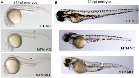
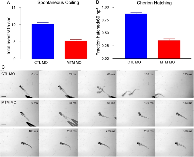
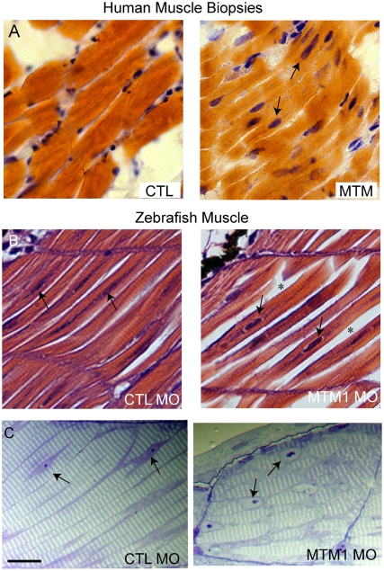
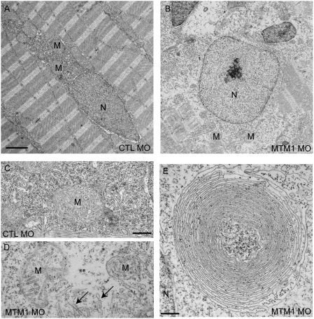
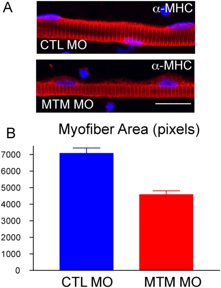
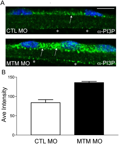
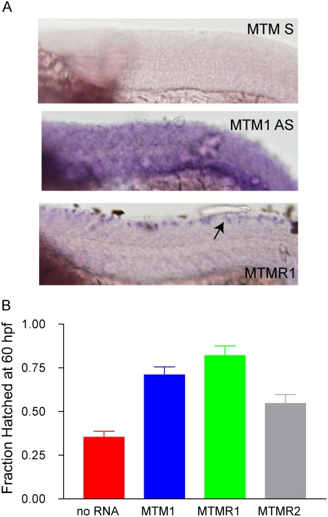
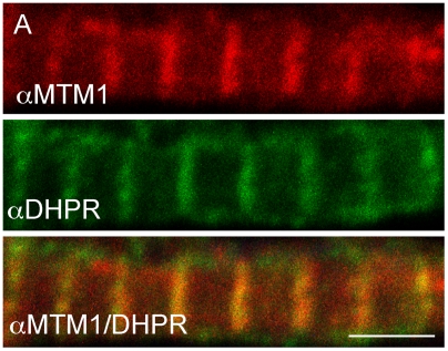
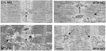
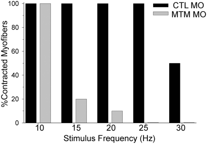
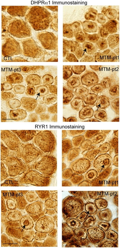
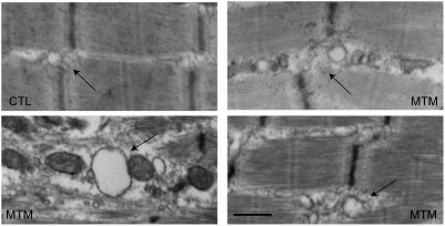
Myotubularin is a lipid phosphatase implicated in endosomal trafficking in vitro, but with an unknown function in vivo. Mutations in myotubularin cause myotubular myopathy, a devastating congenital myopathy with unclear pathogenesis and no current therapies. Myotubular myopathy was the first described of a growing list of conditions caused by mutations in proteins implicated in membrane trafficking. To advance the understanding of myotubularin function and disease pathogenesis, we have created a zebrafish model of myotubular myopathy using morpholino antisense technology. Zebrafish with reduced levels of myotubularin have significantly impaired motor function and obvious histopathologic changes in their muscle. These changes include abnormally shaped and positioned nuclei and myofiber hypotrophy. These findings are consistent with those observed in the human disease. We demonstrate for the first time that myotubularin functions to regulate PI3P levels in a vertebrate in vivo, and that homologous myotubularin-related proteins can functionally compensate for the loss of myotubularin. Finally, we identify abnormalities in the tubulo-reticular network in muscle from myotubularin zebrafish morphants and correlate these changes with abnormalities in T-tubule organization in biopsies from patients with myotubular myopathy. In all, we have generated a new model of myotubular myopathy and employed this model to uncover a novel function for myotubularin and a new pathomechanism for the human disease that may explain the weakness associated with the condition (defective excitation-contraction coupling). In addition, our findings of tubuloreticular abnormalities and defective excitation-contraction coupling mechanistically link myotubular myopathy with several other inherited muscle diseases, most notably those due to ryanodine receptor mutations. Based on our findings, we speculate that congenital myopathies, usually considered entities with similar clinical features but very disparate pathomechanisms, may at their root be disorders of calcium homeostasis.
Conflict of interest statement
The authors have declared that no competing interests exist.
Figures
Similar articles
-
Loss of catalytically inactive lipid phosphatase myotubularin-related protein 12 impairs myotubularin stability and promotes centronuclear myopathy in zebrafish.PLoS Genet. 2013 Jun;9(6):e1003583. doi: 10.1371/journal.pgen.1003583. Epub 2013 Jun 20. PLoS Genet. 2013. PMID: 23818870 Free PMC article.
-
AAV-mediated intramuscular delivery of myotubularin corrects the myotubular myopathy phenotype in targeted murine muscle and suggests a function in plasma membrane homeostasis.Hum Mol Genet. 2008 Jul 15;17(14):2132-43. doi: 10.1093/hmg/ddn112. Epub 2008 Apr 22. Hum Mol Genet. 2008. PMID: 18434328 Free PMC article.
-
Cellular, biochemical and molecular changes in muscles from patients with X-linked myotubular myopathy due to MTM1 mutations.Hum Mol Genet. 2017 Jan 15;26(2):320-332. doi: 10.1093/hmg/ddw388. Hum Mol Genet. 2017. PMID: 28007904
-
Common Pathogenic Mechanisms in Centronuclear and Myotubular Myopathies and Latest Treatment Advances.Int J Mol Sci. 2021 Oct 21;22(21):11377. doi: 10.3390/ijms222111377. Int J Mol Sci. 2021. PMID: 34768808 Free PMC article. Review.
-
X-linked myotubular myopathy.Neuromuscul Disord. 2021 Oct;31(10):1004-1012. doi: 10.1016/j.nmd.2021.08.003. Neuromuscul Disord. 2021. PMID: 34736623 Review.
Cited by
-
Gene therapy in myotubular myopathy: promising progress and future directions.Ann Transl Med. 2015 Apr;3(5):61. doi: 10.3978/j.issn.2305-5839.2015.01.11. Ann Transl Med. 2015. PMID: 25992360 Free PMC article. No abstract available.
-
Phosphoinositide regulation of integrin trafficking required for muscle attachment and maintenance.PLoS Genet. 2011 Feb 10;7(2):e1001295. doi: 10.1371/journal.pgen.1001295. PLoS Genet. 2011. PMID: 21347281 Free PMC article.
-
MTM1-mediated production of phosphatidylinositol 5-phosphate fuels the formation of podosome-like protrusions regulating myoblast fusion.Proc Natl Acad Sci U S A. 2024 Jun 4;121(23):e2217971121. doi: 10.1073/pnas.2217971121. Epub 2024 May 28. Proc Natl Acad Sci U S A. 2024. PMID: 38805272 Free PMC article.
-
Recent advances using zebrafish animal models for muscle disease drug discovery.Expert Opin Drug Discov. 2014 Sep;9(9):1033-45. doi: 10.1517/17460441.2014.927435. Epub 2014 Jun 14. Expert Opin Drug Discov. 2014. PMID: 24931439 Free PMC article. Review.
-
Modeling Human Muscular Dystrophies in Zebrafish: Mutant Lines, Transgenic Fluorescent Biosensors, and Phenotyping Assays.Int J Mol Sci. 2023 May 5;24(9):8314. doi: 10.3390/ijms24098314. Int J Mol Sci. 2023. PMID: 37176020 Free PMC article. Review.
References
-
- Pierson CR, Tomczak K, Agrawal P, Moghadaszadeh B, Beggs AH. X-linked myotubular and centronuclear myopathies. J Neuropathol Exp Neurol. 2005;64:555–564. - PubMed
-
- Dubowitz V, Sewry CA. Muscle BIopsy: A Practical Approach: Saunders. 2006:626.
-
- Laporte J, Hu LJ, Kretz C, Mandel JL, Kioschis P, et al. A gene mutated in X-linked myotubular myopathy defines a new putative tyrosine phosphatase family conserved in yeast. Nat Genet. 1996;13:175–182. - PubMed
-
- Clague MJ, Lorenzo O. The myotubularin family of lipid phosphatases. Traffic. 2005;6:1063–1069. - PubMed
Publication types
MeSH terms
Substances
Grants and funding
LinkOut - more resources
Full Text Sources
Other Literature Sources
Medical
Molecular Biology Databases
