. 2009 Feb;5(2):e1000289.
doi: 10.1371/journal.ppat.1000289. Epub 2009 Feb 6.

Phthiocerol dimycocerosates of M. tuberculosis participate in macrophage invasion by inducing changes in the organization of plasma membrane lipids

Affiliations

Phthiocerol dimycocerosates of M. tuberculosis participate in macrophage invasion by inducing changes in the organization of plasma membrane lipids

Catherine Astarie-Dequeker et al. PLoS Pathog. 2009 Feb.

Abstract

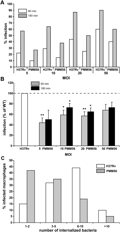
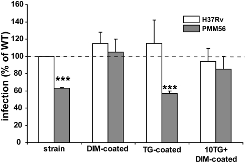
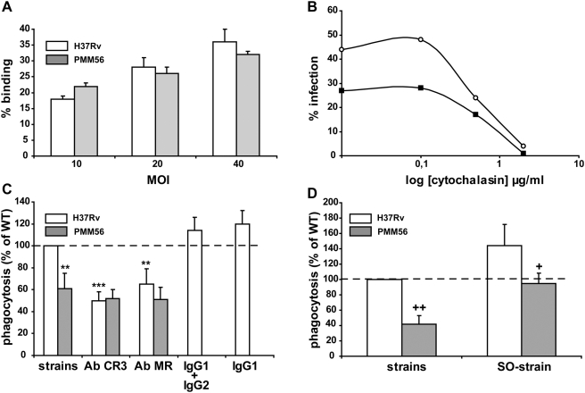
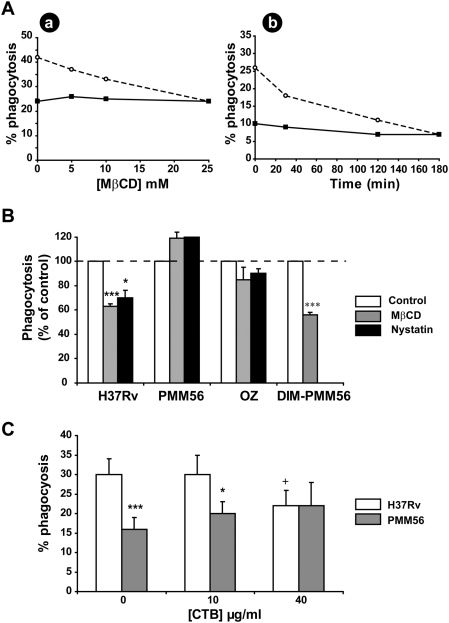
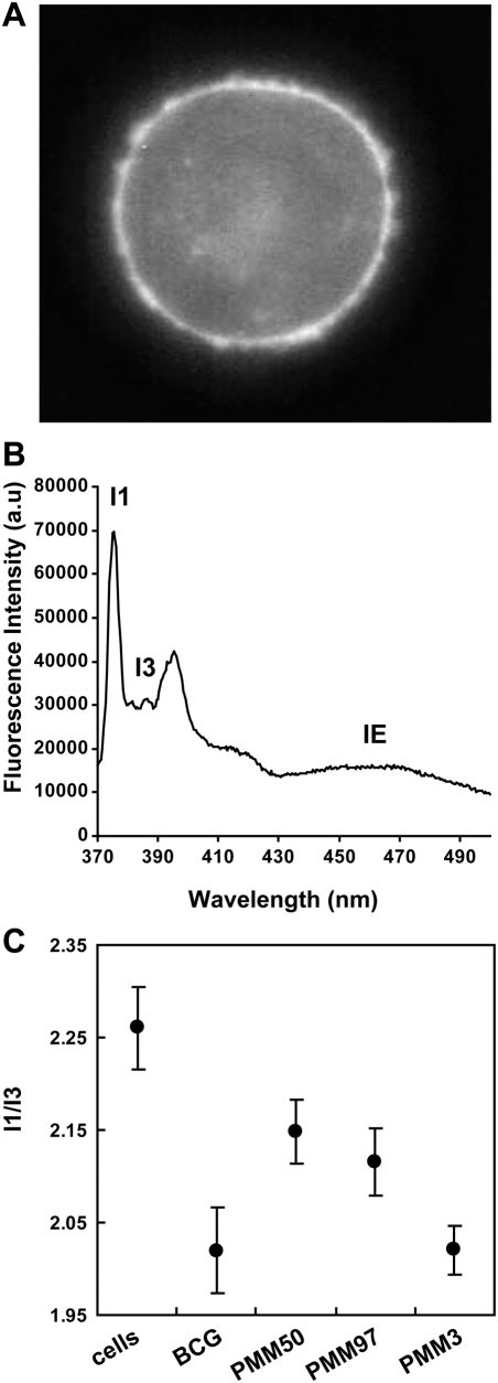
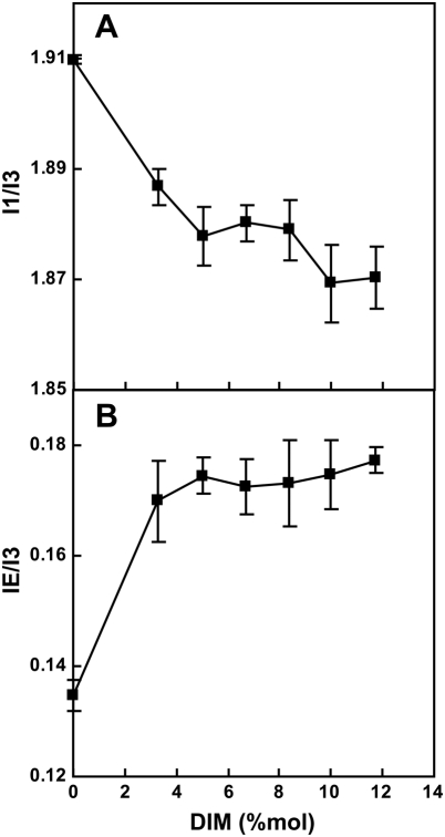
Phthiocerol dimycocerosates (DIM) are major virulence factors of Mycobacterium tuberculosis (Mtb), in particular during the early step of infection when bacilli encounter their host macrophages. However, their cellular and molecular mechanisms of action remain unknown. Using Mtb mutants deleted for genes involved in DIM biosynthesis, we demonstrated that DIM participate both in the receptor-dependent phagocytosis of Mtb and the prevention of phagosomal acidification. The effects of DIM required a state of the membrane fluidity as demonstrated by experiments conducted with cholesterol-depleting drugs that abolished the differences in phagocytosis efficiency and phagosome acidification observed between wild-type and mutant strains. The insertion of a new cholesterol-pyrene probe in living cells demonstrated that the polarity of the membrane hydrophobic core changed upon contact with Mtb whereas the lateral diffusion of cholesterol was unaffected. This effect was dependent on DIM and was consistent with the effect observed following DIM insertion in model membrane. Therefore, we propose that DIM control the invasion of macrophages by Mtb by targeting lipid organisation in the host membrane, thereby modifying its biophysical properties. The DIM-induced changes in lipid ordering favour the efficiency of receptor-mediated phagocytosis of Mtb and contribute to the control of phagosomal pH driving bacilli in a protective niche.


Conflict of interest statement

The authors have declared that no competing interests exist.

Figures

Figure 1
Figure 1. DIM are involved in the infection of human macrophages by Mtb: MDMs were incubated with H37Rv WT or a DIM-less PMM56 mutant and processed for counting by fluorescence microscopy.
(A) Percentage of macrophages having ingested at least one bacterium after 60 or 180 minutes of incubation. Data from one experiment performed in duplicate and representative of three independent experiments. (B) Percentage of macrophages infected by the PMM56 mutant expressed with respect to the percentage of macrophages infected with the WT (H37Rv, set at 100%). The values are means±SEM of three to five separate experiments performed in duplicate. The significance of differences between the mutant and the WT strain was evaluated: *, p<0.05; **, p<0.02. (C) Percentage of infected macrophages after 180 minutes of incubation as a function of the number of ingested WT or the PMM56 mutant. Data from one experiment performed in duplicate and representative of two independent experiments.
Figure 2
Figure 2. DIM contribute directly to macrophage infection by Mtb.
MDMs were incubated for 60 minutes with strains at an MOI of 10: solvent-treated H37Rv WT or the PMM56 mutant uncoated or coated with DIM or triglycerides (TG) or DIM with excess TG. Data are expressed as a percentage infection with respect to the WT (H37Rv; 100%). The values are means±SEM of three separate experiments. We evaluated the significance of difference between the WT strain and the mutant or TG-coated mutant: *** p<0.01.
Figure 3
Figure 3. DIM participate in the control of the receptor-dependent phagocytosis of H37Rv.
(A) MDMs were incubated for 30 minutes at 4°C with the WT or the PMM56 mutant, at various MOI, and binding was determined by assessing immunofluorescence. Data are presented as the percentage of macrophages having bound at least one bacterium; one experiment that is representative of two independent experiments. (B) MDMs were incubated at 37°C for 30 minutes with various concentrations of cytochalasin and were then infected with either H37Rv WT (white circle) or the PMM56 mutant (black square) at an MOI of 10. Data are presented as percentage phagocytosis; one experiment representative of two independent experiments. (C) MDMs were incubated at 37°C for 30 minutes with irrelevant IgG1 or a cocktail of IgG1 and IgG2 or with mAbs directed against CR3 or MR. They were then washed and infected for 60 minutes with H37Rv WT or the PMM56 mutant at an MOI of 10. Data are presented as the percentage of phagocytosis compared to WT value (100% H37Rv). (D) MDMs were infected for 60 minutes with serum-opsonized (SO-strain) or non opsonized strains H37Rv WT or the PMM56 mutant at an MOI of 10. Data are presented as the percentage phagocytosis with respect to WT (H37Rv; 100%). The values presented are means±SEM of three independent experiments. The significance of difference between mAb-treated cell and untreated cells was evaluated: *, p<0.05; **, p<0.02. We also evaluated the significance of difference between mutants and the WT strain: +, p<0.05; ++, p<0.01.
Figure 4
Figure 4. The DIM-dependent phagocytosis of Mtb required plasma membrane cholesterol.
(A) MDMs were initially incubated at 37°C with (a) various concentrations of MβCD for 30 minutes or (b) 10 mM MβCD for various periods of time. They were then washed and infected for 60 minutes with H37Rv WT (white circle) or the PMM56 mutant at an MOI of 10 (black square); one experiment representative of three separate experiments. (B) MDMs left untreated or treated with 10 mM MβCD or 20 µg/ml Nystatin for 30 minutes were infected with H37Rv WT or the PMM56 mutant or the DIM-coated PMM56 mutant at an MOI of 10 for 60 minutes or incubated with the serum-opsonized zymosan (OZ) at an MOI of 25 for 45 minutes. The data are expressed as a percentage phagocytosis with respect to untreated conditions (control; 100%). The values presented are means±SEM for three to four separate experiments. We evaluated the significance between cell subjected to drug pretreatment and untreated cells: *, p<0.05; ***, p<0.01. (C) MDMs were left untreated or treated with various concentrations of CTB for 30 minutes. They were washed and infected for 60 minutes with H37Rv WT or the PMM56 mutant at an MOI of 10. The data are presented as the percentage of phagocytosis. The values are means±SEM for three separate experiments. The significance of the drug effect was assessed by comparing data under treated and untreated conditions: +, p<0.05; the significance of the effect of DIM deficiency was assessed by comparing data for mutants and the WT strain.*, p<0.05, ***p<0.01.
Figure 5
Figure 5. H37Rv associates weakly with plasma membrane domains enriched in GM1 gangliosides.
H37Rv expressing GFP (green fluorescence) were allowed to adhere to MDMs for 30 minutes at 4°C. Cells were then stained with Alexa-647 CTB (blue fluorescence) and processed for analysis by confocal microscopy. Representative single optical x-y planes from confocal microscopy images are shown. (A) Colocalization of GFP-H37Rv and Alexa-647 CTB at the plasma membrane gives a turquoise signal. (B–C) Two examples of an absence of colocalization between GFP-H37Rv and Alexa-647 CTB at the plasma membrane. The data presented are from a single experiment representative of two. Bar: 12 µm.
Figure 6
Figure 6. DIM deficiency induces acidification of Mtb-containing phagosome in a cholesterol-dependent manner.
MDMs were infected for 60 minutes with heat-killed (triangles) or live H37Rv WT (diamonds) or the PMM56 mutant (square) at MOI 10, washed, and further incubated in the presence of serum. At various times after infection, MDMs were processed. (A,B,E) macrophages were added with LysoTracker, fixed, and processed for analysis by microscopy. LysoTracker-positive phagosomes were quantified over a period of 192 h (A) or 2 h after infection with H37Rv, PMM56, and PMM100 DIM-less mutants, or the complemented strain PMM100pMVE (B), or left untreated or treated with 10 mM MβCD before infection (E). (C–D) MDMs were fixed, permeabilized, and immunostained with (C) polyclonal anti-serum against H+-ATPase or (D) mAb against CD63 and processed for fluorescence microscopy and quantification. (C) A representative micrograph of cells analysed by confocal microscopy. Colocalization of H37Rv and H+-ATPase gave a yellow signal. Arrowheads indicated phagosome having occasionally accumulated H+-ATPase in MDMs infected by H37Rv. Bar: 34 µm. The percentage of phagosomes positive for each marker was determined by counting 100 phagosomes from at least ten different fields in duplicate samples. Data are means±SEM of two to four experiments. The significance of the effect was assessed by comparing data for mutants and the WT strain. **, p<0.01, or for the complemented strain and the unmarked mutant. +, p<0.05.
Figure 7
Figure 7. DIM contributes to the decrease in membrane polarity of THP-1 during the interaction with M. bovis BCG.
(A) Py-met-chol was incorporated into the plasma membrane of THP-1, and cells were examined (A) by microscopy on a Zeiss Axioplan II using a DAPI Filter or (B) by spectrofluorimetry. (C) The I 1/I 3 fluorescence ratios were determined in THP-1 cells alone (n = 25) or after incubation of the cells for 30 minutes at room temperature with M. bovis BCG (n = 18), DIM-less mutants (PMM50, n = 15; PMM97, n = 11), or a PGL-less mutant (PMM3, n = 10), at an MOI of 10. Data are means±SD.
Figure 8
Figure 8. Insertion of DIM into model membranes decreases the polarity of the bilayer core by modifying membrane structure.
DIM were incorporated into LUVs containing 1 mol% of Py-met-chol. Changes in the I 1/I 3 (A) and I E/I 3 (B) fluorescence ratios of Py-met-chol in LUVs were examined as a function of DIM mole fraction. Measurements were performed in the fluid state, at 45°C. Data are means±SD of three independent experiments.

References

    1. Daffe M, Draper P. The envelope layers of mycobacteria with reference to their pathogenicity. Adv Microb Physiol. 1998;39:131–203. - PubMed
    1. Andersen R. The chemistry of lipids of tubercle bacilli. Harvey Lect. 1940;35:271–313.
    1. Daffe M, Laneelle MA. Distribution of phthiocerol diester, phenolic mycosides and related compounds in mycobacteria. J Gen Microbiol. 1988;134:2049–2055. - PubMed
    1. Ortalo-Magne A, Lemassu A, Laneelle MA, Bardou F, Silve G, et al. Identification of the surface-exposed lipids on the cell envelopes of Mycobacterium tuberculosis and other mycobacterial species. J Bacteriol. 1996;178:456–461. - PMC - PubMed
    1. Brennan PJ, Nikaido H. The envelope of mycobacteria. Annu Rev Biochem. 1995;64:29–63. - PubMed

Publication types

MeSH terms

LinkOut - more resources