

Roles for transforming growth factor beta superfamily proteins in early folliculogenesis
- PMID: 19197801
- PMCID: PMC2947191
- DOI: 10.1055/s-0028-1108006
Roles for transforming growth factor beta superfamily proteins in early folliculogenesis
Abstract
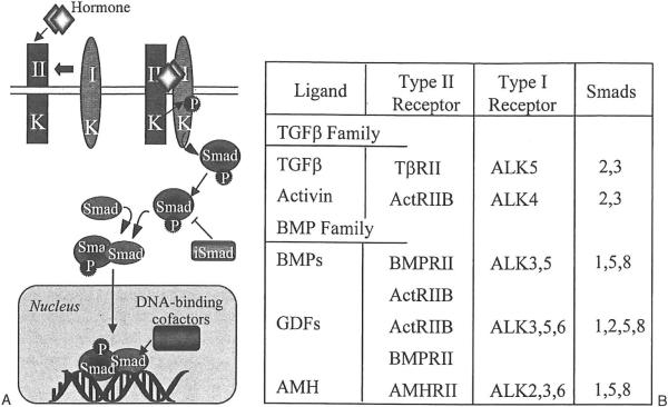
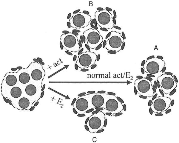
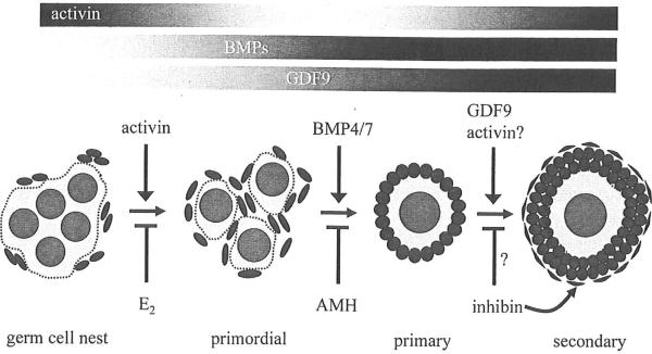
Primordial follicle formation and the subsequent transition of follicles to the primary and secondary stages encompass the early events during folliculogenesis in mammals. These processes establish the ovarian follicle pool and prime follicles for entry into subsequent growth phases during the reproductive cycle. Perturbations during follicle formation can affect the size of the primordial follicle pool significantly, and alterations in follicle transition can cause follicles to arrest at immature stages or result in premature depletion of the follicle reserve. Determining the molecular events that regulate primordial follicle formation and early follicle growth may lead to the development of new fertility treatments. Over the last decade, many of the growth factors and signaling proteins that mediate the early stages of folliculogenesis have been identified using mouse genetic models, in vivo injection studies, and ex vivo organ culture approaches. These studies reveal important roles for the transforming growth factor beta (TGF-beta) superfamily of proteins in the ovary. This article reviews these roles for TGF-beta family proteins and focuses in particular on work from our laboratories on the functions of activin in early folliculogenesis.
Figures
References
-
- Molyneaux KA, Stallock J, Schaible K, Wylie C. Time-lapse analysis of living mouse germ cell migration. Dev Biol. 2001;240(2):488–498. - PubMed
-
- Pepling ME, Spradling AC. Female mouse germ cells form synchronously dividing cysts. Development. 1998;125(17):3323–3328. - PubMed
-
- Pepling ME, Spradling AC. Mouse ovarian germ cell cysts undergo programmed breakdown to form primordial follicles. Dev Biol. 2001;234(2):339–351. - PubMed
-
- Van Wagenen G, Simpson ME. Embryology of the ovary and testis: Homo sapiens and Macaca mulatta. Yale University Press; New Haven, CT: 1965.
-
- McGee EA, Hsueh AJ. Initial and cyclic recruitment of ovarian follicles. Endocr Rev. 2000;21(2):200–214. - PubMed
