. 2009 Mar;39(3):902-11.
doi: 10.1002/eji.200838780.

Severe functional impairment and elevated PD-1 expression in CD1d-restricted NKT cells retained during chronic HIV-1 infection

Affiliations

Severe functional impairment and elevated PD-1 expression in CD1d-restricted NKT cells retained during chronic HIV-1 infection

Markus Moll et al. Eur J Immunol. 2009 Mar.

Abstract

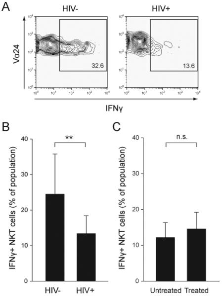
Invariant CD1d-restricted NKT cells play important roles in regulating both innate and adaptive immunity. They are targeted by HIV-1 infection and severely reduced in number or even lost in many infected subjects. Here, we have investigated the characteristics of NKT cells retained by some patients despite chronic HIV-1 infection. NKT cells preserved under these circumstances displayed an impaired ability to proliferate and produce IFN-gamma in response to CD1d-restricted lipid antigen as compared with cells from uninfected control subjects. HIV-1 infection was associated with an elevated expression of the inhibitory programmed death-1 (PD-1) receptor (CD279) on the CD4(-) subset of NKT cells. However, blocking experiments indicated that the functional defects in NKT cells were largely PD-1-independent. Furthermore, the elevated PD-1 expression and the functional defects were not restored by anti-retroviral treatment, and the NKT cell numbers in blood did not recover significantly in response to treatment. The functional phenotype of NKT cells in these patients suggests an irreversible immune exhaustion due to chronic activation in vivo. The data demonstrate a severe functional impairment in the remaining NKT-cell compartment in HIV-1-infected patients, which limits the prospects to mobilize these cells in immunotherapy approaches in patients.


Figures

Figure 1
Figure 1
Some HIV-1 infected patients have NKT cells that persist over time irrespective of ART. (A) Identification of CD1d-restricted NKT cells in peripheral blood. CD1d DimerX-αGalCer binding NKT cells were identified among CD3+ cells by co-expression of the T cell receptor chains Vα24 and Vβ11 as determined by flow cytometry. (B) Longitudinal measurement of NKT cells counts (cells/μl), CD4+ T cell counts (cells/μl) and viral load (copies/ml, horizontal line indicates the limit of detection) in (Group 1, n=9) untreated patients with ongoing viral replication; (Group 2, n=9) patients on ART with no detectable virus; (Group 3, n=10) patients on ART with unstable control of virus; and (Group 4, n=7) patients starting ART after the first blood sample was drawn. Patients with NKT cell counts above the limit of detection are indicated in red. The vertical line in panel (Group 4) indicates the start of treatment. *** indicates P < 0.001, and n.s. indicates non-significant as determined by the paired T-test.
Figure 2
Figure 2
Poor proliferative capacity in NKT cells from HIV-1 infected subjects. The proliferative capacity of NKT cells was assessed by culturing PBMC in triplicates in the presence of antigen (αGalCer, 100 ng/ml), IL-2 (10 ng/ml) and the anti-retroviral drug AZT (10 μM). NKT cell frequencies were determined by FACS analysis at day 0 and 13, and the average fold expansion was calculated for each donor. (A-B) Comparison of the proliferative capacity of NKT cells and CD4- and CD4+ NKT cell subsets from healthy donors (HIV-, n=13) and HIV-1 infected subjects (HIV+, n=21), as well as untreated (n=9) and treated patients (n=17), respectively. NKT cell numbers were assessed before (d0) and after culture for 13 days (d13), and the fold expansion of NKT cells was calculated. (C) Proportion of CD4+ NKT cells before (d0) and after culture for 13 days (d13). Data are shown as mean ± SD. * indicates P < 0.05, *** indicates P < 0.001, and n.s. indicates non-significant as determined by the Mann-Whitney Rank Sum Test (A) and paired T-test (C).
Figure 3
Figure 3
Impaired IFNγ production in residual NKT cells in HIV-1 infected patients. The production of IFNγ in NKT cells was measured at the single cell level by intracellular staining and FACS analysis after stimulation with αGalCer. (A) Representative contour plots showing the expression of IFNγ in NKT cells from a healthy donor (HIV-) and a HIV-1 infected patient (HIV+). (B-C) graphs showing mean percentages of IFNγ-positive NKT cells ± SD from HIV- (n=12) and HIV+ (n=11) subjects, and untreated (n=5) and treated patients (n=9), respectively. Data shown are mean ± SD. ** indicates P < 0.01, and n.s. indicates non-significant as determined by the T-test.
Figure 4
Figure 4
Elevated PD-1 expression in CD4- NKT cells in HIV-1 infected subjects. (A) PD-1 expression was determined in NKT cells and T cells from HIV- and HIV+ subjects by flow cytometry. Representative contour plots including an isotype control are shown. (B) Percentage of PD-1 expressing NKT cells in healthy controls (HIV-, n=12) and HIV-1 infected patients (HIV+, n=11). (C) PD-1+ NKT cells and T cells in HIV+ subjects. (D) Graphs comparing the percentage of PD-1 positive cells in HIV- and HIV+ subjects in CD4- and CD4+ NKT cell subpopulations. (E) Graphs comparing the proliferative capacity of NKT cells and CD4- and CD4+ NKT cell subsets from HIV-1 infected subjects (n=11) in the presence of anti-PD-L1 plus anti-PD-L2 antibodies or isotype control. (F) Expression of IFNγ in NKT cells from HIV-1 infected patients (n=6) in the presence and absence of anti-PD-L1 plus anti-PD-L2 antibodies. Data are shown as mean ± SD. * indicates P < 0.05, ** indicates P < 0.01, *** indicates P < 0.001, and n.s. indicates non-significant as determined by T-test, or alternatively the Mann-Whitney Rank Sum Test in case data failed the normality test.

References

    1. Godfrey DI, Kronenberg M. Going both ways: immune regulation via CD1d-dependent NKT cells. J. Clin. Invest. 2004;114:1379–1388. - PMC - PubMed
    1. Sandberg JK, Ljunggren HG. Development and function of CD1d-restricted NKT cells: influence of sphingolipids, SAP and sex. Trends Immunol. 2005;26:347–349. - PubMed
    1. Fujii S, Shimizu K, Hemmi H, Steinman RM. Innate Valpha14+ natural killer T cells mature dendritic cells, leading to strong adaptive immunity. Immunol. Rev. 2007;220:183–198. - PubMed
    1. Dougan SK, Kaser A, Blumberg RS. CD1 expression on antigen-presenting cells. Curr. Top. Microbiol. Immunol. 2007;314:113–141. - PubMed
    1. Stronge VS, Salio M, Jones EY, Cerundolo V. A closer look at CD1d molecules: new horizons in studying NKT cells. Trends Immunol. 2007;28:455–462. - PubMed

Publication types

MeSH terms