. 2009 Apr 1;115(7):1363-80.
doi: 10.1002/cncr.24156.

Significance of breast lesion descriptors in the ACR BI-RADS MRI lexicon

Affiliations

Significance of breast lesion descriptors in the ACR BI-RADS MRI lexicon

Garima Agrawal et al. Cancer. .

Abstract

In recent years, dynamic contrast-enhanced magnetic resonance imaging (DCE-MRI) has altered the clinical management for women with breast cancer. In March 2007, the American Cancer Society (ACS) issued a new guideline recommending annual MRI screening for high-risk women. This guideline is expected to substantially increase the number of women each year who receive breast MRI. The diagnosis of breast MRI involves the description of morphological and enhancement kinetics features. To standardize the communication language, the Breast Imaging Reporting and Data System (BI-RADS) MRI lexicon was developed by the American College of Radiology (ACR). In this article, the authors will review various appearances of breast lesions on MRI by using the standardized terms of the ACR BI-RADS MRI lexicon. The purpose is to familiarize all medical professionals with the breast MRI lexicon because the use of this imaging modality is rapidly growing in the field of breast disease. By using this common language, a comprehensive analysis of both morphological and kinetic features used in image interpretation will help radiologists and other clinicians to communicate more clearly and consistently. This may, in turn, help physicians and patients to jointly select an appropriate management protocol for each patient's clinical situation.


Figures

Figure 1
Figure 1
Depending on the dominant morphological features, lesions can be separated into focus/foci, mass, and non-mass-like enhancements. A focus can not be further characterized due to the small size. A mass is characterized by shape, margin, and the internal enhancement characteristics. A non-mass-like enhancement is characterized by the overall distribution pattern of enhancement, and further by the internal enhancement characteristics, and whether the enhancement is symmetric or asymmetric between both breasts.
Figure 2
Figure 2
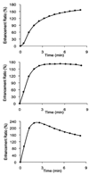
The descriptor for the enhancement kinetics is based on two different phases: the initial phase and the delayed phase. The kinetics showing rapid initial enhancement followed by wash-out or plateau is suspicious of malignancy; while that showing medium or slow initial enhancement followed by persistent enhancement is more likely benign.
Figure 3
Figure 3
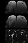
Fibroadenoma in a 30 year-old (y/o) patient. (A) Pre-contrast, axial view T1 weighted image showing a well defined lobulated mass with smooth margin; (B) Contrast-enhanced T1 weighted image at 1.5 minute showing mild enhancement, suggesting a benign lesion. (C) Contrast-enhanced subtraction image clearly showing the enhancement of the lesion.
Figure 4
Figure 4
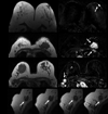
(Left) pre-contrast axial view T1-weighted images, (Right) contrast-enhanced subtraction images. (A) Invasive ductal carcinoma in a 68 y/o patient. Note a round shaped mass with well defined and smooth margin showing strong enhancement; (B) Invasive lobular carcinoma in a 71 y/o patient. A lobulated mass in the right breast showing heterogeneous enhancement; (C) Invasive ductal carcinoma in a 47 y/o patient. Note an irregular shaped mass with spiculated margin and rim enhancement.
Figure 5
Figure 5
(Left) pre-contrast axial T1-weighted images showing hypointense foci; (Right) contrast-enhanced subtraction images showing strong enhancement. (A) Focal fibrocystic changes of the right breast in a 60 y/o patient. Note a single enhancing focus in the right breast; (B) Fibroadenoma in a 62 y/o patient; (C) Intermediate grade pure DCIS in a 79 y/o patient; (D) Invasive ductal carcinoma in a 52 y/o patient. All arrows indicate lesions.
Figure 6
Figure 6
(Left) pre-contrast axial view T1-weighted images, (Right) contrast-enhanced subtraction images. (A) Invasive mammary carcinoma with ductal and lobular features in a 33 y/o patient. Note the linear band-like enhancement in right breast not conforming to ductal structure; (B) Invasive lobular carcinoma with focal lobular carcinoma in situ in a 43 y/o patient. Note ductal clumped pattern of enhancement in the left breast. (C) Invasive ductal carcinoma with multiple foci of ductal carcinoma in situ along with lymphatic invasion of nipple areolar complex in 55 y/o patient. Note the ductal branching pattern of non mass type enhancement merging towards nipple with skin thickening and enhancement. All arrows indicate lesions.
Figure 7
Figure 7
(Left) pre-contrast axial view T1-weighted images, (Right) contrast-enhanced subtraction images. (A) Ductal carcinoma in situ in a 51 y/o patient. Note the segmental clumped pattern of enhancement in the central third aspect of the right breast; (B) Invasive lobular carcinoma in a 50 y/o patient. Note the segmental pattern of the lesion toward the nipple in the left breast; (C) Invasive lobular carcinoma in a 55 y/o patient. Note the spiculated mass in the right breast with linear enhancement extending to the nipple. All arrows indicate lesions.
Figure 8
Figure 8
(Left) pre-contrast axial view T1-weighted images, (Right) contrast-enhanced subtraction images. (A) Fibrocystic change in a 31 y/o patient. Note a patch of faint regional enhancement in the right breast; (B) Invasive ductal carcinoma in the left breast in a 51 y/o patient. Note a regional area of clumped enhancement in the left breast. All arrows indicate lesions.
Figure 9
Figure 9
(Left) pre-contrast axial view T1-weighted images, (Right) contrast-enhanced subtraction images. (A) Invasive ductal carcinoma with skin invasion in a 36 y/o patient. Note the diffuse non mass type enhancement of left breast with involvement of skin as evident by skin thickening and bright enhancement; (B) Extensive ductal carcinoma in-situ with multicentric invasive ductal carcinoma in a 49 y/o patient. Note a large regional non-mass type area of clumped enhancement in the right breast; (C) Bilateral fibrocystic changes in a 45 y/o patient. Note symmetrical diffuse enhancement in the both breast. All arrows indicate lesions.
Figure 10
Figure 10
(A–C, left) pre-contrast T1 weighted axial view image, (A–C, right) contrast-enhanced subtraction images. (A) Paget’s disease of the left nipple with underlying high grade DCIS in a 62 y/o patient. Contrast-enhanced subtraction image shows an asymmetric enlarged left nipple with strong enhancement, which extends to the sub-areolar area; (B) Invasive ductal carcinoma with pectoris muscle invasion in a 50 y/o patient. Note the enhancement of the tumor and the involved muscle in the left breast; (C) Invasive ductal carcinoma with axillary lymph nodes in a 39 y/o patient. Note several round shape hypointense lymph nodes in the right axillary area. These lymph nodes show strong enhancements; (D) same patient as (C), showing lymph nodes on pre-contrast sagittal view T1-weighted images with a smaller field of view. All arrows indicate lesions.
Figure 11
Figure 11
Representative enhancement kinetic curves from three different patients. (A) moderate initial enhancement followed by a persistent enhancement in a fibrocystic change shown in Figure 8A; (B) rapid initial enhancement reaching to a plateau in an invasive lobular cancer shown in Figure 7C; (C) rapid initial enhancement followed by washout in an invasive ductal carcinoma shown in Figure 5D.

References

    1. Much wider use of M.R.I.’s urged for breast exam, Health. The New York Times, By Denise Grady Published; 2007. Mar 28,
    1. Saslow D, Boetes C, Burke W, et al. American Cancer Society Breast Cancer Advisory Group. American Cancer Society guidelines for breast screening with MRI as an adjunct to mammography. CA Cancer J Clin. 2007 Mar–Apr;57(2):75–89. - PubMed
    1. Tillman GF, Orel SG, Schnall MD, et al. Effect of breast magnetic resonance imaging on the clinical management of women with early-stage breast carcinoma. J Clin Oncol. 2002 Aug 15;20(16):3413–3423. - PubMed
    1. Bedrosian I, Mick R, Orel SG, et al. Changes in the surgical management of patients with breast carcinoma based on preoperative magnetic resonance imaging. Cancer. 2003 Aug 1;98(3):468–473. - PubMed
    1. Hylton N. Magnetic resonance imaging of the breast: opportunities to improve breast cancer management. J Clin Oncol. 2005 Mar 10;23(8):1678–1684. - PubMed

Publication types