. 2009 May;50(4):317-27.
doi: 10.1002/em.20462.

Requirement of the Saccharomyces cerevisiae APN1 gene for the repair of mitochondrial DNA alkylation damage

Affiliations

Requirement of the Saccharomyces cerevisiae APN1 gene for the repair of mitochondrial DNA alkylation damage

Karina Acevedo-Torres et al. Environ Mol Mutagen. 2009 May.

Abstract

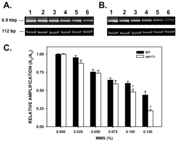
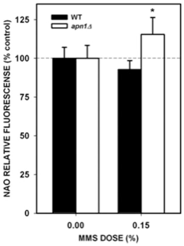
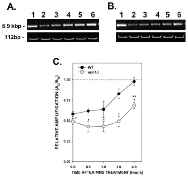
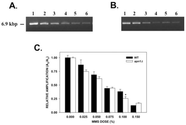
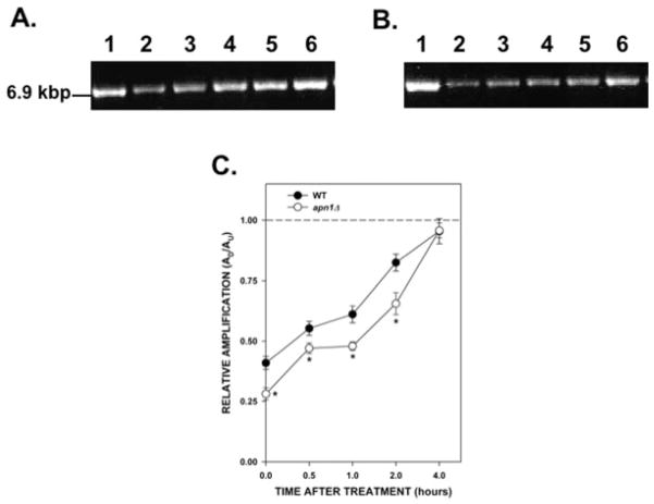
The Saccharomyces cerevisiae APN1 gene that participates in base excision repair has been localized both in the nucleus and the mitochondria. APN1 deficient cells (apn1 Delta) show increased mutation frequencies in mitochondrial DNA (mtDNA) suggesting that APN1 is also important for mtDNA stability. To understand APN1-dependent mtDNA repair processes we studied the formation and repair of mtDNA lesions in cells exposed to methyl methanesulfonate (MMS). We show that MMS induces mtDNA damage in a dose-dependent fashion and that deletion of the APN1 gene enhances the susceptibility of mtDNA to MMS. Repair kinetic experiments demonstrate that in wild-type cells (WT) it takes 4 hr to repair the damage induced by 0.1% MMS, whereas in the apn1 Delta strain there is a lag in mtDNA repair that results in significant differences in the repair capacity between the two yeast strains. Analysis of lesions in nuclear DNA (nDNA) after treatment with 0.1% MMS shows a significant difference in the amount of nDNA lesions between WT and apn1 Delta cells. Interestingly, comparisons between nDNA and mtDNA damage show that nDNA is more sensitive to the effects of MMS treatment. However, both strains are able to repair the nDNA lesions, contrary to mtDNA repair, which is compromised in the apn1 Delta mutant strain. Therefore, although nDNA is more sensitive than mtDNA to the effects of MMS, deletion of APN1 has a stronger phenotype in mtDNA repair than in nDNA. These results highlight the prominent role of APN1 in the repair of environmentally induced mtDNA damage.


Figures

Fig. 1
Fig. 1
Mitochondrial DNA damage induced by MMS. Yeast cells (A, wild type; B, apn1Δ mutant) were treated with increasing concentrations of MMS for 20 min, followed by DNA isolation. QPCR analysis was performed as described in the “Materials and Methods” section. (A, B) Representative gel electrophoresis indicating the expected sizes of the large (6.9 kb) and small (112 bp) mitochondrial DNA fragments. Lanes 1–6: PCR products from yeast cells treated with 0, 0.025, 0.05, 0.075, 0.1, 0.15% MMS, respectively. (C) Relative levels of amplification of the 6.9-kb mitochondrial fragment after normalization with the small mtDNA fragment (n = 3 independent experiments performed in triplicate). Asterisks (*) denote statistical significance (P < 0.05). Error bars represent SEM.
Fig. 2
Fig. 2
Genomic DNA integrity obtained from cells treated with MMS. DNA (0.75 μg) isolated from WT and apn1Δ cells after MMS treatment was analyzed by native and alkaline agarose gel electrophoresis. (A) Native agarose gel electrophoresis. Samples were loaded according to increasing MMS dosage. (B) Alkaline agarose gel electrophoresis. Samples were loaded as in A. The ratio of the intensities of the high-molecular-weight DNA band to the low-molecular-weight DNA smear was determined as described in the “Materials and Methods” section. MW: molecular weight marker containing λ DNA digested with Hind III. DNA from the untreated WT or apn1Δ strains was digested with Bgl I/Hind III and included as low-molecular-weight DNA controls.
Fig. 3
Fig. 3
Estimation of mitochondrial mass after MMS treatment. Yeast cells were treated with 0.15% MMS for 20 min. After MMS treatment, cells were loaded with NAO and the fluorescence intensity was determined as described in the “Materials and Methods” section. The values represent the result of three independent experiments performed in triplicate. Asterisks (*) denote statistical significance (P < 0.05). Error bars represent SEM.
Fig. 4
Fig. 4
Kinetics of repair of mtDNA damage induced by MMS. Yeast cells were treated with 0.1% MMS for 20 min. After MMS inactivation, cells were washed with water, resuspended in fresh YPD media, and incubated for up to 4 hr at 30°C. Aliquots of cells were removed at the indicated times. DNA isolation and QPCR were performed as described. (A) Wild type; (B) apn1Δ mutant. (A, B) Representative gel electrophoresis indicating the expected sizes of the mtDNA fragments. Lane 1: PCR products from untreated yeast cells; lanes 2–6: PCR products from yeast cells after 0, 0.5, 1, 2, and 4 hr after MMS, respectively. (C) Relative levels of amplification of the 6.9-kb mitochondrial fragment after normalization with the small mtDNA fragment (n = 2 independent experiments performed in triplicate). Asterisks (*) denote statistical significance (P < 0.05) between WT (closed circles) and apn1Δ (open circles) cells. Double asterisk (**) denotes statistical significance in the apn1Δ cells between the 0 and 4 hr after treatment time points. Error bars represent SEM.
Fig. 5
Fig. 5
nDNA damage induced by MMS. Relative levels of amplification of a 6.9-kb nDNA fragment (from the PFK2 gene) in yeast cells treated with MMS. Yeast cells (A, wild type; B, apn1Δ mutant) were treated with increasing concentrations of MMS for 20 min, followed by DNA isolation. QPCR analysis was performed as described in the “Materials and Methods” section. (A, B) Representative gel electrophoresis indicating the expected sizes of the 6.9-kb nDNA fragment. Lanes 1–6: PCR products from yeast cells treated with 0, 0.025, 0.05, 0.075, 0.1, 0.15% MMS, respectively; (C) Relative levels of amplification of the 6.9-kb nDNA fragment (n = 3 independent experiments performed in triplicate). Asterisks (*) denote statistical significance (P < 0.05). Error bars represent SEM.
Fig. 6
Fig. 6
Kinetics of repair of nDNA damage induced by MMS. Yeast cells were treated with 0.1% MMS for 20 min. Cells were washed with water, resuspended in fresh YPD media, and incubated for up to 4 hr at 30°C. Aliquots of cells were removed at the indicated times. DNA isolation and QPCR were performed as described. (A) Wild type; (B) apn1Δ mutant. (A, B) Representative gel electrophoresis indicating the expected sizes of the nDNA fragments. Lane 1: PCR products from untreated yeast cells; lanes 2–6: PCR products from yeast cells after 0, 0.5, 1, 2, and 4 hr after MMS, respectively. (C) Relative levels of amplification of the 6.9-kb nDNA fragment. The dashed line represents the amplification of untreated cells, which was set to a value of 1.0. Results are from two independent experiments with each PCR performed in triplicate. Asterisks (*) denote statistical significance (P < 0.05) between WT (closed circles) and apn1Δ cells (open circles). Error bars represent SEM.

References

    1. Alseth I, Korvald H, Osman F, Seeberg E, Bjoras M. A general role of the DNA glycosylase Nth1 in the abasic sites cleavage step of base excision repair in Schizosaccharomyces pombe. Nucleic Acids Res. 2004;32:5119–5125. - PMC - PubMed
    1. Ayala-Torres S, Chen Y, Svoboda T, Rosenblatt J, Van Houten B. Analysis of gene-specific DNA damage and repair using quantitative polymerase chain reaction. Methods. 2000;22:135–147. - PubMed
    1. Bohr VA. Repair of oxidative DNA damage in nuclear and mitochondrial DNA, some changes with aging in mammalian cells. Free Radic Biol Med. 2002;32:804–812. - PubMed
    1. Cai S, Xu Y, Cooper RJ, Ferkowicz MJ, Hartwell JR, Pollok KE, Kelley MR. Mitochondrial targeting of human O6-methylguanine DNA methyltransferase protects against cell killing by chemotherapeutic alkylating agents. Cancer Res. 2005;65:3319–3327. - PubMed
    1. Chattopadhyay R, Wiederhold L, Szczesny B, Boldogh I, Hazra TK, Izumi T, Mitra S. Identification and characterization of mitochondrial abasic (AP)-endonuclease in mammalian cells. Nucleic Acids Res. 2006;34:2067–2076. - PMC - PubMed

Publication types

MeSH terms