

doi: 10.1038/nmeth.1300.
Epub 2009 Feb 8.
Nanomole-scale protein solid-state NMR by breaking intrinsic 1HT1 boundaries
Affiliations
- PMID: 19198596
- PMCID: PMC2649701
- DOI: 10.1038/nmeth.1300
Item in Clipboard
Nanomole-scale protein solid-state NMR by breaking intrinsic 1HT1 boundaries
Nat Methods.
2009 Mar.
Abstract
We present an approach that accelerates protein solid-state NMR 5-20-fold using paramagnetic doping to condense data-collection time (to approximately 0.2 s per scan), overcoming a long-standing limitation on slow recycling owing to intrinsic (1)H T(1) longitudinal spin relaxation. Using low-power schemes under magic-angle spinning at 40 kHz, we obtained two-dimensional (13)C-(13)C and (13)C-(15)N solid-state NMR spectra for several to tens of nanomoles of beta-amyloid fibrils and ubiquitin in 1-2 d.
Figures
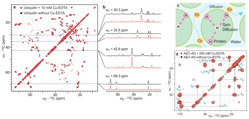
The effectiveness of the PACC approach for hydrated proteins. (a) Comparison of superimposed 2D 13C/13C chemical-shift correlation SSNMR spectra of uniformly 13C-labeled ubiquitin (1.8 mg) in microcrystals in the (black) absence and (red) presence of 10 mM Cu(II)-EDTA respectively obtained in standard and PACC approaches at a 1H frequency of 400.2 MHz, together with (b) slices at selected positions. (c) A proposed mechanism of paramagnetic 1H T1 relaxation enhancement for hydrated protein microcrystals by Cu(II)-EDTA doping. (d) Comparison of 2D 13C/13C SSNMR spectra of fibrillized 13C- and 15N-labeled 40-residue Alzheimer’s β-amyloid (Aβ(1–40); 1.4 mg) that were obtained in the (red) presence and (black) absence of 200 mM Cu-EDTA. All the experiments in Fig. 1 were performed at a spinning speed of 40 kHz with signal acquisitions under low-power TPPM decoupling at rf-fields of 7 kHz. The mixing time for the fpRFDR sequence was 1.6 ms. See Supplementary Methods and Supplementary Figs. 1, 2, 6 online for sample preparations, signal assignments for ubiquitin, pulse sequences, and other details.

2D chemical-shift correlation SSNMR spectra for mass-limited systems by the PACC approach. (a) A 2D 13C/13C correlation SSNMR spectrum obtained by PACC for fibrillized uniformly 13C- and 15N-labeled 42-residue Alzheimer’s β-amyloid (Aβ) peptide (0.4 mg or 80 nmol) doped with 200 mM Cu-EDTA, together with preliminary assignments based on amino-acid types for 13Cα/13Cβ cross peaks (dotted circles) and 13Cα/13CO (dotted squares). The recycle delays were set to 180 ms, which is approximately three times the 1H T1 value (~ 60 ms). The presence of strong cross peaks for Ala-42 (orange arrows) suggest the ordered structure at the C-terminal of Aβ(1–42). (b) Nano-mole scale analysis by 2D 13CO/15N correlation SSNMR of uniformly 13C- and 15N-labeled ubiquitin (22 nmol or 200 μg) in microcrystals doped with 10 mM Cu-EDTA. The experimental time was only 2.7 h with recycle delays of 165 ms (1H T1 ~ 55 ms). After the first cross-polarization, 15N signals were observed during the t1 period (t1max = 12 ms) under low-power TPPM decoupling sequence. The real or imaginary component of the signal was transferred to 13CO by double-quantum 13C-15N ramped cross polarization, in which the rf-intensity for 13C (ωC/2π) was swept from 27 to 23 kHz while that for 15N (ωN/2π) was fixed at 15 kHz during the contact time of 5 ms. See the Supplementary Methods and Supplementary Fig. 1 online for further details about the pulse sequences and sample preparation.

13C T1 relaxation rate enhancement (ΔR1) by 200 mM Cu-EDTA paramagnetic relaxation agent on Aβ(1–40) fibrils. The data were collected for three Aβ samples labeled with uniformly 13C- and 15N-labeled amino acids at selected sites in different schemes as (1) Ala-2, Phe-4, Gly-9, Val-18, (2) Val-18, Phe-20, Ala-21, Ile-31, Gly-33 and (3) Ala-30, Ile-32, Gly-38, Val-39. 13C longitudinal relaxation rates R1 were measured in the presence (R1′) and absence (R1dia) of Cu-EDTA by inversion recovery experiments detected in 1D 13C CPMAS spectra at a spinning speed of 40 kHz at 1H frequency of 400.2 MHz. ΔR1 was obtained from ΔR1 = R1′ − R1dia. The color coding corresponds to the data for the unstructured N-terminal region (yellow: Residue 1–9), the N-terminal β-sheet region (red: Residue 10–22), and the C-terminal β-sheet region (blue: Residue 30–40). The structural model based on ref. in the inset is also color coded as described above with the loop region (Residue 23–29), which is also denoted in yellow. See the Supplementary Methods online for further details about the experiments and the sample preparation.
References
-
- Ernst RR, Bodenhausen G, Wokaun A. Principles of nuclear magnetic resonance in one and two dimensions. Oxford University Press; Oxford: 1987. p. 146.
-
- Weliky DP, Bennett AE, Zvi A, Anglister J, Steinbach PJ, Tycko R. Solid-state NMR evidence for an antibody-dependent conformation of the V3 loop of HIV-1 gp120. Nat Struct Biol. 1999;6:141–145. - PubMed
-
- Igumenova TI, McDermott AE, Zilm KW, Martin RW, Paulson EK, Wand AJ. Assignments of carbon NMR resonances for microcrystalline ubiquitin. J Am Chem Soc. 2004;126:6720–6727. - PubMed
-
- Lange A, Giller K, Hornig S, Martin-Eauclaire MF, Pongs O, Becker S, Baldus M. Toxin-induced conformational changes in a potassium channel revealed by solid-state NMR. Nature. 2006;440:959–962. - PubMed
Publication types
MeSH terms
Substances
Grants and funding
LinkOut - more resources
Full Text Sources
Other Literature Sources
