. 2009 Mar;15(3):325-30.
doi: 10.1038/nm.1916. Epub 2009 Feb 8.

Hypernitrosylated ryanodine receptor calcium release channels are leaky in dystrophic muscle

Affiliations

Hypernitrosylated ryanodine receptor calcium release channels are leaky in dystrophic muscle

Andrew M Bellinger et al. Nat Med. 2009 Mar.

Abstract

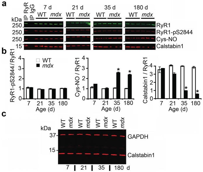
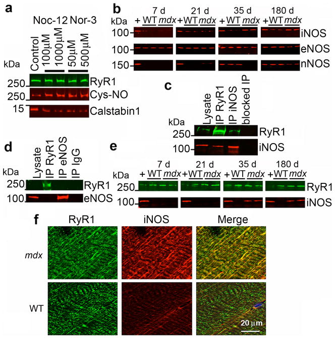
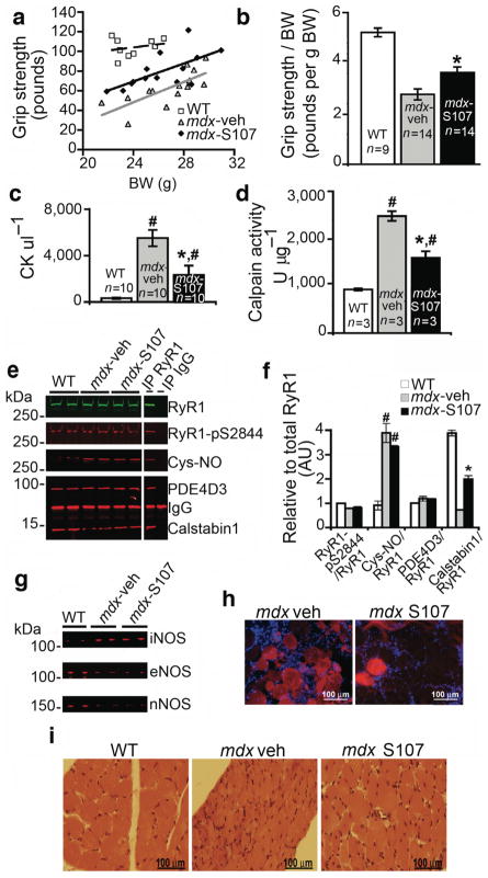
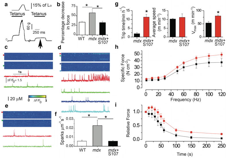
Duchenne muscular dystrophy is characterized by progressive muscle weakness and early death resulting from dystrophin deficiency. Loss of dystrophin results in disruption of a large dystrophin glycoprotein complex, leading to pathological calcium (Ca2+)-dependent signals that damage muscle cells. We have identified a structural and functional defect in the ryanodine receptor (RyR1), a sarcoplasmic reticulum Ca2+ release channel, in the mdx mouse model of muscular dystrophy that contributes to altered Ca2+ homeostasis in dystrophic muscles. RyR1 isolated from mdx skeletal muscle showed an age-dependent increase in S-nitrosylation coincident with dystrophic changes in the muscle. RyR1 S-nitrosylation depleted the channel complex of FKBP12 (also known as calstabin-1, for calcium channel stabilizing binding protein), resulting in 'leaky' channels. Preventing calstabin-1 depletion from RyR1 with S107, a compound that binds the RyR1 channel and enhances the binding affinity of calstabin-1 to the nitrosylated channel, inhibited sarcoplasmic reticulum Ca2+ leak, reduced biochemical and histological evidence of muscle damage, improved muscle function and increased exercise performance in mdx mice. On the basis of these findings, we propose that sarcoplasmic reticulum Ca2+ leak via RyR1 due to S-nitrosylation of the channel and calstabin-1 depletion contributes to muscle weakness in muscular dystrophy, and that preventing the RyR1-mediated sarcoplasmic reticulum Ca2+ leak may provide a new therapeutic approach.


Figures

Figure 1
Figure 1. RyR1 is S-nitrosylated and depleted of calstabin1 in mdx mice
(a) RyR1 was immunoprecipitated from EDL muscle of mdx mice and WT littermates at 7, 21, 35, and 180 days after birth and immunoblotted for RyR1, RyR1 PKA phosphorylated at Ser2844 (RyR1-pS2844), S-nitrosylation of cysteine residues on RyR1 (Cys-NO), and calstabin1 bound to RyR1. Positive and negative control (IgG) immunoprecipitation are shown for 7 day WT hearts. Blots are representative of three independent experiments. (b) Quantification of RyR1 PKA phosphorylation, RyR1 S-nitrosylation, and bound calstabin1 relative to total RyR1. Data presented as mean ± S.E.M. *, P < 0.05, t-test. (c) Immunoblot for total calstabin1 in whole EDL muscle lysate (25 μg) from WT and mdx mice at indicated ages. GAPDH was used as a loading control.
Figure 2
Figure 2. iNOS co-immunoprecipitates and co-localizes with RyR1 and S-nitrosylation of RyR1 depletes the channel of calstabin1
(a) In vitro S-nitrosylation of skeletal SR microsomes with NO donors Nor-3 or Noc-12 results in depletion of calstabin1 from immunoprecipitated RyR1. (b) Immunoblot of expression of three NOS isoforms (iNOS, eNOS, and nNOS) from WT and mdx whole muscle lysates at the indicated ages. (c) Co-immunoprecipitation of RyR1 and iNOS. 50 μg of mdx EDL lysate was used as positive control. RyR1 and iNOS separately immunoprecipitated from 250 μg of mdx muscle lysate and probed for RyR1 and iNOS. Antibody against RyR was pre-incubated with 100-fold excess antigenic peptide prior to immunoprecipitation (blocked IP RyR). (d) Immunoprecipitation-immunoblotting of RyR1 and eNOS from mdx lysate. IgG control immunoprecipitation shown at right. (e) RyR1 was immunoprecipitated from WT and mdx EDL lysates at indicated ages and immunoblotted for RyR1 and iNOS. (f) Immunohistochemistry showing co-localization of RyR1 and iNOS in murine skeletal muscle (EDL) from mdx but not WT mice. Scale bar in lower right panel applies to all six panels.
Figure 3
Figure 3. Preventing calstabin1 depletion from the RyR1 complex with S107 improves grip strength and reduces muscle damage
(a) We determined forelimb grip strength in sedentary mice after two weeks of treatment with S107 administered via an osmotic pump as described in the methods. (mdx-S107, n = 14, black diamond), vehicle (mdx-vehicle, n = 14, grey triangle), WT (n = 9, open square) mice. Data are presented as a scatter plot of absolute grip strength (ponds) versus body weight (BW, g). Least square fit lines are overlaid. (b) Grip strength normalized to BW. *, P < 0.015, t-test with Bonferroni adjustment, mdx-S107 vs mdx-veh. (c) CK levels (#, P < 0.015 vs. WT; *, P < 0.015 mdx-S107 vs. mdx-veh; t-tests with Bonferroni adjustment). (d) EDL tissue calpain activity (#,P < 0.015 vs. WT; *, P < 0.015 mdx-S107 vs. mdx-veh; t-tests with Bonferroni adjustment). (e) RyR1 immunoprecipitated from hind limb EDL muscle immunoblotted for total RyR1, RyR1-pS2844, Cys-NO, PDE4D3 and calstabin1 bound to RyR1. (f) Quantification of (e) showing levels of indicated proteins normalized to the total amount of RyR1 (AU, arbitrary units). Data presented as mean ± S.E.M. (#, P < 0.015 for RyR1-Cys-NO, mdx vs. WT; *, P < 0.015, for calstabin1 binding to RyR1, mdx treated with S107 vs. mdx treated with vehicle). (g) Immunoblot for iNOS, eNOS, and nNOS in EDL whole muscle lysates. (h) Representative images of DAPI stained 10 μm TA sections from mice injected with 100 μl of 1% Evans Blue Dye intraperitoneally 24 hrs prior to sacrifice. S107 treatment was begun at 35 days of age and continued for 4 wks via osmotic pump. (i) Representative H&E stained images from diaphragm.
Figure 4
Figure 4. Preventing RyR1 leak with S107 reduces Ca2+ leak, enhances muscle force, and voluntary exercise in mdx mice
(a) Isometric and eccentric force production in EDL muscle in situ in anesthetized mice (Supplementary Methods, online). Typical recording from an mdx mouse EDL muscle (bottom graph) indicating a decline in force production during tetanus following mechanical stress (arrow). (b) Effect of oral S107 on decrease in force production in mdx mice (S107, 0.25 mg ml−1, in the drinking water for 10 days prior to testing, n = 5 for each group). Spontaneous Ca2+ sparks recorded in EDL muscles from: WT (c), vehicle treated mdx (d), and S107 treated mdx mice (e). Representative ΔF/F0 images (top) and fluorescence time courses (bottom) at different triads (colored arrow heads). (f) Spark frequency (n = 5 mice for each condition, 3–4 fibers per muscle). Data are mean ± SEM (*P < 0.05, WT vs. mdx, vehicle treated mdx vs. mdx plus S107). (g) Effect of oral S107 on mdx mice voluntary exercise. N = 5 animals for each condition, * P < 0.05. (h) Force frequency relationship in EDL muscle stimulated from 1 to 120 Hz (300 ms pulse trains, orange circles are S107 treated mdx mice, black squares are vehicle treated mdx mice, n = 5 for each, P < 0.001 using a two Way Analysis of Variance comparing S107 treated vs. vehicle treated mdx mice). (i) Effect of S107 on fatigue resistance using endurance protocol (30 Hz 300 ms trains every second for 300 s). Orange circles are S107 treated mdx mice, black squares are vehicle treated mdx mice, n = 5 for each, P < 0.001 using a two Way Analysis of Variance comparing S107 treated vs. vehicle treated mdx mice.

Comment in

  • NO may prompt calcium leakage in dystrophic muscle.
    Tidball JG, Villalta SA. Tidball JG, et al. Nat Med. 2009 Mar;15(3):243-4. doi: 10.1038/nm0309-243. Nat Med. 2009. PMID: 19265820 No abstract available.
  • Fixing the leak.
    Crunkhorn S. Crunkhorn S. Nat Rev Drug Discov. 2009 Apr;8(4):277. doi: 10.1038/nrd2857. Nat Rev Drug Discov. 2009. PMID: 19348031 No abstract available.

References

    1. Bodensteiner JB, Engel AG. Intracellular calcium accumulation in Duchenne dystrophy and other myopathies: a study of 567,000 muscle fibers in 114 biopsies. Neurology. 1978;28:439–446. - PubMed
    1. Glesby MJ, Rosenmann E, Nylen EG, Wrogemann K. Serum CK, calcium, magnesium, and oxidative phosphorylation in mdx mouse muscular dystrophy. Muscle Nerve. 1988;11:852–856. - PubMed
    1. Blake DJ, Weir A, Newey SE, Davies KE. Function and genetics of dystrophin and dystrophin-related proteins in muscle. Physiol Rev. 2002;82:291–329. - PubMed
    1. Turner PR, Westwood T, Regen CM, Steinhardt RA. Increased protein degradation results from elevated free calcium levels found in muscle from mdx mice. Nature. 1988;335:735–738. - PubMed
    1. Fong PY, Turner PR, Denetclaw WF, Steinhardt RA. Increased activity of calcium leak channels in myotubes of Duchenne human and mdx mouse origin. Science. 1990;250:673–676. - PubMed

Publication types

MeSH terms