

Review
doi: 10.1038/nrmicro2090.
Epub 2009 Feb 9.
The spike protein of SARS-CoV--a target for vaccine and therapeutic development
Affiliations
- PMID: 19198616
- PMCID: PMC2750777
- DOI: 10.1038/nrmicro2090
Item in Clipboard
Review
The spike protein of SARS-CoV--a target for vaccine and therapeutic development
Nat Rev Microbiol.
2009 Mar.
Abstract
Severe acute respiratory syndrome (SARS) is a newly emerging infectious disease caused by a novel coronavirus, SARS-coronavirus (SARS-CoV). The SARS-CoV spike (S) protein is composed of two subunits; the S1 subunit contains a receptor-binding domain that engages with the host cell receptor angiotensin-converting enzyme 2 and the S2 subunit mediates fusion between the viral and host cell membranes. The S protein plays key parts in the induction of neutralizing-antibody and T-cell responses, as well as protective immunity, during infection with SARS-CoV. In this Review, we highlight recent advances in the development of vaccines and therapeutics based on the S protein.
Figures
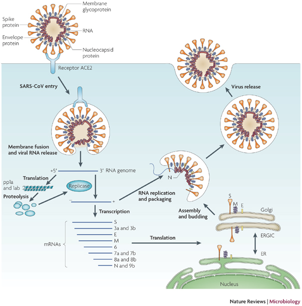
Severe acute respiratory syndrome-coronavirus (SARS-CoV) enters target cells through an endosomal pathway,,,,. S protein first binds to the cellular receptor angiotensin-converting enzyme 2 (ACE2), and the ACE2–virus complex is then translocated to endosomes, where S protein is cleaved by the endosomal acid proteases (cathepsin L) to activate its fusion activity. The viral genome is released and translated into viral replicase polyproteins pp1a and 1ab, which are then cleaved into small products by viral proteinases. Subgenomic negative-strand templates are synthesized from discontinuous transcription on the plus-strand genome and serve as templates for mRNA synthesis. The full-length negative-strand template is made as a template for genomic RNA. Viral nucleocapsids are assembled from genomic RNA and N protein in the cytoplasm, followed by budding into the lumen of the ERGIC (endoplasmic reticulum (ER)–Golgi intermediate compartment). Virions are then released from the cell through exocytosis.
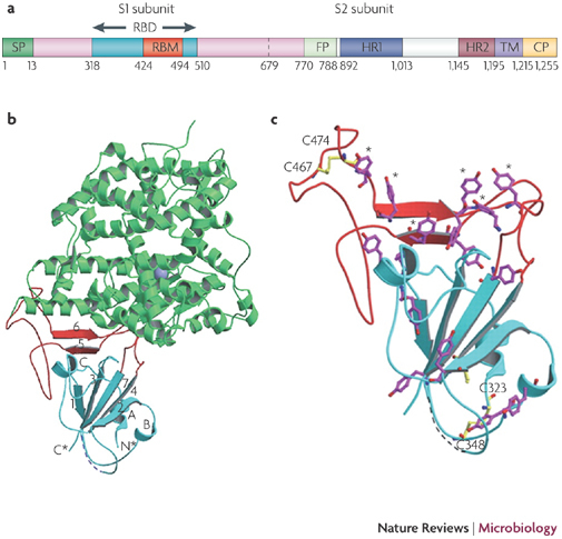
a | Schematic of the S protein,,,. The residue numbers of each region represent their positions in the S protein of severe acute respiratory syndrome-coronavirus (SARS-CoV). b | Crystal structures of the RBD complexed with the receptor. RBD (the core structure is cyan and the loop RBM is red) interacts with the receptor angiotensin-converting enzyme 2 (ACE2; green). A five-stranded anti-parallel β-sheet (β1–β4 and β7) that connects with three short α-helices (αA–αC) constitutes the core, whereas a two-stranded β-sheet (β5 and β6) forms the loop. N* and C* represent the amino and carboxyl termini of the RBD, respectively. c | The RBD tyrosine (magenta) and cysteine (yellow) residue distribution. The asterisks represent six ACE2-contacting tyrosines on the RBD, and two disulphide bonds are shown to link C323 to C348 and C467 to C474. CP, cytoplasm domain; FP, fusion peptide; HR, heptad repeat; RBD, receptor-binding domain; RBM, receptor-binding motif; SP, signal peptide; TM, transmembrane domain. Parts b and c are adapted, with permission, from Ref. © (2005) American Association for the Advancement of Science.
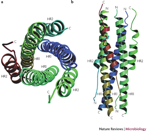
The fusion core is a six-helix bundle with three HR2 α-helices packed in an oblique antiparallel manner against the hydrophobic grooves on the surface of the central HR1 trimer,. A top (a) and side (b) view is shown of the severe acute respiratory syndrome-coronavirus (SARS-CoV) S protein six-helix bundle fusion core structure formed by the HR1 and HR2 domains in the S2 subunit. C, carboxyl; N, amino. Figure adapted, with permission, from Ref. © (2004) American Society for Biochemistry and Molecular Biology.
References
Publication types
MeSH terms
Substances
Grants and funding
LinkOut - more resources
Full Text Sources
Other Literature Sources
Medical
Miscellaneous
