Review
. 2010 May;25(5):831-41.
doi: 10.1007/s00467-009-1116-y. Epub 2009 Feb 7.

Physiopathology and etiology of stone formation in the kidney and the urinary tract

Affiliations
Review

Physiopathology and etiology of stone formation in the kidney and the urinary tract

Andrew P Evan. Pediatr Nephrol. 2010 May.

Abstract

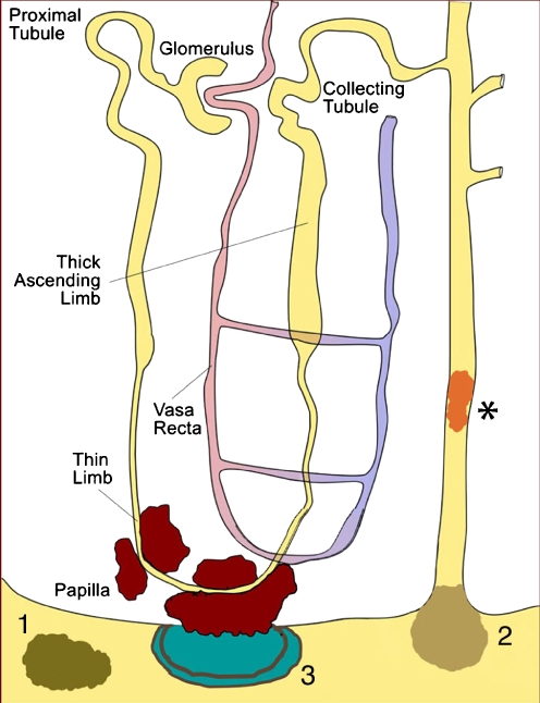
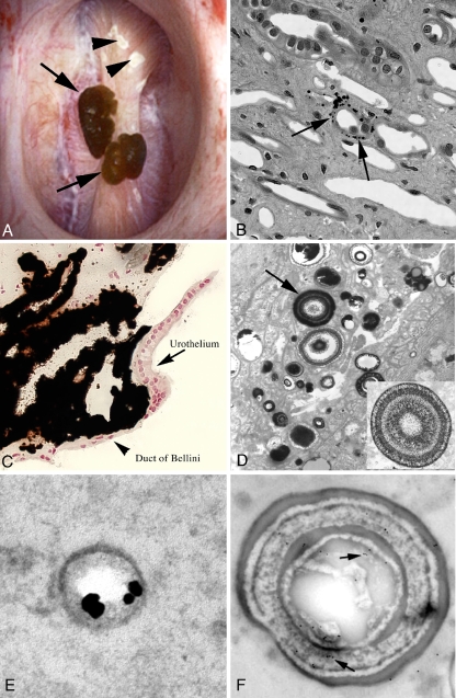
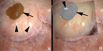
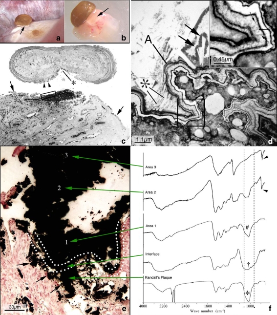
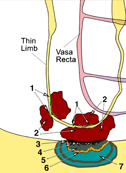
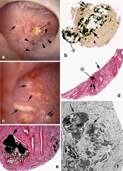
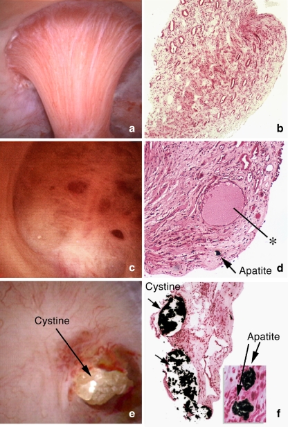
All stones share similar presenting symptoms, and urine supersaturation with respect to the mineral phase of the stone is essential for stone formation. However, recent studies using papillary biopsies of stone formers have provided a view of the histology of renal crystal deposition which suggests that the early sequence of events leading to stone formation differs greatly, depending on the type of stone and on the urine chemistry leading to supersaturation. Three general pathways for kidney stone formation are seen: (1) stones fixed to the surface of a renal papilla at sites of interstitial apatite plaque (termed Randall's plaque), as seen in idiopathic calcium oxalate stone formers; (2) stones attached to plugs protruding from the openings of ducts of Bellini, as seen in hyperoxaluria and distal tubular acidosis; and (3) stones forming in free solution in the renal collection system, as in cystinuria. The presence of hydroxyapatite crystals in either the interstitial or tubule compartment (and sometimes both) of the renal medulla in stone formers is the rule and has implications for the initial steps of stone formation and the potential for renal injury.


Figures

Fig. 1
Fig. 1
Location of staghorn and non-staghorn kidney stones. Staghorn stones fill various amounts of the renal collection system. Non-staghorn stones can be very variable in size and can be found in a major or minor calyx, in the renal pelvis or at different sites along the ureters (proximal, middle or distal). Stones can also be found in the urinary bladder
Fig. 2
Fig. 2
Illustration of three pathways for kidney stone formation and growth. The first pathway (1) represents ‘free particle’ formation, either in the collection system of the kidney or along the nephron (asterisk). The second pathway (2) requires crystal nuclei to form in the lumen of a nephron at sites of cell injury, which results in crystal attachment and growth. In this illustration, crystal attachment occurred at the opening of a duct of Bellini, and a plug of crystalline material projects into a minor calyx. The third pathway (3) suggests that crystals in the urine can become attached to a site of exposed crystalline deposits of interstitial calcium phosphate following loss of the normal urothelial covering of the renal papilla
Fig. 3
Fig. 3
Endoscopic and histologic images of Randall’s plaque seen in an idiopathic calcium oxalate stone former. a A papilla with two attached stones (arrows) and several sites of Randall’s plaque (arrowheads). b Light microscopy shows the initial sites of crystal deposits (arrows) in the basement membrane of the thin loops of Henle. These individual deposits collect in the interstitial space (c) all the way to the urothelium (arrow), embedded in a dense coating of matrix. The individual deposits (arrow, d) are multi-laminated spheres of alternating layers of crystal (electron lucent) and matrix (electron dense). These spheres have both osteopontin (e) and the heavy chain of the inter-alpha-trypsin molecule (arrow, f)
Fig. 4
Fig. 4
Correspondence between tissue plaque and attachment site on the same stone. a Endoscopic image of CaOx stone (arrow) on a papilla from an idiopathic calcium oxalate stone former. Multiple sites of Randall’s plaque (arrowheads) are easily seen. Following removal of the attached stone, it was imaged by micro-computed tomography so that the attachment site of calcium phosphate could be localized and matched to the site of Randall’s plaque in the papilla tissue. b The stone (arrow) was ghosted so that the site of calcium phosphate could be readily seen (arrowhead) and placed over the tissue site of calcium phosphate so that it could be documented that each stone had been attached and attached to a site of Randall’s plaque
Fig. 5
Fig. 5
Ultrastructural and μ-FTIR features of the attachment site of a kidney stone from an idiopathic calcium oxalate stone former. a Endoscopic view of a 0.5 mm stone seen attached to a papilla tip at the site of a Randall’s plaque (arrow). b The same stone, seen by light microscopy, with underlying tissue (arrow) after biopsy. c The same stone–tissue complex as in b but after demineralization. Note that the stone is separated from the underlying tissue (rectangular box). Some tissue is still stuck to the stone (asterisk). The arrowheads show a region of cellular debris. The arrows point to areas on the papilla that still have a urothelial covering; these cells are lost at the stone–tissue junction. d High magnification transmission electron micrograph of the tissue attachment site. The region of Randall’s plaque (lower right) is seen covered by a multi-layered ribbon-like structure with crystalline and matrix material, which is highlighted (arrows) in the insert (upper right). The region A shows small (asterisk) and large (arrows) crystals embedded in the outer (urine) side of the ribbon. By μ-FTIR (e and f), the mineral in the Randall’s plaque (arrows in e) is shown to be apatite (asterisk in f), amorphous apatite (dagger in f) at the tissue–stone interface (outlined by dotted lines), back to apatite (number sign in f) in the stone area closest to the interface and then progressing from a mixture of apatite to just CaOx (arrowheads in f)
Fig. 6
Fig. 6
Schematic representation of stone development in idiopathic calcium oxalate stone formers. The sequence of steps are as follows: 1 apatite deposits develop in the basement membrane of the thin loops of Henle; 2 these apatite deposits extend into the interstitial space and are embedded in matrix, forming islands of interstitial plaque termed Randall’s plaque; 3 these areas of interstitial plaque are exposed to the urine due to a loss of urothelial covering; 4 urine proteins and ions coat the exposed interstitial plaque; 5 a layer of amorphous apatite forms on top of the interstitial plaque, and this new mineral layer is coated with urine matrix molecules; 6 a layer of biological apatite with matrix coating forms on the amorphous apatite; 7 a layer of both apatite and CaOx forms, and, at the outer margin of this small stone, only CaOx is found
Fig. 7
Fig. 7
Endoscopic and histologic images showing three distinct papillary patterns of crystal deposits in brushite patients. a Two of the three crystalline patterns are demonstrated: (1) small irregular white areas of suburothelial plaque (arrow), termed Randall’s plaque, and (2) yellowish crystalline deposit at the opening of a duct of Bellini. Note a defect in the side of this papilla marked by arrowheads. b Biopsy through a brushite papilla revealing an enormously dilated inner medullary collecting duct/duct of Bellini (single arrow), with Yasue-positive mineral protruding from the opening of this duct (asterisk). Double arrow is a site of Randall’s plaque. c Sites of Randall’s plaque (double arrow) (the first pattern) and the third crystalline pattern, which is a yellowish deposit in the inner medullary collecting ducts forming a spoke and wheel-like pattern (single arrows). These yellowish deposits are clearly seen in the collecting ducts (asterisk) in d. The arrow points out the urothelium, while the double arrow is at the site of a Randall’s plaque. The brushite papillae have large regions of disrupted to destroyed papillae that are filled with Yasue-positive mineral (e, arrow and asterisk). This same area is seen in f, but it is now stained with hematoxylin and eosin (H&E) to show the extensive interstitial fibrosis around these damaged papillary ducts (arrows)
Fig. 8
Fig. 8
Endoscopic and histologic images from a cystinuric stone former. Papillary morphology varies from normal (a and b) to flattened and deformed (c and d). d A loop of Henle, filled with apatite deposits, and a grossly dilated inner medullary collecting duct (asterisk). Cystine plugs are seen protruding from the dilated mouths of ducts of Bellini (e). Medullary tubules of cystinuric patients may be filled with either cystine at the ducts of Bellini or apatite along inner medullary collecting ducts or loops of Henle (f)

References

    1. Pearle MS, Calhourn EA, Curhan GC. Urologic diseases in America project: urolithiasis. J Urol. 2005;173:848–857. doi: 10.1097/01.ju.0000152082.14384.d7. - DOI - PubMed
    1. Stamatelou KK, Francis ME, Jones CA, Curhan GC. Time trends in reported prevalence of kidney stones in the United Sates: 1976-1994. Kidney Int. 2003;63:1817–1923. doi: 10.1046/j.1523-1755.2003.00917.x. - DOI - PubMed
    1. Mandel NS, Mandel I, Fryjoff K, Rejniak T, Mandel G. Conversion of calcium oxalate to calcium phosphate with recurrent stone episodes. J Urol. 2003;169:2026–2029. doi: 10.1097/01.ju.0000065592.55499.4e. - DOI - PubMed
    1. Parks JH, Worcester EM, Coe FL, Evan AP, Lingeman JE. Clinical implications of abundant calcium phosphate in routinely analyzed kidney stones. Kidney Int. 2004;66:777–785. doi: 10.1111/j.1523-1755.2004.00803.x. - DOI - PubMed
    1. Parks JH, Coe FL, Evan AP, Worcester EM. Urine pH in renal calcium stone formers who do and do not increase stone phosphate content with time. Nephrol Dial Transplant. 2009;24:130–136. doi: 10.1093/ndt/gfn420. - DOI - PMC - PubMed

Publication types