Review
. 2009 Mar 3;48(8):1663-74.
doi: 10.1021/bi802324w.

Designer DNA nanoarchitectures

Affiliations
Review

Designer DNA nanoarchitectures

Chenxiang Lin et al. Biochemistry. .

Abstract

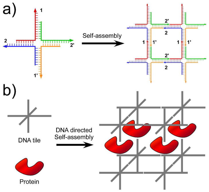
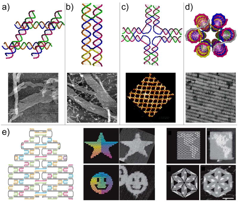
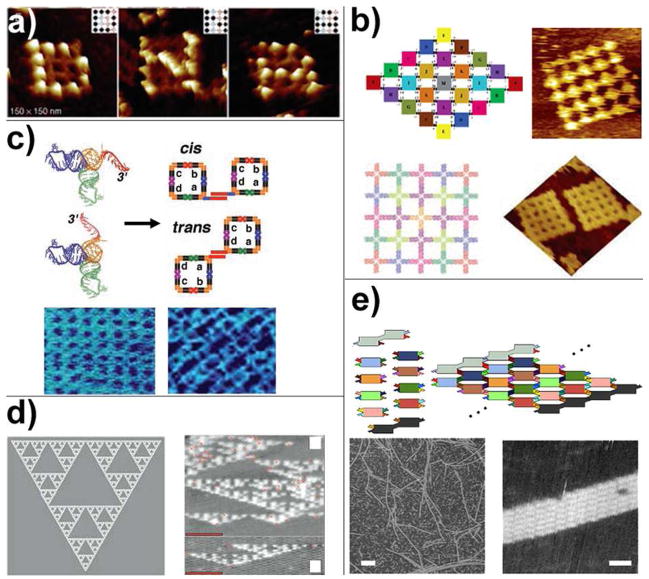
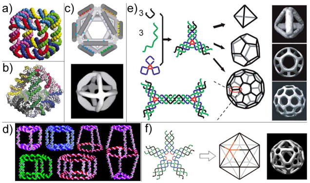
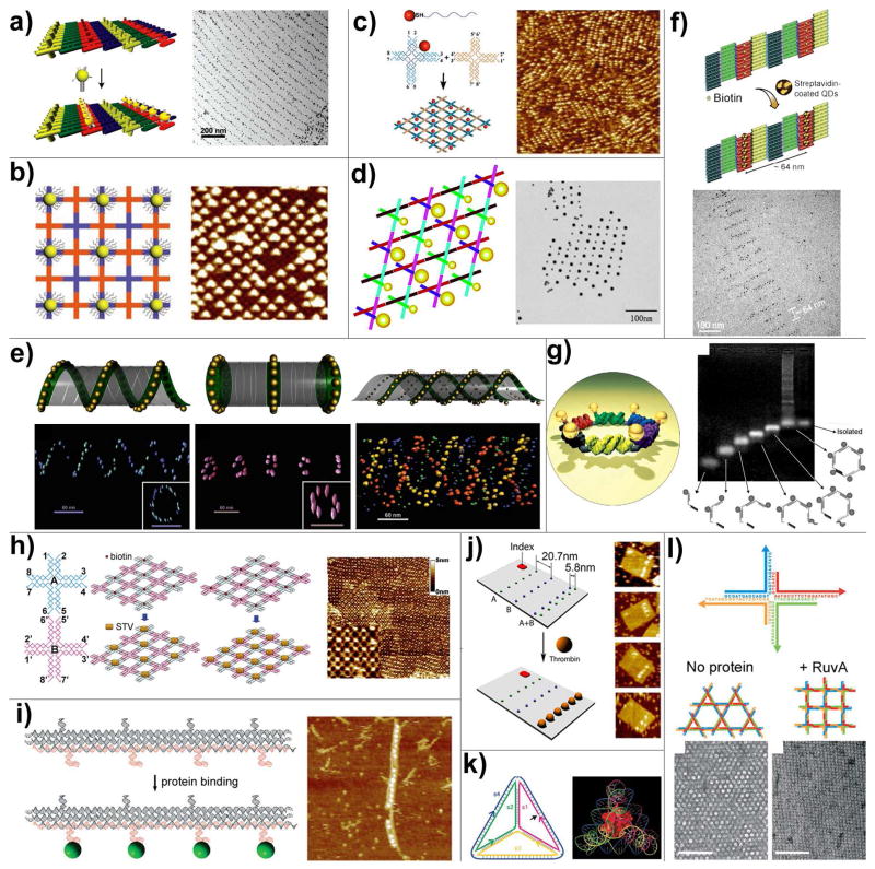
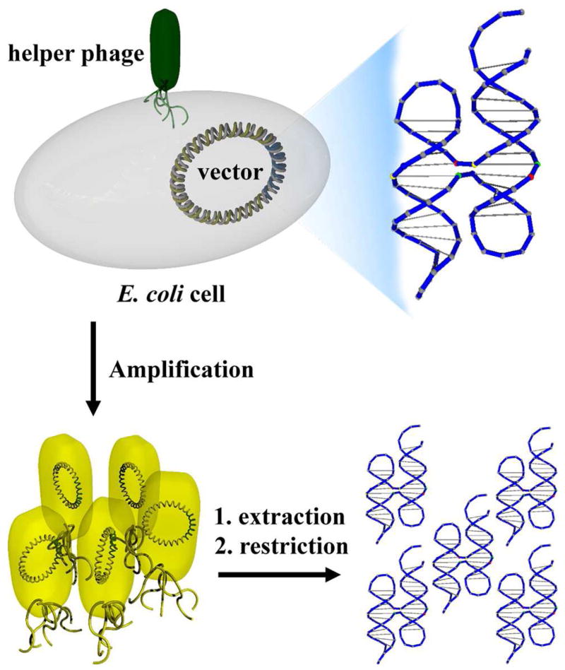
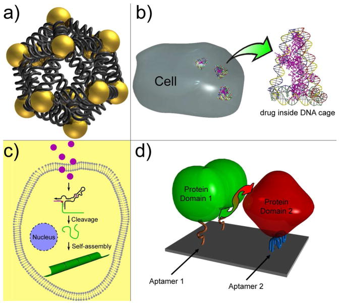
Naturally existing biological systems, from the simplest unicellular diatom to the most sophisticated organ such as the human brain, are functional self-assembled architectures. Scientists have long been dreaming about building artificial nanostructures that can mimic such elegance in nature. Structural DNA nanotechnology, which uses DNA as a blueprint and building material to organize matter with nanometer precision, represents an appealing solution to this challenge. On the basis of the knowledge of helical DNA structure and Watson-Crick base pairing rules, scientists have constructed a number of DNA nanoarchitectures with a large variety of geometries, topologies, and periodicities with considerably high yields. Modified by functional groups, those DNA nanostructures can serve as scaffolds to control the positioning of other molecular species, which opens opportunities to study intermolecular synergies, such as protein-protein interactions, as well as to build artificial multicomponent nanomachines. In this review, we summarize the principle of DNA self-assembly, describe the exciting progress of structural DNA nanotechnology in recent years, and discuss the current frontier.


Figures

Figure 1
Figure 1
Principle and application of DNA self-assembly, as proposed by Ned Seeman. (a) Principle of DNA self-assembly: combining branched DNA nanostructures with sticky-ends to form 2D arrays. Arabic numbers indicate base-paring strategies between sticky-ends (1 is complementary to 1′, etc.) (b) Protein crystallization templated by DNA 3D self-assembly.
Figure 2
Figure 2
Models of some representative DNA tiles and their assemblies into periodic 2D arrays. (a) Parallelogram DNA tile formed by joining four Holliday junctions in parallel. (b) Double helix (DX) tile formed through strand exchange between two DNA duplexes. (c) A cross-shaped tile with four arms (4×4 tile); each arm represents a four-arm junction. (d) Six-helix bundle tube tile viewed from the end of the tube. (a)–(d) Representative AFM images of the 2D arrays were shown below the corresponding cartoon models. (e) DNA origami. Left: principle of DNA origami: folding long ssDNA into shapes by multiple helper strands. Middle: Star and smiley face DNA origami tiles self-assembled by folding a 7-kb ssDNA with over 200 helper strands. Right: Hairpin loops (white dots) can be introduced to certain helper strands to accurately display designated geometries on the fully addressable origami tiles.
Figure 3
Figure 3
Programmable connectivity between DNA tiles. (a) a sixteen-tile finite-sized DNA array made of cross-shaped tiles. Streptavidins were attached to certain tiles to display letters “D”, “N” and “A” on the arrays. (b) Taking advantage of the geometric symmetry, finite-sized DNA arrays can be assembled from minimal number of unique tiles. Shown here are a two-fold and a four-fold symmetric 25-tile arrays constructed using 13 and 7 unique tiles, respectively. (c) Selective combinations of tecto-RNAs can self-assemble into tecto-squares with different sticky-tail conformations, which further self-assemble into finite and infinite arrays with various cavity and periodicity. (d) Algorithm self-assembly of Sierpinski triangle DNA sheet. (e) Nucleated assembly of fixed-width DNA ribbon.
Figure 4
Figure 4
DNA self-assembled 3D nanoarchitectures. (a) Model of a DNA cube. (b) Model of a DNA tetrahedron. (c) Model (top) and cryo-EM image (bottom) of a DNA octahedron. (d) A library of 3D prisms and cubes are assembled from cyclic and single-stranded DNA molecules with organic vertices. (e) DNA tetrahedron, dodecahedron and buckyball each self-assembled from a single symmetric three-point star tile. (f) DNA icosahedron self-assembled from a five-point star tile.
Figure 5
Figure 5
DNA directed assembly of multi-component nanoarrays. (a) Organization of 5 nm AuNPs on DNA DX lattices. (b) Periodic 5 nm AuNP nanoarrays with well controlled interparticle distances templated by 2D DNA nanogrids. (c) DNA mono-modified 5 nm AuNPs directly participate in the self-assembly process and yield periodic nanoparticle arrays. (d) 2D periodic array of 5- and 10-nm AuNPs generated by incorporating DNA mono-modified AuNPs into robust triangle-shaped DNA motifs. (e) Controlled self-assembly of DNA tubules through integration of AuNPs. The assembly results in 3D nanoparticle architectures such as single-spiral tube (left), stacking ring tube (middle) and interlocking double-spiral tube (right). The schematic views are placed above corresponding electron tomographic images. (f) Quantum dots organized on DNA DX lattices through biotin-streptavidin interaction. (g) Discrete hexagonal AuNP array displayed on a DNA hexagon consisting of six non-identical molecules each with two ssDNA arms linked by an organic molecule. (h) Programmable streptavidin 2D arrays formed on biotinylated DNA lattices. (i) 1D thrombin array assembled by incorporating anti-thrombin aptamers into linear TX DNA array. (j) Selective binding of thrombin proteins to the bivalent anti-thrombin aptamers displayed on the surface of rectangular origami arrays. (k) A cytochrome c protein trapped inside a DNA tetrahedron cage. (l) The binding of RuvA to Holliday junction tiles alters the assembly product from Kagome-type lattice (left) to square-planar lattice (right).
Figure 6
Figure 6
Schematic illustration of the in vivo replication of DNA Holliday junction structure. The DNA junction was inserted into a phagemid vector, transformed into E. coli cells, and then replicated in the presence of helper phage. The single-stranded phagemid DNA molecules were packed and secreted into the culture medium. After simple post-treatment like DNA extraction and restriction, high copy numbers of cloned nanostructures can be obtained as a result of the exponential replication of phagemid vectors in bacteria cells.
Figure 7
Figure 7
Perspective applications of DNA nanotechnology. (a) Assembly of discrete 3D nanoparticle structure to study the inter-paticle plasmonic effect. Shown here is a DNA icosahedron with nanoparticle at each vertex. (b) DNA mediated drug delivery. (c) Small molecule triggered in vivo DNA self-assembly for medical applications. For example, the binding between taken up molecule and an allosteric DNAzyme can trigger the cleavage of the substrate DNA. The released DNA segment can then self-assemble into nanotube, which can be visualized as a pathogen marker or directly lead to cell death. (d) DNA nanochip displaying two aptamers can bring two protein subunits into proximity and therefore induce protein-protein interaction.

References

    1. Odom TW, Huang JL, Kim P, Lieber CM. Structure and electronic properties of carbon nanotubes. J Phys Chem B. 2000;104:2794–2809.
    1. Theobald JA, Oxtoby NS, Phillips MA, Champness NR, Beton PH. Controlling molecular deposition and layer structure with supramolecular surface assemblies. Nature. 2003;424:1029–1031. - PubMed
    1. Columbo G, Soto P, Gazit E. Peptide self-assembly at the nanoscale: a challenging target for computational and experimental biology. Trends Biotechnol. 2007;25:211–218. - PubMed
    1. Seeman NC. DNA in a material world. Nature. 2003;421:427–431. - PubMed
    1. Seeman NC. Nucleic acid junctions and lattices. J Theor Biol. 1982;99:237–247. - PubMed

Publication types