. 2009 Mar;8(3):1304-14.
doi: 10.1021/pr800658c.

Proteomic analysis of human parotid gland exosomes by multidimensional protein identification technology (MudPIT)

Affiliations

Proteomic analysis of human parotid gland exosomes by multidimensional protein identification technology (MudPIT)

Mireya Gonzalez-Begne et al. J Proteome Res. 2009 Mar.

Abstract

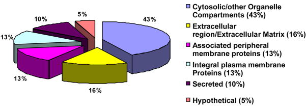
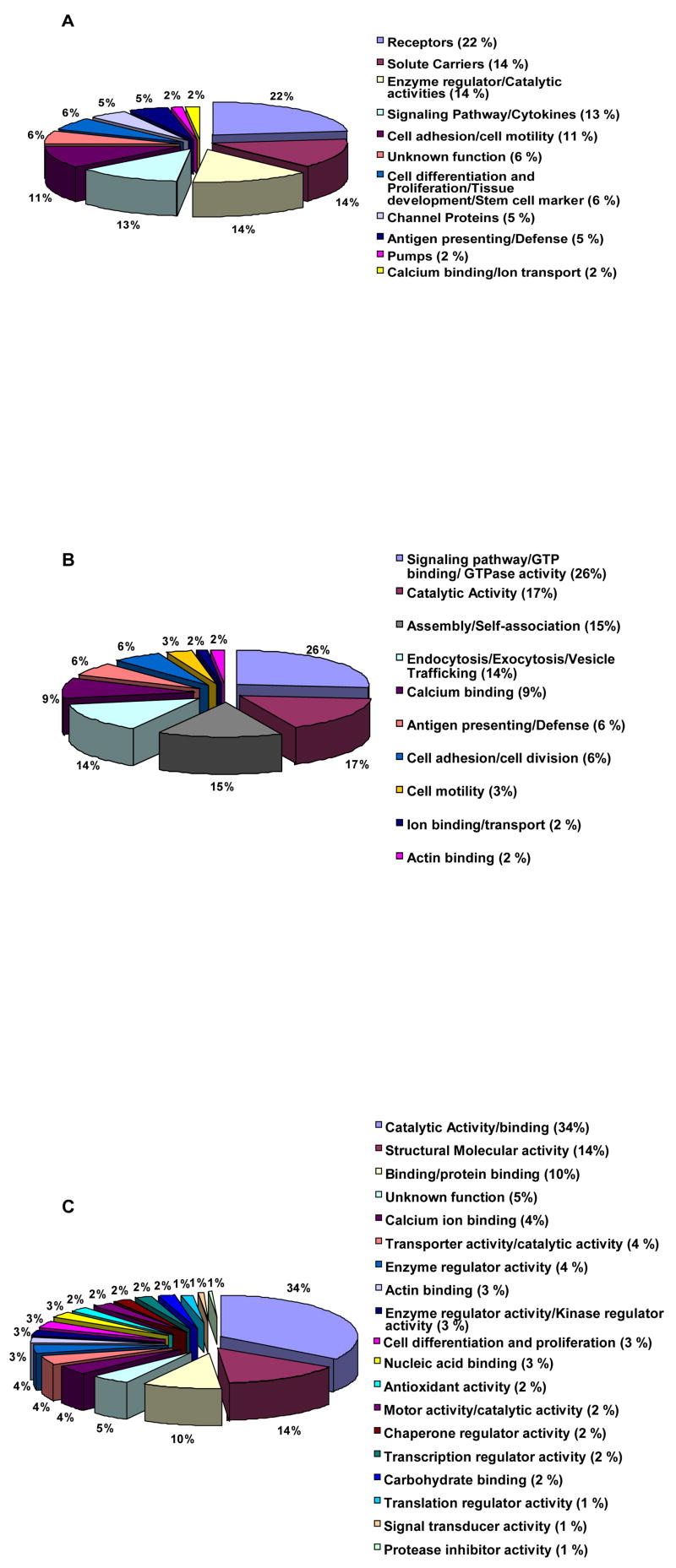
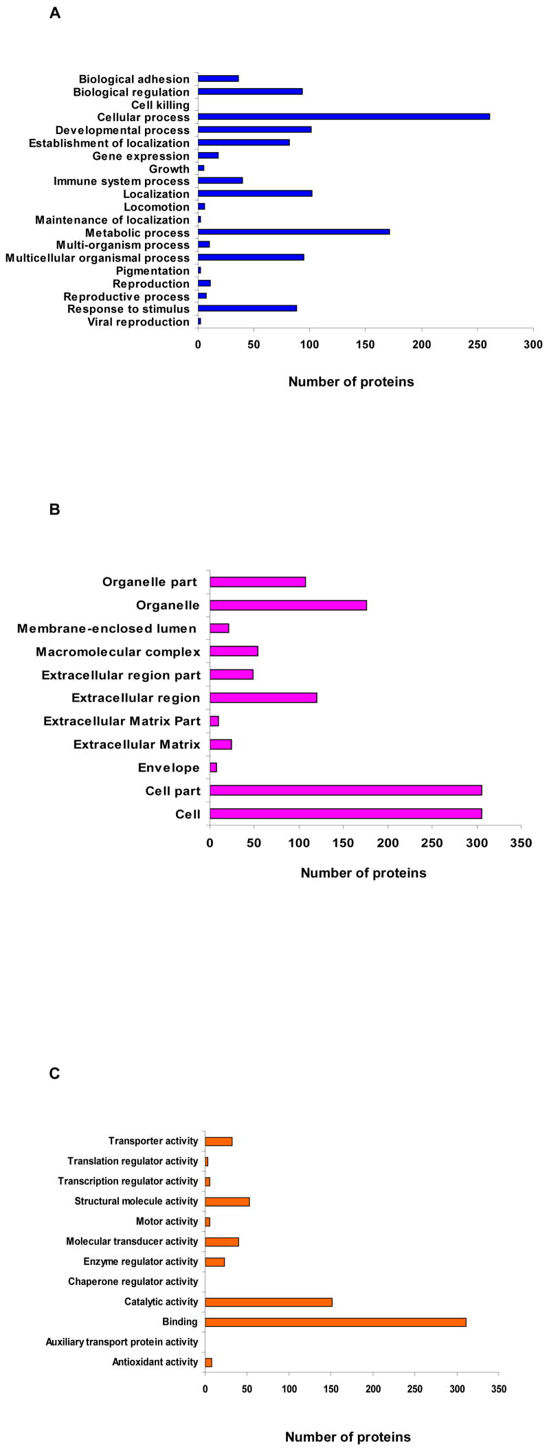
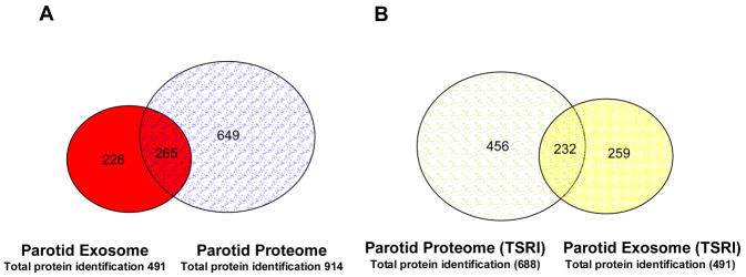
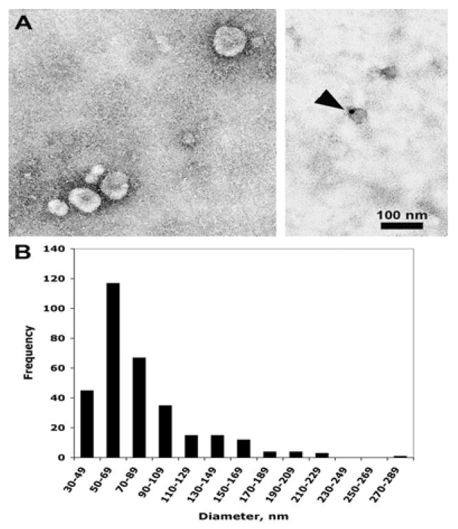
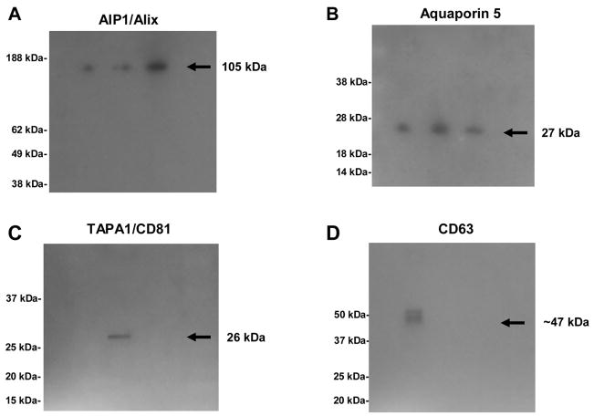
Human ductal saliva contributes over a thousand unique proteins to whole oral fluids. The mechanism by which most of these proteins are secreted by salivary glands remains to be determined. The present study used a mass spectrometry-based, shotgun proteomics approach to explore the possibility that a subset of the proteins found in saliva are derived from exosomes, membrane-bound vesicles of endosomal origin within multivesicular endosomes. Using MudPIT (multidimensional protein identification technology) mass spectrometry, we catalogued 491 proteins in the exosome fraction of human parotid saliva. Many of these proteins were previously observed in ductal saliva from parotid glands (265 proteins). Furthermore, 72 of the proteins in parotid exosomes overlap with those previously identified as urinary exosome proteins, proteins which are also frequently associated with exosomes from other tissues and cell types. Gene Ontology (GO) and KEGG pathway analyses found that cytosolic proteins comprise the largest category of proteins in parotid exosomes (43%), involved in such processes as phosphatidylinositol signaling system, calcium signaling pathway, inositol metabolism, protein export, and signal transduction, among others; whereas the integral plasma membrane proteins and associated/peripheral plasma membrane proteins (26%) were associated with extracellular matrix-receptor interaction, epithelial cell signaling, T-cell and B-cell receptor signaling, cytokine receptor interaction, and antigen processing and presentation, among other biological functions. In addition, these putative saliva exosomal proteins were linked to specific diseases (e.g., neurodegenerative disorders, prion disease, cancers, type I and II diabetes). Consequently, parotid glands secrete exosomes that reflect the metabolic and functional status of the gland and may also carry informative protein markers useful in the diagnosis and treatment of systemic diseases.


Figures

Figure 1
Figure 1. Annotation of identified parotid exosome proteins by subcellular location
Protein classification was obtained by using UniProtKB/Swiss-Prot, EBI and UniProtKB/TrEMBL databases. The majority of exosomal proteins were allocated to the cytosolic compartment, whereas the smallest number was identified as hypothetical.
Figure 2
Figure 2. Protein annotation using UniProtKB/Swiss-Prot, EBI and UniProtKB/TrEMBL databases
A. Annotation of identified parotid exosome integral plasma membrane proteins by function. Twenty-two % of the proteins identified were receptors and the minority (2%) was represented by pumps and/or calcium binding/ion transport proteins. B. Annotation of identified parotid exosome peripheral membrane proteins by function. The bulk of parotid exosomal proteins was associated with proteins involved in signaling pathways, while lesser amounts of proteins were linked to ion binding/transport and actin binding activities. C. Annotation of identified parotid exosome cytosolic proteins by function. Thirty-four % of the cytosolic proteins were associated with catalytic activities whilst only 0.5% with protease inhibitor activity.
Figure 3
Figure 3. Gene Ontology annotation of parotid exosome proteins
A. GO annotation by biological process. Allocation of parotid exosome proteins by biological process showed that the greatest number of these proteins (261) was allocated to cellular process and the smallest (1) to cell killing. B. GO annotation by cellular component. Allocation of parotid exosome proteins by cell component demonstrated that the majority belonged to the cell part and cell (306 each), while the minority resided in the envelope (7). C. GO annotation by molecular function. Allocation of parotid exosome proteins by molecular function confirmed that the highest number of proteins (311) was associated with binding activities and the lowest (1 each) with auxiliary transport protein activity and chaperone regulator activity.
Figure 4
Figure 4. Comparisons between parotid exosome and parotid salivary proteomes
A. Venn diagram showing the overlap of proteins between the parotid exosome and the parotid salivary proteomes. Twenty-three % of the proteins were found in both proteomes, whereas 20% were unique parotid exosome proteins and 57% were unique to the parotid saliva. B. Venn diagram showing the overlap of proteins between the parotid exosome and the parotid salivary proteomes from the same donor. Twenty-five % proteins were found in both proteomes, whereas 27% were unique parotid exosome proteins and 48% were unique to the parotid saliva.
Figure 5
Figure 5. Immunoelectron microscopy of parotid exosomes
A. Electron microscopy of parotid exosomes. Left panel, ammonium molybdate negative stain; right panel, immunogold labeling with anti-TSG101 (50 μg/ml). The arrowhead indicates a labeled exosome. B. Frequency distribution of parotid exosomes by diameter. A total of 318 exosomes were measured.
Figure 6
Figure 6. Western blot analyses of parotid exosome proteins isolated from human parotid saliva
Panel A: AIP1/Alix parotid exosome protein (n=3) was detected using an anti AIP1/Alix polyclonal antibody from Abcam (Cambridge, MA). The expected molecular weight of this protein was around ~ 105 kDa. Panel B: Aquaporin-5 parotid exosomal protein (n=3) was detected with a polyclonal antibody kindly provided by Dr. Anil Menon (Department of Molecular Genetics, Biochemistry and Microbiology; University of Cincinnati, OH). This antibody recognized a protein of ~27 kDa. Panel C: TAPA1/CD81 and Panel D: CD63 (non-reducing condition) parotid-exosome proteins involved in biogenesis process were detected using monoclonal antibodies from Abcam (Cambridge, MA). The corresponding molecular weights of these proteins were ~ 26 kDa and ~ 47 kDa respectively.

Similar articles

Cited by

References

    1. Bennick A. J Oral Biosci. 2007;49:24–26.
    1. Denny P, Hagen FK, Hardt M, Liao L, Yan W, Arellanno M, Bassilian S, Bedi GS, Boontheung P, Cociorva D, Delahunty CM, Denny T, Dunsmore J, Faull KF, Gilligan J, Gonzalez-Begne M, Halgand F, Hall SC, Han X, Henson B, Hewel J, Hu S, Jeffrey S, Jiang J, Loo JA, Ogorzalek Loo RR, Malamud D, Melvin JE, Miroshnychenko O, Navazesh M, Niles R, Park SK, Prakobphol A, Ramachandran P, Richert M, Robinson S, Sondej M, Souda P, Sullivan MA, Takashima J, Than S, Wang J, Whitelegge JP, Witkowska HE, Wolinsky L, Xie Y, Xu T, Yu W, Ytterberg J, Wong DT, Yates JR, 3rd, Fisher SJ. J Proteome Res. 2008;7:1994–2006. - PMC - PubMed
    1. Oppenheim FG, Salih E, Siqueira WL, Zhang W, Helmerhorst EJ. Ann N Y Acad Sci. 2007;1098:22–50. - PubMed
    1. Tabak LA. Pediatr Dent. 2006;28:110–117. discussion 192–118. - PubMed
    1. Vitorino R, Lobo MJ, Ferrer-Correira AJ, Dubin JR, Tomer KB, Domingues PM, Amado FM. Proteomics. 2004;4:1109–1115. - PubMed

Publication types

LinkOut - more resources