. 2009 Mar 3;48(8):1691-701.
doi: 10.1021/bi801710g.

Context-dependent substrate recognition by protein farnesyltransferase

Affiliations

Context-dependent substrate recognition by protein farnesyltransferase

James L Hougland et al. Biochemistry. .

Abstract

Prenylation is a posttranslational modification whereby C-terminal lipidation leads to protein localization to membranes. A C-terminal "Ca(1)a(2)X" sequence has been proposed as the recognition motif for two prenylation enzymes, protein farnesyltransferase (FTase) and protein geranylgeranyltransferase type I. To define the parameters involved in recognition of the a(2) residue, we performed structure-activity analysis which indicates that FTase discriminates between peptide substrates based on both the hydrophobicity and steric volume of the side chain at the a(2) position. For nonpolar side chains, the dependence of the reactivity on side chain volume at this position forms a pyramidal pattern with a maximal activity near the steric volume of valine. This discrimination occurs at a step in the kinetic mechanism that is at or before the farnesylation step. Furthermore, a(2) selectivity is also affected by the identity of the adjacent X residue, leading to context-dependent substrate recognition. Context-dependent a(2) selectivity suggests that FTase recognizes the sequence downstream of the conserved cysteine as a set of two or three cooperative, interconnected recognition elements as opposed to three independent amino acids. These findings expand the pool of proposed FTase substrates in cells. A better understanding of the molecular recognition of substrates performed by FTase will aid in both designing new FTase inhibitors as therapeutic agents and characterizing proteins involved in prenylation-dependent cellular pathways.


Figures

Figure 1
Figure 1
Structure of a peptide substrate bound to FTase illustrating the a2 residue binding site. The a2 residue of the peptide substrate KKKSKTKCVIM (green) is surrounded by residues W102β (orange), W106β (red), and Y361β (yellow) within the active site of FTase. The a2 residue also contacts the isoprenoid tail of the FPP analogue inhibitor FPT-II (purple). Figure derived from PDB ID 1D8D and adapted from (10).
Figure 2
Figure 2
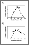
Reactivity of FTase with dansyl-GCVa2S peptides correlates with the hydrophobicity and steric volume of the a2 residue. (a) Correlation of ΔGtransfer, indicative of amino acid hydrophobicity (42), with steric volume (43). For amino acids that lie on the solid black line (G, A, S, P, T, V, I, L, M, F, and W; filled squares) hereafter referred to as the “nonpolar” amino acids, hydrophobicity is proportional to steric volume (slope = 0.021 ± 0.002, R2 = 0.91). A second group of more polar amino acids (D, E, N, Q, H, Y, K, R, and C; open squares) also show a correlation between hydrophobicity and volume (slope = 0.028 ± 0.004, R2 = 0.91) although they fall on a different line. Three amino acids (C,K,R) appear to be outliers. (b) Correlation between the relative reactivity of FTase with dansyl-GCVa2S peptides and the hydrophobicity of the a2 residue. The solid (a2 = G, A, S, T, P, and V; slope = 1.1 ± 0.4, R2 = 0.63) and dashed (a2 = V, M, I, L, F, and W; slope = −1.5 ± 0.5, R2 = 0.72) lines are linear fits to the relative reactivities of FTase with the “nonpolar” amino acids. The dotted line (slope = 4.8 ± 0.9, R2 = 0.89) is a linear fit to the reactivities of FTase with a subset of the polar amino acids (D, E, N, and Q). The ΔΔG(kcat/KMpeptide) values are calculated relative to dansyl-GCVGS as described in Materials and Methods from kcat/KMpeptide values reported in Table 1. Symbols are identical to those described in (a). (c) Correlation between FTase reactivity with Dansyl-GCVa2S peptides and the steric volume of the a2 residue. The solid and dotted lines are linear fits to the relative reactivities of FTase with the “nonpolar” amino acids described in (a). The solid line (encompassing “nonpolar” amino acids with volumes ≤140 Å3) has a slope of 0.033 ± 0.003 kcal/(mol*Å3) (R2 = 0.97) and the dotted line (including “nonpolar” amino acids with volumes ≥140 Å3) has a slope of −0.031 ± 0.003 kcal/(mol*Å3) (R2 = 0.96). The ΔΔG(kcat/KMpeptide) values are calculated relative to dansyl-GCVGS as described in Materials and Methods from kcat/KMpeptide values reported in Table 1. Symbols are identical to those described in (a).
Figure 3
Figure 3
The identity of the X residue affects a2 selectivity. For all four peptide panels, ΔΔG(kcat/KMpeptide) for ten “nonpolar” peptides are plotted versus residue volume (filled squares); proline-containing peptides are shown as an open triangle. A subset of a2 residues (G, A, V, M, F, W) are labeled. Linear fits were performed within the low volume (solid line) and high volume (dotted line) regions of the plot. (a) Dansyl-GCVa2A peptide panel. The solid line has a slope of 0.026 ± 0.004 kcal/(mol*Å3) (R2 = 0.93) and the dotted line has a slope of −0.021 ± 0.003 kcal/(mol*Å3) (R2 = 0.92). (b) Dansyl-GCVa2S peptide panel (duplicated from Figure 1c). (c) Dansyl-GCVa2Q peptide panel. The solid line has a slope of 0.018 ± 0.004 kcal/(mol*Å3) (R2 = 0.92) and the dotted line has a zero slope within error. (d) Dansyl-GCVa2M peptide panel. The solid line has a slope of 0.020 ± 0.003 kcal/(mol*Å3) (R2 = 0.96) and the dotted line has a zero slope within error. The ΔΔG(kcat/KMpeptide) values are calculated relative to peptides containing glycine at the a2 position as described in Materials and Methods from kcat/KMpeptide values reported in Table 1.
Figure 4
Figure 4
The effect of altering the X residue on reactivity of FTase with peptides. (a) Reactivities of FTase with dansyl-GCVa2Q peptides compared to dansyl-GCVa2A peptides. For each residue, the relative reactivity is calculated as log [ (kcat/KMdansylGCVa2Q)/(kcat/KMdansylGCVa2A)]. (b) Reactivities of FTase with dansyl-GCVa2Q peptides compared to dansyl-GCVa2S peptides. For each a2 residue, the relative reactivity is calculated as log [ (kcat/KMdansylGCVa2Q)/(kcat/KMdansylGCVa2S)].
Figure 5
Figure 5
Correlation of the single-turnover rate constant for farnesylation catalyzed by FTase with the volume of the a2 residue for dansyl-GCVa2S and dansyl-GCVa2M peptides. For both peptide panels, −ΔΔG(kSTO) for ten “nonpolar” peptides are plotted versus the a2 residue volume (filled squares). A subset of a2 residues (G, A, V, M, F, W) are labeled. (a) Dansyl-GCVa2S peptide panel. The solid line has a slope of 0.030 ± 0.006 kcal/(mol*Å3) (R2 = 0.91) and the dotted line has a slope of −0.032 ± 0.005 kcal/(mol*Å3) (R2 = 0.91) (b) Dansyl-GCVa2M peptide panel. The solid line has a slope of 0.03 ± 0.01 kcal/(mol*Å3) (R2 = 0.82) and the dotted line has a slope of −0.008 ± 0.003 kcal/(mol*Å3) (R2 = 0.63). The −ΔΔG(kSTO) values are calculated relative to peptides containing glycine at the a2 position, as described in Materials and Methods, using the kSTO values reported in Table 2.
Figure 6
Figure 6
Crystallographic contacts observed between the X residue of the Ca1a2X sequence and the FTase active site. Proposed hydrogen bond contacts are shown as dashed lines between heteroatoms, and distances were measured between heteroatoms involved in the proposed hydrogen bond. In each case, the peptide substrates are complexed with FTase and the FPP analogue inhibitor FPT-II. Peptide substrate residues are denoted with a “sub” subscript. Figures are adapted from (10). (a) Complex of a KKKSKTKCVIM substrate with FTase and FPT-II (PDB ID 1D8D). The substrate methionine sulfur is positioned to form hydrogen bonds with the side chain hydroxyl of S99β (3.2 Å) and side chain indole nitrogen of W102β (3.5 Å). (b) Complex of a DDPTASACNIQ substrate with FTase and FPT-II (PDB ID 1TN6). The amide oxygen of the glutamine side chain in the peptide substrate is proposed to hydrogen bond with the side chain indole nitrogen of W102β (2.8 Å) and side chain hydroxyl of S99β (3.5 Å), and the amide nitrogen is near the side chain hydroxyl of S99β (3.4 Å) and the backbone carbonyl oxygen of A98β (2.9 Å). (c) Complex of a GCVLS substrate with FTase and FPT-II (PDB ID 1TN8). The substrate serine hydroxyl group is positioned to form a hydrogen bond with the backbone carbonyl oxygen of A98β through a water molecule (blue sphere), with distances of 2.7 Å from the serine hydroxyl group oxygen to the water molecule oxygen and 3.0 Å from the water to the backbone carbonyl oxygen of A98β.
Figure 7
Figure 7
Substitution of the S99β and W102β side chains with alanine does not alter a2 selectivity in the dansyl-GCVa2M peptide panel. Relative reactivities for dansyl-GCVa2M peptides with W102βA (filled squares) and S99βA (open triangles) mutant FTases are plotted versus the steric volume of the residue; ΔΔG(kcat/KMpeptide) values are calculated relative to dansyl-GCVGM as described in Materials and Methods from kcat/KMpeptide values reported in Table 2. ΔΔG(kcat/KMpeptide) values for reaction of dansyl-GCVa2M peptides (solid line with filled circles) and dansyl-GCVa2S peptides (dotted line with open circles) with WT FTase are plotted for comparison; ΔΔG(kcat/KMpeptide) values for peptide reactivity with WT FTase are all calculated relative to reactivity with dansyl-GCVGM.
Scheme 1
Scheme 1
Scheme 2
Scheme 2

References

    1. Zhang FL, Casey PJ. Protein prenylation: Molecular mechanisms and functional consequences. Ann Rev Biochem. 1996;65:241–269. - PubMed
    1. Benetka W, Koranda M, Eisenhaber F. Protein prenylation: An (almost) comprehensive overview on discovery history, enzymology, and significance in physiology and disease. Monatsh Chem. 2006;137:1241–1281.
    1. Casey PJ, Seabra MC. Protein prenyltransferases. J Biol Chem. 1996;271:5289–5292. - PubMed
    1. Casey PJ. Lipid modifications of g proteins. Curr Opin Cell Biol. 1994;6:219–225. - PubMed
    1. Marshall CJ. Protein prenylation: A mediator of protein-protein interactions. Science. 1993;259:219–225. - PubMed

Publication types