. 2009 Feb;29(4):718-26.
doi: 10.1111/j.1460-9568.2009.06633.x. Epub 2009 Feb 6.

Neurokinin 1 receptor-expressing projection neurons in laminae III and IV of the rat spinal cord have synaptic AMPA receptors that contain GluR2, GluR3 and GluR4 subunits

Affiliations
Free PMC article

Neurokinin 1 receptor-expressing projection neurons in laminae III and IV of the rat spinal cord have synaptic AMPA receptors that contain GluR2, GluR3 and GluR4 subunits

Andrew J Todd et al. Eur J Neurosci. 2009 Feb.
Free PMC article

Abstract

alpha-Amino-3-hydroxy-5-methyl-4-isoxazolepropionic acid receptors (AMPArs), which mediate fast excitatory glutamatergic transmission, are tetramers made from four subunits (GluR1-4 or GluRA-D). Although synaptic AMPArs are not normally detected by immunocytochemistry in perfusion-fixed tissue, they can be revealed by using antigen retrieval with pepsin. All AMPAr-positive synapses in spinal cord are thought to contain GluR2, while the other subunits have specific laminar distributions. GluR4 can be alternatively spliced such that it has a long or short cytoplasmic tail. We have reported that <10% of AMPAr-containing synapses in lamina II have the long form of GluR4, and that these are often arranged in dorsoventrally orientated clusters. In this study, we test the hypothesis that GluR4-containing receptors are associated with dorsal dendrites of projection neurons in laminae III and IV that express the neurokinin 1 receptor (NK1r). Immunostaining for NK1r was carried out before antigen retrieval, and sections were then reacted to reveal GluR2 and either GluR4 (long form), GluR3 or GluR1. All NK1r-positive lamina III/IV neurons had numerous GluR2-immunoreactive puncta in their dendritic plasma membranes, and virtually all (97%) of the puncta tested were labelled (usually strongly) with the GluR4 antibody. Sizes of puncta varied, but many were elongated and they were significantly larger than nearby puncta that were not associated with the NK1r cells. None of the GluR2 puncta on these cells was positive for GluR1, while 85% were GluR3-immunoreactive. These results show that synaptic AMPArs on the dendrites of the lamina III/IV NK1r projection neurons contain GluR2, GluR3 and GluR4, but not GluR1 subunits.


Figures

Fig. 1
Fig. 1
Staining for GluR2 and GluR4 in spinal cord sections from GluR4 knockout (Gria4−/−) and wild-type (Gria4+/+) mice. These confocal images show parts of the ventral horn from sections that had been reacted with antibodies against GluR2 and GluR4 after antigen retrieval with pepsin. Punctate staining for GluR2, which represents synaptic active zones at glutamatergic synapses, is visible in both cases. In the wild-type mouse, there is also punctate staining for GluR4, and this is extensively colocalized with the GluR2 staining. GluR4-immunoreactivity is absent from the knockout tissue. Both sets of images are projections of five optical sections at 0.3 μm z-separation. Scale bar: 5 μm.
Fig. 2
Fig. 2
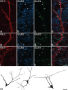
AMPAr subunit-immunoreactive puncta on dendrites of lamina III/IV neurokinin 1 receptor (NK1r)-expressing cells. (a–l) Projected confocal images obtained from sections stained for GluR2 and either GluR4 (a–d), GluR3 (e–h) or GluR1 (i–l). In each case, the section was initially reacted with anti-NK1r (TSA method), and then underwent pepsin treatment and immunoreaction for the corresponding AMPAr subunits. Parts of the dendritic trees of three neurons are shown in red (a, e, i). In each case, there are several GluR2-immunoreactive puncta that appear to be located within the dendritic membrane (b, d, f, h, j, l), and some of these are indicated with arrows. (a–d) The GluR2 puncta on this dendrite are also immunoreactive for GluR4. Although there are numerous GluR2 puncta throughout this field, there are very few GluR4-positive puncta, other than those associated with the dendrite of the NK1r-immunoreactive cell. (e–g) GluR2 puncta on this dendrite show very weak GluR3-immunoreactivity, compared with many of the surrounding puncta. (i–l) Although many of the GluR2-positive puncta in this field are GluR1-immunoreactive, those on the dendrite are not. (m) Drawings of the three cells that show the locations of the regions of dendrites illustrated in (a–l). The confocal images are projections of nine (a–d), four (e–h) or three (i–l) optical sections at 0.5 μm z-separation. Scale bars: 10 μm (a–l) and 50 μm (m).
Fig. 3
Fig. 3
Sizes of AMPAr-positive puncta on the neurokinin 1 receptor (NK1r)-immunoreactive lamina III/IV cells compared with the sizes of other puncta in laminae II and III. The histogram shows the frequency distribution for the GluR4-positive puncta that were identified on the lamina III/IV NK1r-immunoreactive cells (green bars, n=775 puncta on 15 cells), and of GluR2-positive/GluR4-negative puncta in laminae II and III that were not associated with NK1r-immunoreactive dendrites (blue bars, n=600 puncta). For further details, see text.
Fig. 4
Fig. 4
Scatter plots showing the relation between the size of GluR4 puncta on the dendrites of the NK1r-immunoreactive lamina III/IV cells and their distance from the soma. Puncta on dendrites in the superficial (I–II) and deep (III–V) laminae are plotted separately.

Similar articles

Cited by

References

    1. Alvarez FJ, Dewey DE, Harrington DA, Fyffe REW. Cell-type specific organization of glycine receptor clusters in the mammalian spinal cord. J. Comp. Neurol. 1997;379:150–170. - PubMed
    1. Alvarez FJ, Villalba RM, Zerda R, Schneider SP. Vesicular glutamate transporters in the spinal cord, with special reference to sensory primary afferent synapses. J. Comp. Neurol. 2004;472:257–280. - PubMed
    1. Antal M, Fukazawa Y, Eordogh M, Muszil D, Molnar E, Itakura M, Takahashi M, Shigemoto R. Numbers, densities, and colocalization of AMPA- and NMDA-type glutamate receptors at individual synapses in the superficial spinal dorsal horn of rats. J. Neurosci. 2008;28:9692–9701. - PMC - PubMed
    1. Beyer B, Deleuze C, Letts VA, Mahaffey CL, Boumil RM, Lew TA, Huguenard JR, Frankel WN. Absence seizures in C3H/HeJ and knockout mice caused by mutation of the AMPA receptor subunit Gria4. Hum. Mol. Genet. 2008;17:1738–1749. - PMC - PubMed
    1. Bleazard L, Hill RG, Morris R. The correlation between the distribution of the NK1 receptor and the actions of tachykinin agonists in the dorsal horn of the rat indicates that substance P does not have a functional role on substantia gelatinosa (lamina II) neurons. J. Neurosci. 1994;14:7655–7664. - PMC - PubMed

Publication types

Substances

LinkOut - more resources