. 2009 Feb 10;19(3):253-9.
doi: 10.1016/j.cub.2008.12.044.

Neutrophils establish rapid and robust WAVE complex polarity in an actin-dependent fashion

Affiliations

Neutrophils establish rapid and robust WAVE complex polarity in an actin-dependent fashion

Arthur Millius et al. Curr Biol. .

Abstract

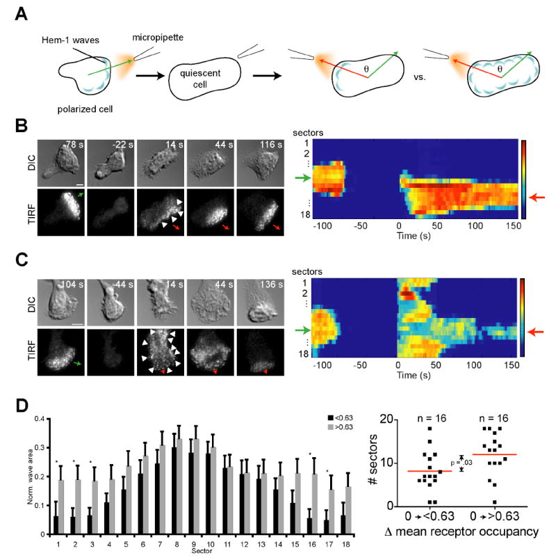
Asymmetric intracellular signals enable cells to migrate in response to external cues. The multiprotein WAVE (also known as SCAR or WASF) complex activates the actin-nucleating Arp2/3 complex [1-4] and localizes to propagating "waves," which direct actin assembly during neutrophil migration [5, 6]. Here, we observe similar WAVE complex dynamics in other mammalian cells and analyze WAVE complex dynamics during establishment of neutrophil polarity. Earlier models proposed that spatially biased generation [7] or selection of protrusions [8] enables chemotaxis. These models require existing morphological polarity to control protrusions. We show that spatially biased generation and selection of WAVE complex recruitment also occur in morphologically unpolarized neutrophils during development of their first protrusions. Additionally, several mechanisms limit WAVE complex recruitment during polarization and movement: Intrinsic cues restrict WAVE complex distribution during establishment of polarity, and asymmetric intracellular signals constrain it in morphologically polarized cells. External gradients can overcome both intrinsic biases and control WAVE complex localization. After latrunculin-mediated inhibition of actin polymerization, addition and removal of agonist gradients globally recruits and releases the WAVE complex from the membrane. Under these conditions, the WAVE complex no longer polarizes, despite the presence of strong external gradients. Thus, actin polymer and the WAVE complex reciprocally interact during polarization.


Figures

Figure 1
Figure 1. Propagating waves of the WAVE complex are mechanistically conserved in other mammalian cells and represent a dynamic quantitative polarity readout in neutrophils
(A) Representative TIRF timelapse sequences for a B16F10 fibroblast cell migrating on fibronectin expressing Abi1 (a component of the WAVE complex) tagged with YFP. Similar to HL-60 cells, propagating waves of the WAVE complex are observed at the leading edge (Movie S1). (B) Representative Abi1-YFP TIRF timelapse sequences for a migrating B16F10 fibroblast exposed to 10 μM latrunculin at 0 s. Similar to HL-60 cells, B16F10 cells exhibit an enrichment of WAVE complex near the membrane following latrunculin treatment, suggesting a role for actin polymer in WAVE complex recycling (Movie S2). (C) Representative brightfield and Hem1-YFP TIRF timelapse sequences for a HL-60 cell executing a turn in response to a change in the direction of the agonist gradient (Movie S3). (D) Corresponding heatmap shows wave response. Green arrow indicates initial up-gradient direction; red arrow indicates final up-gradient direction. Bars, 5 μm.
Figure 2
Figure 2. Cells establish Hem-1 wave asymmetry through either focused generation or uniform generation followed by selection
(A) Illustration of experimental setup. An agonist gradient was applied to a cell and then removed. This process was necessary to ensure quiescence because cells adhered to a coverslip often exhibited polarity and motility even in the absence of chemoattractant. Cells were classified as quiescent when they lost all wave dynamics and any obvious morphological front and back. The micropipette was repositioned at a different angle and the gradient reapplied at t = 0 s. Therefore, all cells start with a mean receptor occupancy of 0 for this figure. The angle difference and interval between agonist applications did not affect WAVE complex distribution, nor did the cell retain memory of the original micropipette position after the micropipette was turned back on (Fig. S2). (B and C) Representative DIC and Hem-1-YFP TIRF timelapse sequences and corresponding heatmaps show that cells exhibit a focused (B, Movie S4) or uniform (C, Movie S5) distribution of waves. Note that wave asymmetry is apparent in the absence of any obvious morphological differences (arrowheads). Green arrows indicate initial up-gradient direction; red arrows indicate final up-gradient direction. Bars, 5 μm. (D) Bar graph (left) of a 20 s average of wave response immediately after gradient reapplication. Black bars show response for cells with mean receptor occupancy (post-stimulation) of <0.63. Gray bars indicate cells with mean receptor occupancy (post-stimulation) of >0.63. Error bars are s.e.m. Asterisks indicate statistically significant differences between means of each sector (p <0.05, Student’s t-test). Dot plot (right) shows a statistically significant difference (p = .03, Student’s t-test) between the mean width (red line) of the distributions as defined in Fig. S1.
Figure 3
Figure 3. Directional bias limits wave generation in response to small increases in mean receptor occupancy
(A and B) Initially quiescent cells were subjected to spatially uniform mean receptor occupancy increases from 0 to 0.1 (at t = 0 s), which produced focused waves (A, Movie S6), or 0 to 0.7 (at t = 0 s), which produced a spatially uniform distribution of Hem-1 waves that ultimately collapsed into a focused distribution (B, Movie S7). Bars, 5 μm. (C) Dot plot shows a statistically significant difference (p = .001, Student’s t-test) between the mean width (red line) of the distributions. (D and E) Representative timelapse images of cells with prepolarized WAVE complex distributions responding to spatially uniform increases in mean receptor occupancy from (D) 0.61 to 0.73 (n = 6, Movie S8) or (E) 0.39 to 0.73 (n = 8, Movie S9). (F) Dot plot shows a statistically significant difference (p = 0.002, Student’s t-test) between the changes in wave width for small versus large increases in mean receptor occupancy (note that mean receptor occupancies are statistically different even after removing the outlier for the 0.61 to 0.73 increase). These data suggest that intrinsic directional bias can maintain the asymmetric distribution of Scar/WAVE over a limited range of agonist concentrations in both quiescent and prepolarized cells.
Figure 4
Figure 4. Actin polymer is required for establishment of Hem-1 wave asymmetry
(A) Transient fMLP pulses (red trace) induce transient Hem-1-YFP (black trace) accumulation at the membrane. An initially migrating cell was subjected to 20 μM latrunculin treatment to depolymerize the actin cytoskeleton (40 s). This induced Hem-1-YFP recruitment even in the absence of external stimuli. Subsequent fMLP pulses from a micropipette (160 and 300 s) induced further recruitment. When the agonist was removed, Hem-1-YFP quickly disappeared from the membrane. (B) Selected TIRF and DIC images (Movie S10) from the traces shown in (A). Red arrows indicate direction of gradient pulses. Arrowheads indicate areas of significant Hem-1-YFP accumulation at the membrane. Note the broad distribution following each agonist pulse. Bar, 5 μm. (C) Dot plot of a 20 s average of wave response immediately after an agonist pulse for cells untreated (-lat) and treated with 20 μM latrunculin (+lat). There is a statistically significant difference between the mean width (red lines) of the two populations (p = .002, Student’s t-test). (D) Untreated cells that showed an initially broad wave distribution after an agonist pulse were compared to latrunculin-treated cells. The wave distribution in untreated cells converged into a focused distribution, whereas the wave distribution in latrunculin-treated cells did not converge. Error bars are s.e.m. Inset shows statistical significance between the difference in mean sectors (red lines) of the two populations (p = .002, Student’s t-test).

References

    1. Machesky LM, Insall RH. Scar1 and the related Wiskott-Aldrich syndrome protein, WASP, regulate the actin cytoskeleton through the Arp2/3 complex. Curr Biol. 1998;8:1347–1356. - PubMed
    1. Kunda P, Craig G, Dominguez V, Baum B. Abi, Sra1, and Kette control the stability and localization of SCAR/WAVE to regulate the formation of actin-based protrusions. Curr Biol. 2003;13:1867–1875. - PubMed
    1. Steffen A, Rottner K, Ehinger J, Innocenti M, Scita G, Wehland J, Stradal TE. Sra-1 and Nap1 link Rac to actin assembly driving lamellipodia formation. The EMBO journal. 2004;23:749–759. - PMC - PubMed
    1. Stradal TE, Scita G. Protein complexes regulating Arp2/3-mediated actin assembly. Current opinion in cell biology. 2006;18:4–10. - PubMed
    1. Weiner OD, Rentel MC, Ott A, Brown GE, Jedrychowski M, Yaffe MB, Gygi SP, Cantley LC, Bourne HR, Kirschner MW. Hem-1 complexes are essential for Rac activation, actin polymerization, and myosin regulation during neutrophil chemotaxis. PLoS Biol. 2006;4:e38. - PMC - PubMed

Publication types