. 2009 Feb 10;19(3):223-8.
doi: 10.1016/j.cub.2009.01.033.

A likely pathway for formation of mobile group I introns

Affiliations

A likely pathway for formation of mobile group I introns

Richard P Bonocora et al. Curr Biol. .

Abstract

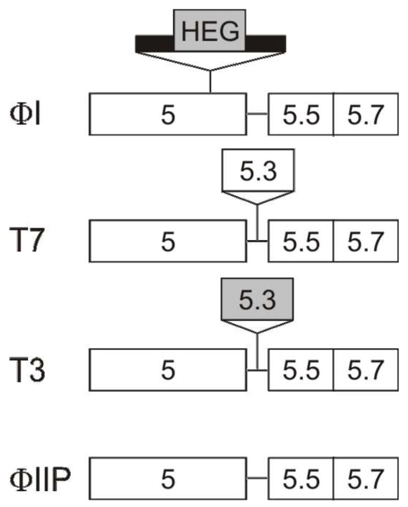
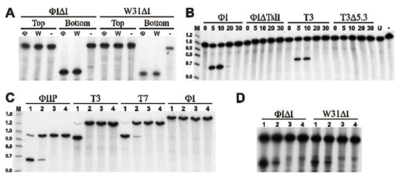
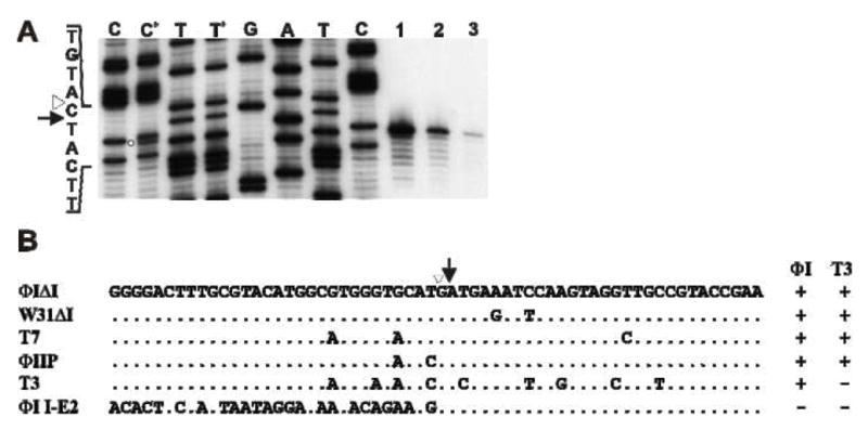
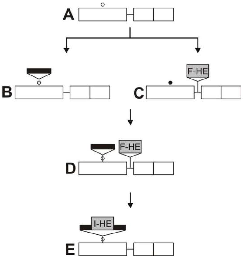
Mobile group I introns are RNA splicing elements that have been invaded by endonuclease genes. These endonucleases facilitate intron mobility by a unidirectional, duplicative gene-conversion process known as homing [1]. Survival of the invading endonuclease depends upon its ability to promote intron mobility. Therefore, the endonuclease must either quickly change its cleavage specificity to match the site of intron insertion, or it must already be preadapted to cleave this sequence. Here we show that the group I intron in the DNA polymerase gene of T7-like bacteriophage PhiI is mobile, dependent upon its intronic HNH homing endonuclease gene, I-TslI. We also show that gene 5.3 of phage T3, located adjacent to its intronless DNA polymerase gene, is a homologous homing endonuclease gene whose protein product initiates efficient spread of gene 5.3 into empty sites in related phages. Both of these endonucleases cleave intronless DNA polymerase genes at identical positions. This shared feature between an intronic and free-standing endonuclease is unprecedented. Based on this evidence, we propose that introns and their homing endonucleases evolve separately to target the same highly conserved sequences, uniting afterwards to create a composite mobile element.


Figures

Figure 1
Figure 1. Schematic representation of the gene 5.3 locus of T7-like phages
Names of representative phages are on the left. Gene names are indicated in the boxes. Thick line indicates intron sequences. Shaded boxes indicate ORFs with HNH motifs.
Figure 2
Figure 2. Endonuclease activity of I-TslI and F-TslI
A. In vitro synthesized I-TslI from ΦI (Φ) and W31 (W), or mock translation products (−) were incubated with 60-mer target DNAs representing the fused exons of the ΦIΔI or W31ΔI DNA polymerase genes. Substrate DNAs (indicated at the top of the figure) are 5′ end-labeled on the coding (top) or template (bottom) strand, as indicated. Reaction products were separated by electrophoresis on a 6% denaturing polyacrylamide gel. B. Endonuclease activity in phage infected cells. Extracts from phage-infected cells (noted above the line) were prepared at various times after infection (noted above each lane). Bottom strand labeled DNA was amplified from ΦIIP. C. and D. Substrate specificity of I-TslI and F-TslI. Extracts prepared 10 min post-infection from phage ΦI (lane 1), T3 (lane 2), ΦIΔTsl (lane 3) and T3Δ5.3 (lane 4) were incubated with substrate DNAs (indicated above the line) generated by (C) PCR from phages or (D) oligonucleotides representing the fused exons from ΦI and W31 (indicated above the line). Only the bottom strand is 5′-end labeled.
Figure 3
Figure 3. I-TslI and F-TslI cleave at the identical location
A. Cleavage site mapping. DNA containing the IIS was amplified by PCR directly from pT7Δ5.3 (using primers ΦI5.0 and 5′-end labeled ΦI5.0rev) and incubated with in vitro synthesized ΦI I-TslI (lane 1), ΦI-infected cell extract (lane 2) and T3-infected cell extract (lane 3) for 15 min. Reaction products were separated by denaturing polyacrylamide gel electrophoresis, next to sequencing reactions generated with the same labeled primer and template DNA used in the PCR. Lanes C+ and T+ contain a mixture of the contents of lane 2 with the C and T sequencing reactions, respectively. The open circle indicates the ΦI cleavage product in the C+ lane. The site of intron insertion is indicated by an open triangle and the position of cleavage is indicated by an arrow. B. Summary of cleavage specificity. The sequences (top/coding strand) of target DNAs from T7-like phages, surrounding the site of intron insertion, are aligned with the ΦI and W31 fused exon sequences. Results of cleavage by the ΦI and T3 extracts are indicated on the right. Identical positions are indicated by dots; only sequence differences are shown. The site of intron insertion is indicated by an open triangle and the position of cleavage on the bottom strand is indicated by an arrow.
Figure 4
Figure 4. Proposed pathway for establishment of intron homing in the DNA polymerase gene of T7-like phages
A. Ancestral gene arrangement prior to insertions, exemplified by phage ΦII, with location of a highly conserved, functionally importantsequence indicated by an open circle (○). B. Insertion of a self splicing group I intron (black bar) into the highly conserved sequence in gene 5. C. A free-standing endonuclease gene (F-TslI) is inserted between genes 5 and 5.5, as in phage T3. The altered target sequence, resistant to F-TslI, is indicated by a closed circle (●). The endonuclease gene is propagated by intronless homing. D. Phage containing both the intron and the endonuclease gene, generated by homologous recombination during mixed infection between phages B and C. The intron and the endonuclease gene are propagated together by collaborative homing. E. Insertion of the endonuclease gene into the intron, creating a mobile intron.

References

    1. Belfort M, Derbyshire V, Parker MM, Cousineau B, Lambowitz AM. Mobile Introns: Pathways and Proteins. In: Craig N, editor. Mobile DNA II. Washington, DC: ASM Press; 2002.
    1. Biniszkiewicz D, Cesnaviciene E, Shub DA. Self-splicing group I intron in cyanobacterial initiator methionine tRNA: evidence for lateral transfer of introns in bacteria. Embo J. 1994;13:4629–4635. - PMC - PubMed
    1. Bonocora RP, Shub DA. A novel group I intron-encoded endonuclease specific for the anticodon region of tRNA(fMet) genes. Mol Microbiol. 2001;39:1299–1306. - PubMed
    1. Orlowski J, Boniecki M, Bujnicki JM. I-Ssp6803I: the first homing endonuclease from the PD-(D/E)XK superfamily exhibits an unusual mode of DNA recognition. Bioinformatics. 2007;23:527–530. - PubMed
    1. Zhao L, Bonocora RP, Shub DA, Stoddard BL. The restriction fold turns to the dark side: a bacterial homing endonuclease with a PD-(D/E)-XK motif. EMBO J. 2007;26:2432–2442. - PMC - PubMed

Publication types

MeSH terms

Substances

LinkOut - more resources