Review
. 2009 Mar;19(3):99-110.
doi: 10.1016/j.tcb.2009.01.001. Epub 2009 Feb 4.

Guidance from above: common cues direct distinct signaling outcomes in vascular and neural patterning

Affiliations
Review

Guidance from above: common cues direct distinct signaling outcomes in vascular and neural patterning

Maria V Gelfand et al. Trends Cell Biol. 2009 Mar.

Abstract

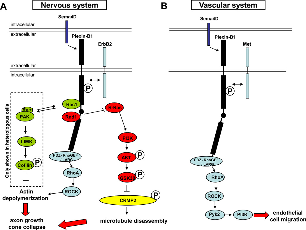
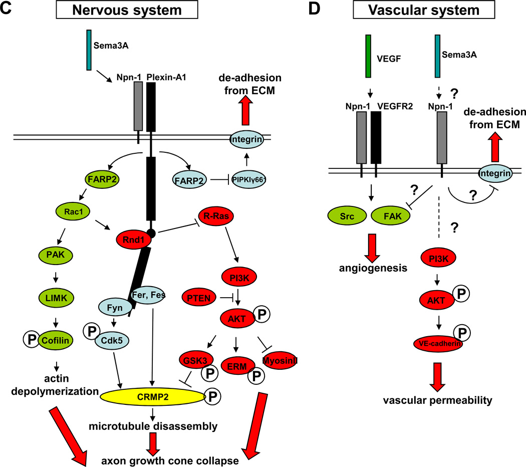
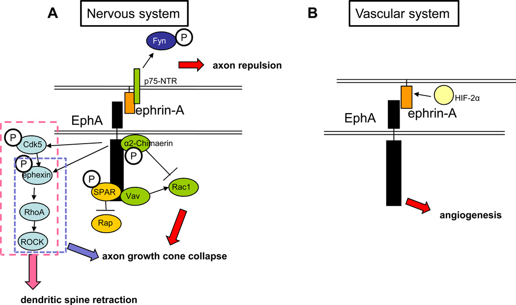
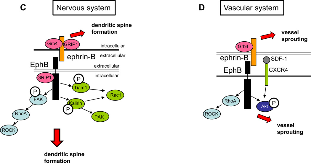
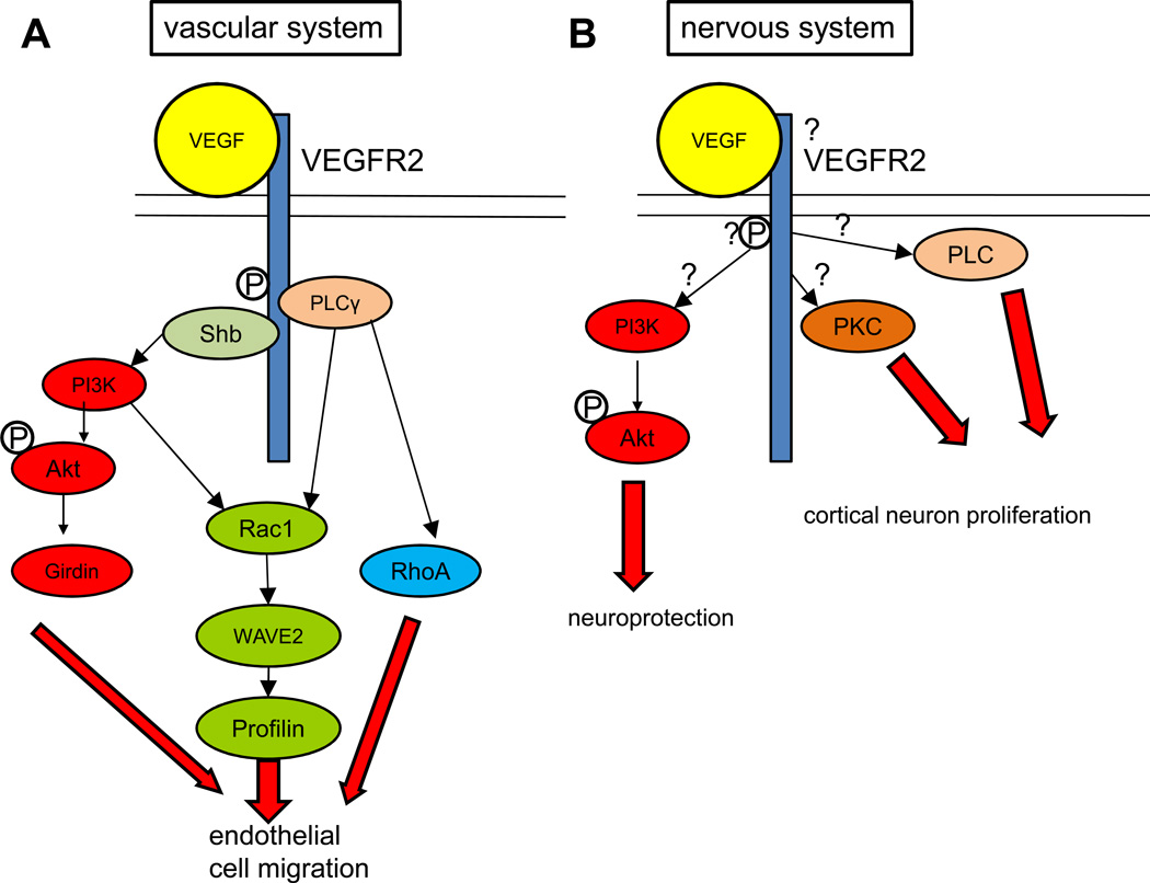
The nervous and vascular systems are both exquisitely branched and complicated systems and their proper development requires careful guidance of nerves and vessels. The recent realization that common ligand-receptor pairs are used in guiding the patterning of both systems has prompted the question of whether similar signaling pathways are used in both systems. This review highlights recent progress in our understanding of the similarities and differences in the intracellular signaling mechanisms downstream of semaphorins, ephrins and vascular endothelial growth factor in neurons and endothelial cells during neural and vascular development. We present evidence that similar intracellular signaling principles underlying cytoskeletal regulation are used to control neural and vascular guidance, although the specific molecules used in neurons and endothelial cells are often different.


Figures

Figure 1
Figure 1. Sema4D and Sema3A signaling in the nervous and vascular systems
Plexin-B1 and Plexin-A1 are shown in black, with the intracellular C1 and C2 domains represented by black rectangles and the linker region in between as a black circle. A: Sema4D signaling in the nervous system. Proteins in the R-Ras pathway are shown in red: in the presence of Sema4D, Rnd1 is recruited to Plexin-B1. Plexin-B1 R-RasGAP activity is activated and downregulates the active form of R-Ras. The decrease of active R-Ras inhibits PI3K-Akt activity, decreasing GSK3β phosphorylation and activing it. GSK3β then phosphorylates and deactivates CRMP2 and causes microtubule disassembly. Proteins in the RhoA pathway are shown in blue: in the presence of Sema4D, receptor tyrosin kinase ErbB2 binds and subsequently phosphorylates Plexin-B1 (as indicated by the double-headed arrow) and then activates PDZ-RhoGEF/LARG, which associates with Plexin-B1. PDZ-RhoGEF/LARG activates RhoA, causing actin depolymerization through ROCK. Proteins in the Rac1 pathway are shown in green: Upon Sema4D binding, activated Plexin-B1 competes for active Rac1 with PAK. The shift on the equilibrium between Plexin-B1- and PAK- bound Rac1 results in decrease of PAK activity, LIMK activity, and Cofillin phosphorylation, thus causing actin depolymerization. This pathway so far has only been shown in heterologous cells, as indicated by the dashed box. Both the actin depolymerization and microtubule disassembly lead to axon growth cone collapse. B: Sema4D signaling in the vascular system. Proteins in the RhoA pathway are shown in blue: in the presence of Sema4D, the receptor tyrosine kinase Met binds and phosphorylates Plexin-B1 (as indicated by the double-headed arrow) and then activates PDZ-RhoGEF, which activates RhoA and leads to endothelial cell migration through the ROCK, Pyk2, and PI3K pathway. It is not clear how this pathway affects actin dynamics or microtubule dynamics in vascular system. C: Sema3A signaling in the nervous system. Rac1 regulating proteins are shown in green: in the presence of Sema3A, FARP2 is released from PlexinA1 and actives Rac1. Rac1 then activates PAK and LIMK and, as a result, phosphorylates Cofilin, which finally causes actin depolymerization. R-Ras regulating proteins are shown in red: in the presence of Sema3A, Rac1 facilitates Rnd1 recruitment to PlexinA1, which induces PlexinA1’s R-RasGAP activity and downregulates active R-Ras. Decrease of active R-Ras downregulates PI3K-Akt activity and leads to axon growth cone collapse through 3 different pathways: reduced phorporylatesGSK3β, reduced phosphorylation of ERM, and activation of myosinII. Kinases are shown in blue: in the presence of Sema3A, FARP2 inhibits PIPKIγ661 and suppresses integrin-induced adhesion. Fer and Fes are activated upon Sema3A binding to Plexin-A1 and phosphorylate and inactivate CRMP2, which leads to microtubule disassembly. Fyn is also activated after its binding to PlexinA1 and inactivates CRMP2 by phosphorylating and activating Cdk5. Both actin depolymerization and microtubule disassembly lead to axon growth cone collapse. D: Sema3A signaling in the vascular system. Sema3A, through an unknown mechanism (possibly through Npn-1 and/or a co-receptor, shown as a dashed line and question mark), inhibits VEGF-induced activation of Src and FAK and contributes to angiogenesis. Sema3A may also function through Npn-1 to inhibit integrin-mediated adhesion of endothelial cells to the ECM. Sema3A can induce VE-cadherin phosphorylation and causes vascular permeability through unknown mechanisms (indicated by question marks), in which PI3K-Akt is involved.
Figure 2
Figure 2. Ephrin-Eph signaling in the nervous and vascular systems
A: ephrin-A-EphA signaling in the nervous system. The Rap pathway is shown in yellow: EphA activation leads to tyrosine phosphorylation of SPAR, a RapGAP that binds to EphA4 through its PDZ domain. SPAR then inactivates Rap. The RhoA pathway is shown in light blue: Upon ephrin-A treatment, EphA recruits and phosphorylates Cdk5, which in turn phosphorylates and activates a RhoGEF, ephexin, activating RhoA. RhoA then activates ROCK, leading to dendritic spine retraction (as shown by the dashed pink box). The dashed blue box indicates the part of this pathway that has been shown to be important for growth cone collapse. The Rac pathway is shown in green: Activation of EphA receptors leads to binding and phosphorylation of the RacGAP α2-chimaerin, which then inactivaes Rac1 in cortical neurons, leading to growth cone collapse. In retinal axons, ephrin binding to EphA receptors leads to activation of the Rho family GEF Vav2, which then activates Rac1. Vav2-mediated activation of Rac1 leads to endocytosis of the membrane, eventually leading to growth cone collapse. In reverse signaling, ephrin-A associates with the p75-NTR receptor. This complex works to phosphorylate Fyn and cause axon repulsion from EphA-expressing cells when EphA binds ephrin-A. B: ephrin–A-EphA signaling in the vascular system. There is limited information on how this signaling regulates angiogenesis, but ephrin-A expression is known to be regulated by HIF-2α expression in surrounding tissue and signaling through EphA in the vascular system can lead to angiogenesis in tumor models. C: ephrin–B-EphB signaling in the nervous system. EphB is recruited to the membrane by GRIP1, a multi-PDZ domain scaffolding protein. The Rac1 pathway is shown in green: Rac1 is activated by the GEFs Kalirin and Tiam1. Both Tiam1 and Kalirin bind to and are phosphorylated by the activated receptor and then activate Rac1, leading to increased dendrite morphogenesis. The RhoA pathway is shown in blue: EphA activation leads to phosphorylation of FAK, which then activates RhoA, activating ROCK and leading to increased dendrite morphogenesis. For reverse signaling through the ephrin-B ligand, several adaptor proteins are required. GRIP1 binds to ephrin-B3 and is perhaps involved in clustering it at the synapse. The SH2-SH3 domain containing adaptor protein Grb4 binds ephrin-Bs and links them with multiple downstream regulators and enhancing dendrite morphogenesis in the ligand-expressing cell. D: ephrin-B–EphB signaling in the vascular system. EphB activation leads to the activation of RhoA and ROCK. In addition, EphB signaling enhances SFD/CXCR4 signaling as seen by increased phosphorylation of Akt. Both pathways lead to endothelial cell migration and sprouting. For reverse signaling, ephrin-B binds the SH2-SH3 domain containing adaptor protein Grb4, leading to vessel sprouting, presumably through multiple downstream regulators recruited by Grb4.
Figure 3
Figure 3. VEGF-VEGFR2 signaling in the nervous and vascular systems
A: VEGF signaling in the vascular system: The PI3K-Akt pathway is shown in red: VEGF binding to VEGFR2 causes autophosphorylation of the receptor. Phosphorylation of the receptor at tyrosine 1175 causes the recruitment of the adaptor protein Shb, which then activates PI3K. PI3K phosphorylates Akt, which acts through substrates like the actin binding protein Girdin to regulate endothelial cell migration. The Rac1 pathway is shown in green: PI3K also activates Rac1, which regulates migration through the effectors WAVE2 and the actin binding protein Profilin. Phosphorylation of Tyr1175 also causes activation of PLCγ, which activates the small GTPases Rac1 and RhoA to regulate endothelial cell migration. B: VEGF-VEGFR2 signaling in the nervous system: Many of the same proteins used by VEGFR2 signaling in the vascular system are also implicated to act in the nervous system, but the evidence for this is less clear (indicated by question marks). PI3K inhibitors abrogate VEGFs neuroprotective effect, while PLC and PKC inhibitors prevented the neurotrophic effect exerted by VEGF on cortical neurons.

Similar articles

Cited by

References

    1. Carmeliet P, Tessier-Lavigne M. Common mechanisms of nerve and blood vessel wiring. Nature. 2005;436(7048):193–200. - PubMed
    1. Gerhardt H, et al. VEGF guides angiogenic sprouting utilizing endothelial tip cell filopodia. J Cell Biol. 2003;161(6):1163–1177. - PMC - PubMed
    1. Freitas C, Larrivee B, Eichmann A. Netrins and UNC5 receptors in angiogenesis. Angiogenesis. 2008;11(1):23–29. - PubMed
    1. Legg JA, et al. Slits and Roundabouts in cancer, tumour angiogenesis and endothelial cell migration. Angiogenesis. 2008;11(1):13–21. - PubMed
    1. Jones CA, Li DY. Common cues regulate neural and vascular patterning. Curr Opin Genet Dev. 2007;17(4):332–336. - PMC - PubMed

Publication types