. 2009:452:297-324.
doi: 10.1016/S0076-6879(08)03619-7.

Methods to monitor chaperone-mediated autophagy

Affiliations

Methods to monitor chaperone-mediated autophagy

Susmita Kaushik et al. Methods Enzymol. 2009.

Abstract

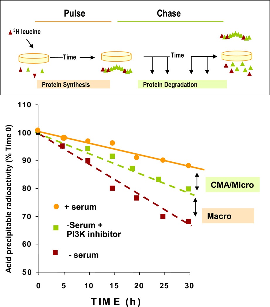
Chaperone-mediated autophagy (CMA) is a selective type of autophagy responsible for the lysosomal degradation of soluble cytosolic proteins. In contrast to other forms of autophagy where cargo is sequestered and delivered to lysosomes through membrane fusion/excision, CMA substrates reach the lysosomal lumen after direct translocation across the lysosomal membrane. CMA is part of the cellular quality control systems and as such, essential for the cellular response to stress. CMA activity decreases with age, likely contributing to the accumulation of altered proteins characteristic in tissues from old organisms. Furthermore, impairment of CMA underlies the pathogenesis of certain human pathologies such as neurodegenerative disorders. These findings have drawn renewed attention to CMA and a growing interest in the measurement of changes in CMA activity under different physiological and pathological conditions. In this chapter we review the different experimental approaches used to assess CMA activity both in cells in culture and in different organs from animals.


Figures

Figure 1
Figure 1. Measurement of long-lived protein degradation
Top: Confluent cells in culture are incubated with a radiolabeled amino acid for 48 h and after extensive washing the amount of acid-soluble radioactivity (amino acids and small peptides) released into the medium at different times is determined. Bottom: Typical example of rates of degradation of long-lived proteins in cultured cells due to CMA, micro- or macroautophagy. CMA activity is calculated as the increase in protein degradation during serum deprivation sensitive to lysosomal protease inhibition and insensitive to the effect of PI3-K type III inhibitors.
Figure 2
Figure 2. Intracellular redistribution of CMA-active lysosomes
A. Indirect immunofluorescence for LAMP-2A in cultured mouse fibroblasts maintained in the presence/absence of serum. Bar; 10 µm. B. Mean distance of the fluorescent puncta (lysosomes) to the nucleus C. Graph representing the intracellular distribution of fluorescent puncta with respect to the nucleus in the two indicated conditions. Values are mean + standard error of 4 different cells in each condition.
Figure 3
Figure 3. In vitro assays for the direct quantification of CMA
A. Incubation of intact or broken lysosomes with radiolabeled CMA substrates allows quantification of the amount of protein processed into soluble amino acids (degradation). In intact lysosomes, substrates need to bind to the lysosomal membrane and translocate before they can be degraded. B-C. Incubation of CMA substrates with intact lysosomes treated or not with protease inhibitors allows determination of the amount of substrate bound to the lysosomal membrane via immunoblot against the substrate after collecting the lysosomes by centrifugation. The amount of substrate translocated into the lysosomal lumen can be calculated in lysosomes treated with protease inhibitors after degradation of the substrate bound to the cytosolic side of the lysosomal membrane with an exogenous protease (B) or by subtracting the amount of “bound” substrate from the total amount of substrate associated with the protease-inhibited lysosomes (C).

References

    1. Agarraberes F, et al. An intralysosomal hsp70 is required for a selective pathway of lysosomal protein degradation. J Cell Biol. 1997;137:825–834. - PMC - PubMed
    1. Aniento F, et al. Uptake and degradation of glyceraldehyde-3- phosphate dehydrogenase by rat liver lysosomes. J Biol Chem. 1993;268:10463–10470. - PubMed
    1. Auteri J, et al. Regulation of intracellular protein degradation in IMR- 90 human diploid fibroblasts. J Cell Physiol. 1983;115:159–166. - PubMed
    1. Bandyopadhyay U, et al. The chaperone-mediated autophagy receptor organizes in dynamic protein complexes at the lysosomal membrane. Mol Cell Biol. 2008 [e-pub ahead of publication] - PMC - PubMed
    1. Brown CR, et al. The Vid vesicle to vacuole trafficking event requires components of the SNARE membrane fusion machinery. J. Biol. Chem. 2003;278:25688–25699. - PubMed

Publication types

Substances

LinkOut - more resources