. 2009 Mar 26;27(15):2177-87.
doi: 10.1016/j.vaccine.2009.01.088. Epub 2009 Feb 6.

Effects of Signal 3 during CD8 T cell priming: Bystander production of IL-12 enhances effector T cell expansion but promotes terminal differentiation

Affiliations

Effects of Signal 3 during CD8 T cell priming: Bystander production of IL-12 enhances effector T cell expansion but promotes terminal differentiation

Weiguo Cui et al. Vaccine. .

Abstract

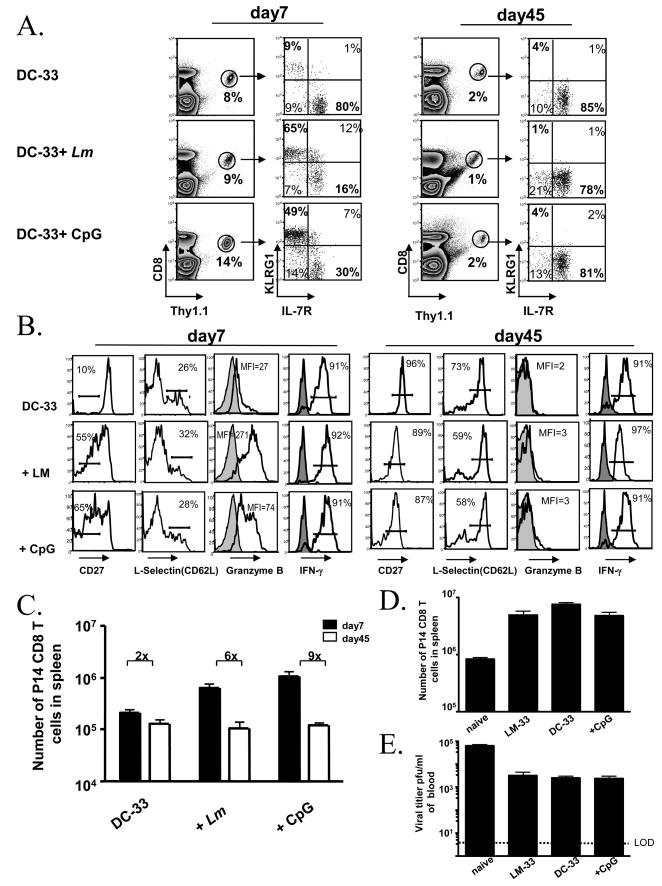
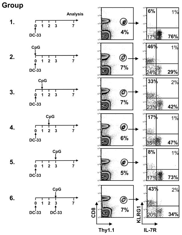
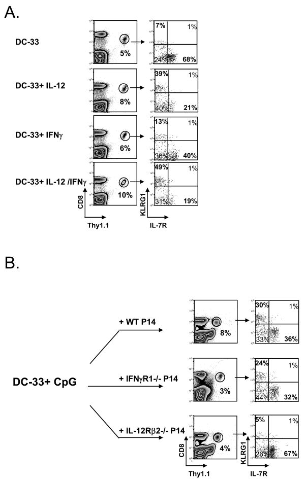
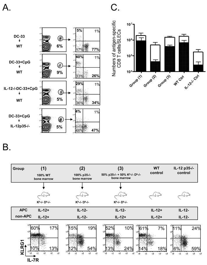
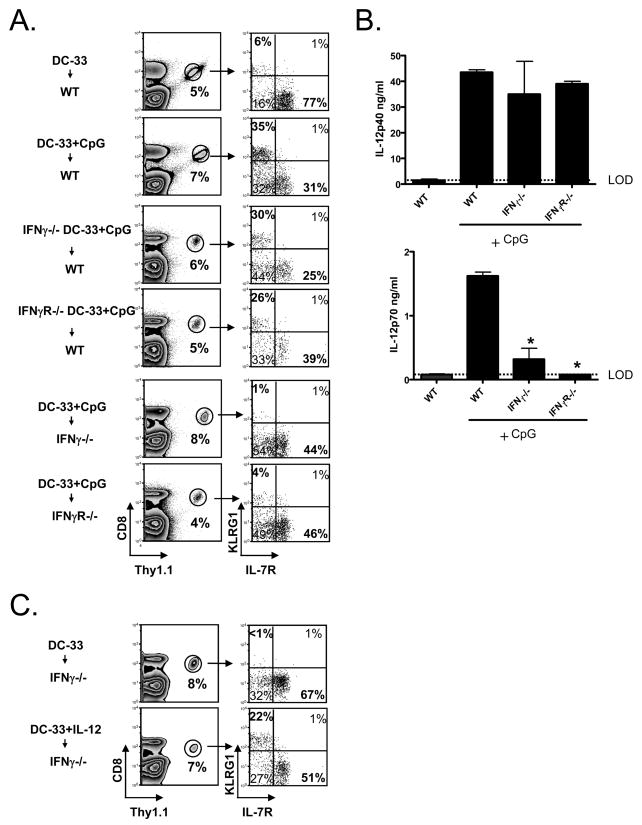
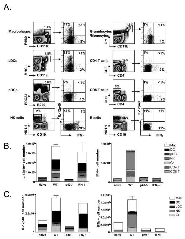
Adjuvants are commonly used in vaccines to augment immune response, but how the inflammatory cytokines elicited by adjuvants directly influence effector and memory CD8 T cell differentiation remains poorly characterized. Here, we used a peptide-pulsed dendritic cell (DC) vaccination model to examine the role of primary cytokines, IL-12 and IFNgamma, elicited by CpG-B adjuvant on CD8 T cell priming and memory CD8 T cell development. During DC vaccination, simultaneous exposure to antigen and a heterologous Listeria infection, CpG-B or IL-12 enhanced a portion of the effector CD8 T cells to expand and differentiate to a larger extent. Simultaneously, this also decreased their ability to become long-lived memory CD8 T cells. However, development of memory CD8 T cells and their precursors was largely unaffected by the additional inflammatory cytokines. Moreover, IL-12 production by the antigen-presenting cell (APC) was not required during DC+CpG vaccination or Listeria infection, but rather 'bystander' macrophages and DCs appeared to be the physiologically relevant cellular sources of this cytokine. Furthermore, IFNgamma induced by CpG was required in vivo for optimal production of IL-12, which in turn, influenced effector CD8 T cell longevity. Together, these findings demonstrate the importance of an interconnected multicellular network between APCs, naïve T cells and bystander cells of the innate immune system that regulate effector and memory CD8 T cell development during vaccination.


Conflict of interest statement

Conflict-of-interest disclosure: The authors declare no competing financial interests.

Figures

Figure 1
Figure 1. Inflammation enhanced effector CD8 T cell clone expansion and potency, but also reduced their memory potential during DC immunization
(A and B) Naïve P14 chimeric mice were immunized with LPS-matured GP33–41-loaded bone marrow derived DCs (DC-33) alone or with heterologous Lm infection or CpG-B ODN. (A) FACS plots show expression of KLRG1 and IL-7R on splenic Thy1.1+ P14 CD8 T cells days 7 and 45 post-immunization. The percentage of P14 CD8 T cells within the total CD8 T cell population is indicated on contour plots. (B) Day 7 and 45 post immunization the P14 CD8 T cells were analyzed for expression of CD27, CD62L (L-selectin), Granzyme B and IFNγ. Histogram plots show expression of CD27, CD62L, Granzyme B directly ex vivo. For Granzyme B staining, CD44lo naïve CD8 T cells (shaded) are shown as controls. IFNγ production was assessed with (open) or without (shaded) 5 hr GP33–41 peptide stimulation in vitro. All plots are gated on donor Thy1.1+ P14 CD8 T cells. (C) Bar graphs show the number of splenic Thy1.1+ P14 CD8 T cells in (A) days 7 and 45 post-immunization. (D and E) Equal numbers of P14 naïve or memory CD8 T cells generated by DC-33 immunization alone, DC-33+CpG or by LM-33 infection were transferred into naïve mice and infected with LCMV clone13. Bar graphs show splenic numbers of memory CD8 T cells on day 8 p.i. (D) and viral titers on day 5 p.i. (E). LOD represents level of detection. Data are representative of at least two independent experiments.
Figure 2
Figure 2. Antigen and inflammation need to be coupled to induce SLEC formation
Six groups of P14 chimeric mice were immunized with DC-33, one group was left untreated as control. Each other group was treated once with CpG at different days after immunization (day 0, 1, 2, or 3), or treated with DC-33+CpG on day 3 post-immunization. FACS plots show expression of KLRG1 and IL-7R on splenic Thy1.1+ P14 CD8 T cells 7 days post immunization. Data are representative of two independent experiments.
Figure 3
Figure 3. IL-12, but not IFNγ, is sufficient to induce SLEC formation during DC immunization
(A) WT P14 chimeric mice were immunized with- DC-33 alone or with IL-12, IFNγor IL-12+IFNγ and analyzed 7 days later. (B) Small numbers of WT, IFNγR1−/− and IL-12Rβ2−/− P14 CD8 T cells were transferred into naïve mice that were then immunized with DC-33+ CpG. P14 CD8 T cells and were analyzed for expression of KLRG1 and IL-7R seven days later. Data are representative of three independent experiments.
Figure 4
Figure 4. Production of IL-12 from non-APCs is sufficient to induce KLRG1hi IL-7Rlo SLECs during DC-33+CpG immunization and Lm infection
(A) WT P14 chimeric mice were immunized with WT DC-33 alone or with WT or IL-12p35−/− DC-33 + CpG, and IL-12p35−/− P14 chimeric mice was immunized with WT DC-33+CpG. P14 CD8 T cells were analyzed for expression of KLRG1 and IL-7R seven days later. (B and C) Kb−/− Db−/− mice were lethally irradiated and reconstituted with the following types of bone marrow cells: 100% WT (group 1), 100% IL-12p35−/− (group 2) or a 50%:50% mixture of IL-12p35−/− and Kb−/− Db−/−. The top panel outlines these different groups of mice and summarizes the cell types capable of producing IL-12 in each case. Unmanipulated WT or IL-12p35−/− mice were also used as controls. Approximately two months after reconstitution, ~1×10⁴ Thy1.1+ P14 CD8 T cells were transferred into the different groups of mice and they were infected with ΔActA-Lm-33. Seven days later, splenic Thy1.1+ P14 effector CD8 T cells were analyzed for KLRG1 and IL-7R expression (B), and numbers of P14 CD8T cells (open bar) and IL-7RloKLRG1hi SLECs (solid bar) were calculated and plotted in the graph (C). Numbers are pooled from three independent experiments.
Figure 5
Figure 5. IFNγ is required for optimal IL-12p70 production, thus indirectly induced SLEC formation in vivo during CpG-B treatment
(A) WT P14 chimeric mice were immunized with WT, IFNγ−/− or IFNγR1−/− DC-33+CpG, and IFN−/− or IFNγR1−/− P14 chimeric mice were immunized with WT DC-33+CpG. P14 CD8 T cells were analyzed seven days later. (B) Bar graphs show the concentration of IL-12p40 (top panel) and IL-12p70 (bottom) in the serum of WT, IFNγ −/− and IFNγR1−/− 6h after CpG injection as measured by ELISA. (C) IFNγ −/− mice containing P14 CD8 T cells were immunized with DC-33 alone or plus recombinant IL-12p70 (1ug/mouse). After 7 days, splenic Thy1.1+ P14 CD8 T cells were analyzed for KLRG1 and IL-7R. LOD=level of detection. Asterisk denotes P<0.05. Data are representative of at least two independent experiments.
Figure 6
Figure 6. Identification of the IL-12 and IFNγ producing cells during DC vaccination and Lm infection
WT, IL-12p40−/− and IFNγ −/− mice were immunized with Lm for 24hrs or CpG for 6 hrs and then the splenocytes were cultured in BFA for an additional 6 hrs and stained for surface markers and intracellular IL-12p40 and IFNγ. (A) Left contour plots show the gating criteria used to identify the different cell populations and right dot plots show the expression of IL-12p40 and IFNγ in these cells 24hrs after Lm infection. Note: similar types of cells, albeit at lower frequencies, produced these cytokines after 6hrs of CpG treatment (data not shown). (B and C) Bar graphs indicate numbers of IL-12- and IFNγ-producing cells in the spleens of WT, IL-12p40−/− and IFNγ −/−mice after Lm infection (B) or CpG immunization (C).

Comment in

References

    1. Pulendran B, Ahmed R. Translating innate immunity into immunological memory: implications for vaccine development. Cell. 2006 Feb 24;124(4):849–63. - PubMed
    1. Kaech SM, Wherry EJ, Ahmed R. Effector and memory T-cell differentiation: implications for vaccine development. Nature reviews. 2002 Apr;2(4):251–62. - PubMed
    1. Letvin NL. Correlates of immune protection and the development of a human immunodeficiency virus vaccine. Immunity. 2007 Sep;27(3):366–9. - PubMed
    1. Schwarz K, Meijerink E, Speiser DE, Tissot AC, Cielens I, Renhof R, et al. Efficient homologous prime-boost strategies for T cell vaccination based on virus-like particles. European journal of immunology. 2005 Mar;35(3):816–21. - PubMed
    1. Pashine A, Valiante NM, Ulmer JB. Targeting the innate immune response with improved vaccine adjuvants. Nature medicine. 2005 Apr;11(4 Suppl):S63–8. - PubMed

Publication types

MeSH terms