

BldG and SCO3548 interact antagonistically to control key developmental processes in Streptomyces coelicolor
- PMID: 19201788
- PMCID: PMC2668407
- DOI: 10.1128/JB.01695-08
BldG and SCO3548 interact antagonistically to control key developmental processes in Streptomyces coelicolor
Abstract
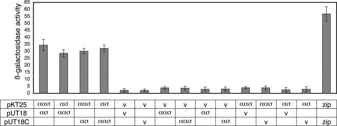
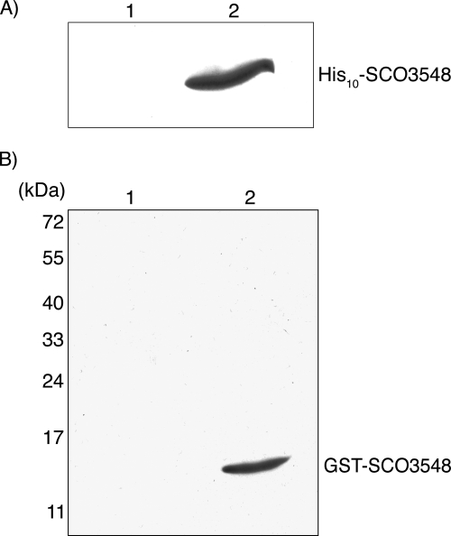
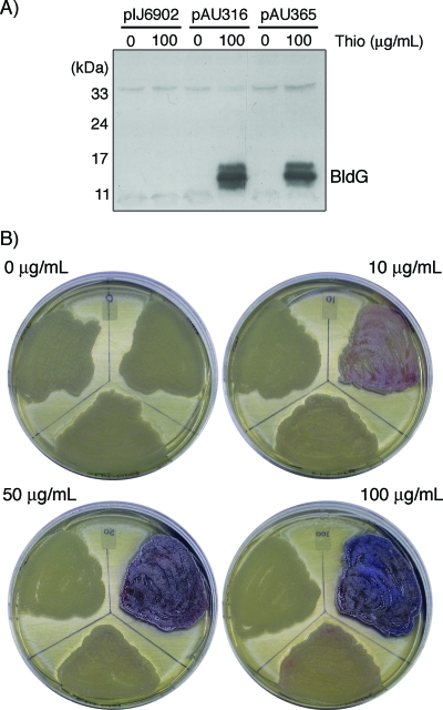
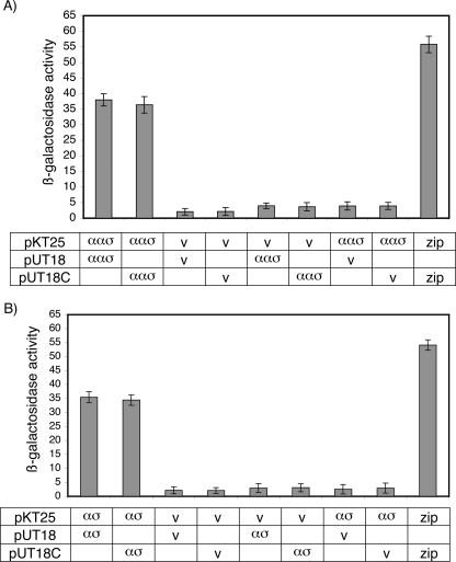
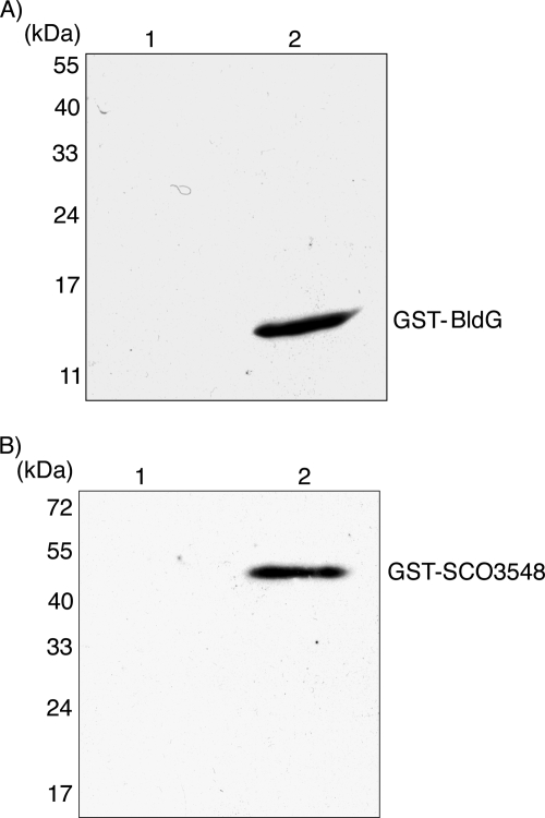
The similarity of BldG and the downstream coexpressed protein SCO3548 to anti-anti-sigma and anti-sigma factors, respectively, together with the phenotype of a bldG mutant, suggests that BldG and SCO3548 interact as part of a regulatory system to control both antibiotic production and morphological differentiation in Streptomyces coelicolor. A combination of bacterial two-hybrid, affinity purification, and far-Western analyses demonstrated that there was self-interaction of both BldG and SCO3548, as well as a direct interaction between the two proteins. Furthermore, a genetic complementation experiment demonstrated that SCO3548 antagonizes the function of BldG, similar to other anti-anti-sigma/anti-sigma factor pairs. It is therefore proposed that BldG and SCO3548 form a partner-switching pair that regulates the function of one or more sigma factors in S. coelicolor. The conservation of bldG and sco3548 in other streptomycetes demonstrates that this system is likely a key regulatory switch controlling developmental processes throughout the genus Streptomyces.
Figures
Similar articles
-
The anti-anti-sigma factor BldG is involved in activation of the stress response sigma factor σ(H) in Streptomyces coelicolor A3(2).J Bacteriol. 2010 Nov;192(21):5674-81. doi: 10.1128/JB.00828-10. Epub 2010 Sep 3. J Bacteriol. 2010. PMID: 20817765 Free PMC article.
-
The σ(F)-specific anti-sigma factor RsfA is one of the protein kinases that phosphorylates the pleiotropic anti-anti-sigma factor BldG in Streptomyces coelicolor A3(2).Gene. 2014 Apr 1;538(2):280-7. doi: 10.1016/j.gene.2014.01.041. Epub 2014 Jan 23. Gene. 2014. PMID: 24462756
-
Pleiotropic anti-anti-sigma factor BldG is phosphorylated by several anti-sigma factor kinases in the process of activating multiple sigma factors in Streptomyces coelicolor A3(2).Gene. 2020 Sep 10;755:144883. doi: 10.1016/j.gene.2020.144883. Epub 2020 Jun 18. Gene. 2020. PMID: 32565321
-
Expression of ccaR, encoding the positive activator of cephamycin C and clavulanic acid production in Streptomyces clavuligerus, is dependent on bldG.Antimicrob Agents Chemother. 2005 Apr;49(4):1529-41. doi: 10.1128/AAC.49.4.1529-1541.2005. Antimicrob Agents Chemother. 2005. PMID: 15793135 Free PMC article.
-
RNA degradation and the regulation of antibiotic synthesis in Streptomyces.Future Microbiol. 2010 Mar;5(3):419-29. doi: 10.2217/fmb.10.14. Future Microbiol. 2010. PMID: 20210552 Review.
Cited by
-
The anti-anti-sigma factor BldG is involved in activation of the stress response sigma factor σ(H) in Streptomyces coelicolor A3(2).J Bacteriol. 2010 Nov;192(21):5674-81. doi: 10.1128/JB.00828-10. Epub 2010 Sep 3. J Bacteriol. 2010. PMID: 20817765 Free PMC article.
-
A Novel AdpA Homologue Negatively Regulates Morphological Differentiation in Streptomyces xiamenensis 318.Appl Environ Microbiol. 2019 Mar 22;85(7):e03107-18. doi: 10.1128/AEM.03107-18. Print 2019 Apr 1. Appl Environ Microbiol. 2019. PMID: 30683747 Free PMC article.
-
Signals and regulators that govern Streptomyces development.FEMS Microbiol Rev. 2012 Jan;36(1):206-31. doi: 10.1111/j.1574-6976.2011.00317.x. Epub 2011 Dec 2. FEMS Microbiol Rev. 2012. PMID: 22092088 Free PMC article. Review.
-
Identification of a predicted partner-switching system that affects production of the gene transfer agent RcGTA and stationary phase viability in Rhodobacter capsulatus.BMC Microbiol. 2014 Mar 19;14:71. doi: 10.1186/1471-2180-14-71. BMC Microbiol. 2014. PMID: 24645667 Free PMC article.
-
Proteomics analysis of global regulatory cascades involved in clavulanic acid production and morphological development in Streptomyces clavuligerus.J Ind Microbiol Biotechnol. 2016 Apr;43(4):537-55. doi: 10.1007/s10295-016-1733-y. Epub 2016 Jan 20. J Ind Microbiol Biotechnol. 2016. PMID: 26790415
References
-
- Alper, S., A. Dufour, D. A. Garsin, L. Duncan, and R. Losick. 1996. Role of adenosine nucleotides in the regulation of a stress-response transcription factor in Bacillus subtilis. J. Mol. Biol. 260165-177. - PubMed
-
- Alper, S., L. Duncan, and R. Losick. 1994. An adenosine nucleotide switch controlling the activity of a cell type-specific transcription factor in B. subtilis. Cell 77195-205. - PubMed
-
- Aravind, L., and E. V. Koonin. 2000. The STAS domain—a link between anion transporters and anti-sigma-factor antagonists. Curr. Biol. 10R53-R55. - PubMed
-
- Beaucher, J., S. Rodrigue, P. E. Jacques, I. Smith, R. Brzezinski, and L. Gaudreau. 2002. Novel Mycobacterium tuberculosis anti-σ factor antagonists control σF activity by distinct mechanisms. Mol. Microbiol. 451527-1540. - PubMed
Publication types
MeSH terms
Substances
LinkOut - more resources
Full Text Sources
Medical
Molecular Biology Databases
