. 2009 Apr;191(8):2656-67.
doi: 10.1128/JB.01689-08. Epub 2009 Feb 6.

Activation of the Campylobacter jejuni FlgSR two-component system is linked to the flagellar export apparatus

Affiliations

Activation of the Campylobacter jejuni FlgSR two-component system is linked to the flagellar export apparatus

Stephanie N Joslin et al. J Bacteriol. 2009 Apr.

Abstract

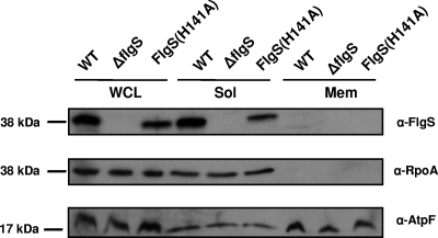
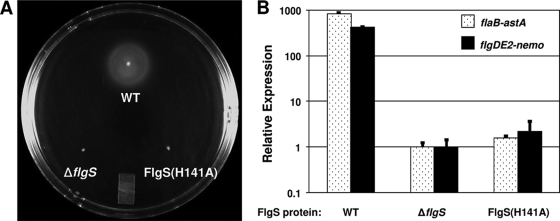
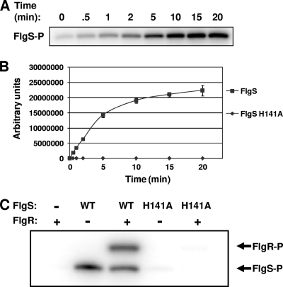
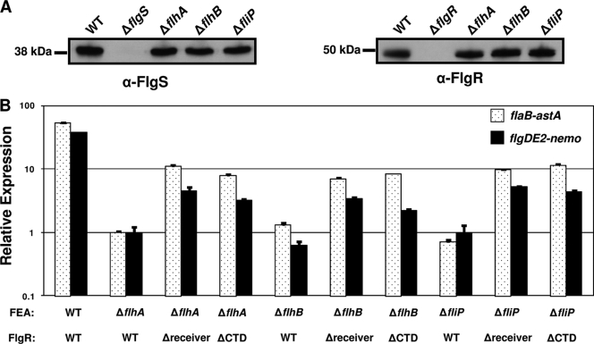
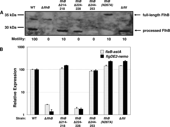
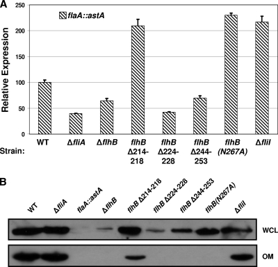
Activation of sigma(54)-dependent gene expression essential for formation of flagella in Campylobacter jejuni requires the components of the inner membrane-localized flagellar export apparatus and the FlgSR two-component regulatory system. In this study, we characterized the FlgS sensor kinase and how activation of the protein is linked to the flagellar export apparatus. We found that FlgS is localized to the C. jejuni cytoplasm and that His141 of FlgS is essential for autophosphorylation, phosphorelay to the cognate FlgR response regulator, motility, and expression of sigma(54)-dependent flagellar genes. Mutants with incomplete flagellar export apparatuses produced wild-type levels of FlgS and FlgR, but they were defective for signaling through the FlgSR system. By using genetic approaches, we found that FlgSR activity is linked to and downstream of the flagellar export apparatus in a regulatory cascade that terminates in expression of sigma(54)-dependent flagellar genes. By analyzing defined flhB and fliI mutants of C. jejuni that form flagellar export apparatuses that are secretion incompetent, we determined that formation of the apparatus is required to contribute to the signal sensed by FlgS to terminate in activation of expression of sigma(54)-dependent flagellar genes. Considering that the flagellar export apparatuses of Escherichia coli and Salmonella species influence sigma(28)-dependent flagellar gene expression, our work expands the signaling activity of the apparatuses to include sigma(54)-dependent pathways of C. jejuni and possibly other motile bacteria. This study indicates that these apparatuses have broader functions beyond flagellar protein secretion, including activation of essential two-component regulatory systems required for expression of sigma(54)-dependent flagellar genes.


Figures

FIG. 1.
FIG. 1.
Localization and stability of FlgS proteins in C. jejuni. Wild-type strain C. jejuni 81-176 Smr (DRH212) (WT), 81-176 Smr ΔflgS (DRH460), and 81-176 Smr flgS(H141A) (DRH1323) were grown, and protein samples were obtained from the WCL, the soluble fraction (Sol), and the insoluble membrane fraction (Mem) after sonication. Anti-FlgS Rab11 antiserum (α-FlgS) was used to detect FlgS proteins (25). Anti-RpoA M59 antiserum (α-RpoA) and anti-AtpF M3 antiserum (α-AtpF) were used to detect the soluble cytoplasmic RpoA protein and the insoluble inner membrane protein AtpF, respectively (4).
FIG. 2.
FIG. 2.
Phenotypic analyses of C. jejuni wild-type and flgS(H141A) mutant strains. (A) Motility phenotypes of C. jejuni strains producing wild-type or mutant FlgS proteins in MH semisolid agar 24 h after inoculation. The strains used included wild-type strain 81-176 Smr (DRH212) (WT), 81-176 Smr ΔflgS (DRH460), and 81-176 Smr flgS(H141A) (DRH1323). (B) Arylsulfatase assays for analysis of expression of flaB::astA and flgDE2::nemo in C. jejuni 81-176 derivatives producing wild-type and FlgS mutant proteins. The results are the results of a typical assay in which each strain was tested in triplicate. The values reported for each strain are the average arylsulfatase activity ± standard deviation relative to the level of expression of each transcriptional fusion in 81-176 Smr ΔastA ΔflgS, which was defined as 1 arylsulfatase unit. For expression of flaB::astA, the strains used included wild-type strain DRH665 (81-176 Smr ΔastA flaB::astA) (WT), DRH939 (81-176 Smr ΔastA ΔflgS flaB::astA), and SNJ958 [81-176 Smr ΔastA flgS(H141A) flaB::astA]. For expression of flgDE2::nemo, the strains used included wild-type strain DRH533 (81-176 Smr ΔastA flgDE2::nemo) (WT), DRH936 (81-176 Smr ΔastA ΔflgS flgDE2::nemo), and SNJ956 [81-176 Smr ΔastA flgS(H141A) flgDE2::nemo]. The FlgS proteins produced by the strains are indicated below the graph.
FIG. 3.
FIG. 3.
Autophosphorylation of FlgS proteins and phosphorelay to FlgR. (A and B) Analysis of autophosphorylation of His6-FlgS and His6-FlgS(H141A) over time after incubation of proteins with [γ-32P]ATP. (A) Representative gel analyzed by autoradiography from an FlgS autophosphorylation assay. (B) Relative quantification of autophosphorylation of FlgS proteins as determined by densitometry after autoradiography of gels. Three separate FlgS and FlgS(H141A) autophosphorylation assays were performed, and the results of these assays were averaged. The amount of incorporation of 32P is expressed in arbitrary units based on the densitometric analysis. (C) Analysis of phosphorelay to His6-FlgR from His6-tagged FlgS or FlgS(H141A) protein. FlgS proteins were preincubated with [γ-32P]ATP before addition of His6-FlgR. A representative gel analyzed by autoradiography from a phosphotransfer assay is shown. The presence (+) or absence (−) of FlgR and the FlgS protein used in each reaction are indicated above the lanes. WT, wild type.
FIG. 4.
FIG. 4.
Production of FlgS and FlgR and activity of FlgR proteins in FEA mutants of C. jejuni. (A) Production of FlgS and FlgR proteins in mutants of C. jejuni lacking one component of the FEA. WCLs of wild-type and C. jejuni mutant strains were prepared for immunoblot analysis. Anti-FlgS Rab11 (α-FlgS) and anti-FlgR (α-FlgR) Rab13 antisera were used to detect the FlgS (left gel) and FlgR (right gel) proteins (25). The strains used for analysis included wild-type strain DRH212 (81-176 Smr) (WT), DRH460 (81-176 Smr ΔflgS), DRH737 (81-176 Smr ΔflgR), DRH946 (81-176 Smr ΔflhA), SNJ471 (81-176 Smr ΔflhB), and DRH1065 (81-176 Smr ΔfliP). (B) Arylsulfatase assays for analysis of expression of flaB::astA and flgDE2::nemo in the C. jejuni 81-176 Smr wild-type strain and mutant strains lacking a component of the FEA and producing wild-type and FlgR mutant proteins. The results are the results of a typical assay in which each strain was tested in triplicate. The values reported for each strain are the average arylsulfatase activity ± standard deviation relative to the level of expression of each transcriptional fusion in 81-176 Smr ΔastA ΔflhA, which was defined as 1 arylsulfatase unit. For expression of flaB::astA, the strains used included (from left to right) wild-type strain DRH665, DRH1049, SNJ112, SNJ273, DRH1827, SNJ109, SNJ1021, DRH1178, SNJ261, and SNJ1015. For expression of flgDE2::nemo, the strains used included (from left to right) wild-type strain DRH533, DRH1021, SNJ115, SNJ274, DRH1827, SNJ113, SNJ1017, DRH1204, SNJ358, and SNJ1012. The FEA mutation and the type of FlgR protein produced in each strain are indicated below the graph. WT, wild type.
FIG. 5.
FIG. 5.
Phenotypic analyses of C. jejuni strains with formed but secretion-impaired FEA complexes. (A) Immunoblot analysis of FlhB proteins and motility phenotypes of C. jejuni wild-type and flhB or fliI mutant strains. Total membrane proteins were isolated from wild-type and mutant strains of C. jejuni. Equal amounts of proteins from the strains were analyzed. Anti-FlhB Rab476 antiserum was used to detect the FlhB proteins. The arrows indicate the positions of the 37-kDa full-length, unprocessed FlhB protein and the 30-kDa processed FlhB protein. The motility phenotypes of wild-type and mutant strains are indicated below the blot. The diameter of the motile ring around the point of inoculation in MH semisolid agar was measured after 36 h of incubation at 37°C under microaerobic conditions. The level of motility of each mutants is expressed relative to the level of motility of the wild-type strain, which was defined as 100%. The strains used for both analyses included (from left to right) wild-type strain DRH461 (WT), DRH1734, SNJ464, SNJ428, SNJ475, and SNJ438. (B) Arylsulfatase assays for analysis of expression of flaB::astA and flgDE2::nemo in C. jejuni 81-176 Smr wild-type or mutant strains containing a secretion-impaired FEA. The results are the results of a typical assay in which each strain was tested in triplicate. The values reported for each strain are the average arylsulfatase activity ± standard deviation relative to the level of expression of each transcriptional fusion in wild-type strain 81-176 Smr ΔastA, which was defined as 100 arylsulfatase units. For expression of flaB::astA, the strains used included (from left to right) wild-type strain DRH665 (WT), DRH1830, SNJ467, SNJ434, SNJ508, SNJ442, and SNJ422. For expression of flgDE2::nemo, the strains used included wild-type strain DRH533 (WT), DRH1827, SNJ466, SNJ433, SNJ504, SNJ439, and SNJ457. The type of mutation in the FEA of each strain is indicated below the graph.
FIG. 6.
FIG. 6.
Analysis of flaA expression and FlaA secretion mediated by the FEA. (A) Arylsulfatase assays for analysis of expression of flaA::astA in the C. jejuni 81-176 Smr wild-type strain or strains with a secretion-impaired FEA. The results are the results of a typical assay in which each strain was tested in triplicate. The values for each strain are the average arylsulfatase activity ± standard deviation relative to the level of expression of each transcriptional fusion in wild-type strain 81-176 Smr ΔastA, which was defined as 100 arylsulfatase units. For expression of flaA::astA, the strains used included (from left to right) wild-type strain DRH655 (WT), DRH1070, SNJ365, SNJ427, SNJ1033, SNJ1034, SNJ1038, and SNJ1042. The type of mutation in each strain is indicated below the graph. (B) Immunoblot analysis of FlaA production in WCLs and secretion to the outer membrane of wild-type and FEA mutant strains. WCL and outer membrane (OM) fractions were isolated from wild-type and mutant strains of C. jejuni. Anti-FlaA LL-1 antiserum was used to detect the FlaA proteins (42). The strains used included (from left to right) wild-type strain DRH212 (WT), DRH724, DRH655, SNJ471, SNJ464, SNJ428, SNJ475, SNJ438, and DRH2257.

Similar articles

Cited by

References

    1. Akerley, B. J., and D. J. Lampe. 2002. Analysis of gene function in bacterial pathogens by GAMBIT. Methods Enzymol. 358100-108. - PubMed
    1. Arora, S. K., B. W. Ritchings, E. C. Almira, S. Lory, and R. Ramphal. 1997. A transcriptional activator, FleQ, regulates mucin adhesion and flagellar gene expression in Pseudomonas aeruginosa in a cascade manner. J. Bacteriol. 1795574-5581. - PMC - PubMed
    1. Baker, M. D., P. M. Wolanin, and J. B. Stock. 2006. Signal transduction in bacterial chemotaxis. Bioessays 289-22. - PubMed
    1. Bingham-Ramos, L. K., and D. R. Hendrixson. 2008. Characterization of two putative cytochrome c peroxidases of Campylobacter jejuni involved in promoting commensal colonization of poultry. Infect. Immun. 761105-1114. - PMC - PubMed
    1. Black, R. E., M. M. Levine, M. L. Clements, T. P. Hughes, and M. J. Blaser. 1988. Experimental Campylobacter jejuni infection in humans. J. Infect. Dis. 157472-479. - PubMed

Publication types

Substances

LinkOut - more resources