. 2009 Apr;191(8):2753-63.
doi: 10.1128/JB.01818-08. Epub 2009 Feb 6.

Combinatorial regulation by a novel arrangement of FruA and MrpC2 transcription factors during Myxococcus xanthus development

Affiliations

Combinatorial regulation by a novel arrangement of FruA and MrpC2 transcription factors during Myxococcus xanthus development

Sheenu Mittal et al. J Bacteriol. 2009 Apr.

Abstract

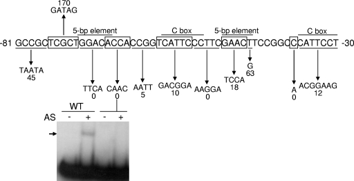
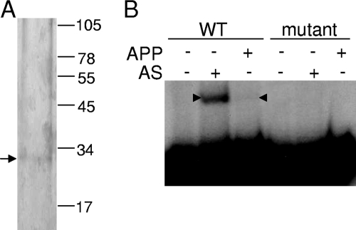
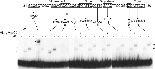
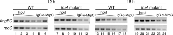
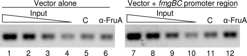
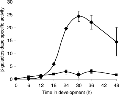
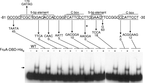
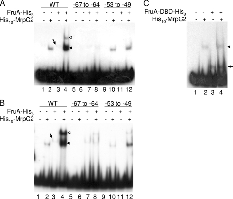
Myxococcus xanthus is a gram-negative soil bacterium that undergoes multicellular development upon nutrient limitation. Intercellular signals control cell movements and regulate gene expression during the developmental process. C-signal is a short-range signal essential for aggregation and sporulation. C-signaling regulates the fmgA gene by a novel mechanism involving cooperative binding of the response regulator FruA and the transcription factor/antitoxin MrpC2. Here, we demonstrate that regulation of the C-signal-dependent fmgBC operon is under similar combinatorial control by FruA and MrpC2, but the arrangement of binding sites is different than in the fmgA promoter region. MrpC2 was shown to bind to a crucial cis-regulatory sequence in the fmgBC promoter region. FruA was required for MrpC and/or MrpC2 to associate with the fmgBC promoter region in vivo, and expression of an fmgB-lacZ fusion was abolished in a fruA mutant. Recombinant FruA was shown to bind to an essential regulatory sequence located slightly downstream of the MrpC2-binding site in the fmgBC promoter region. Full-length FruA, but not its C-terminal DNA-binding domain, enhanced the formation of complexes with fmgBC promoter region DNA, when combined with MrpC2. This effect was nearly abolished with fmgBC DNA fragments having a mutation in either the MrpC2- or FruA-binding site, indicating that binding of both proteins to DNA is important for enhancement of complex formation. These results are similar to those observed for fmgA, where FruA and MrpC2 bind cooperatively upstream of the promoter, except that in the fmgA promoter region the FruA-binding site is located slightly upstream of the MrpC2-binding site. Cooperative binding of FruA and MrpC2 appears to be a conserved mechanism of gene regulation that allows a flexible arrangement of binding sites and coordinates multiple signaling pathways.


Figures

FIG. 1.
FIG. 1.
Effects of mutations on fmgBC promoter activity in vivo and on DNA binding in vitro. The top part of the figure shows a summary of mutational effects on developmental fmgB-lacZ expression (67). The wild-type fmgBC upstream sequence is alternately boxed or underlined to indicate changed sequences, which are shown below the downward arrows. The number beneath each mutant sequence indicates the maximum β-galactosidase activity during development, expressed as a percentage of the maximum activity observed for the wild-type promoter. The bottom part shows EMSAs performed with 32P-labeled fmgBC DNA (12 nM) spanning from positions −104 to −29 and proteins in the AS fraction (0.7 μg/μl). The arrow indicates the shifted complex produced by incubating the wild-type (WT) DNA fragment with the AS fraction. No complex was observed with a DNA fragment bearing the indicated mutation at positions −67 to −64.
FIG. 2.
FIG. 2.
DNA-affinity purification of protein that binds to the fmgBC promoter region. (A) Sodium dodecyl sulfate-polyacrylamide gel electrophoresis of protein purified from the AS fraction using fmgBC DNA (positions −104 to −29). The arrow indicates the major species in the APP after staining with silver. The numbers indicate the migration positions of molecular mass (in kilodaltons) standards. (B) EMSAs with 32P-labeled fmgBC DNA (12 nM) spanning from positions −104 to −29 and proteins in the AS fraction or the APP. Arrowheads indicate the shifted complexes produced with the wild-type (WT) DNA fragment. No complex was observed with a DNA fragment bearing the ACCA to CAAC mutation at positions −67 to −64 (mutant).
FIG. 3.
FIG. 3.
Comparison of purified His10-MrpC2 and the AS fraction for binding to the fmgBC promoter region. EMSAs with 32P-labeled fmgBC DNA (2 nM) spanning from positions −104 to −29, wild-type (WT), or mutant as indicated and His10-MrpC2 (1 μM) or the AS fraction (0.7 μg/μl). An asterisk indicates the mutation from positions −67 to −64 that impairs shifted complex formation. Brackets indicate the shifted complexes produced by the AS fraction and His10-MrpC2 upon addition to wild-type fmgBC DNA and to most of the mutant DNA fragments. The mutation from positions −81 to −77 causes a novel shifted complex to form with the AS fraction (arrowhead). The image is a composite from three experiments, and in each experiment the wild-type fmgBC DNA served as a control, and the signal intensity of the shifted complexes was comparable to that shown. The results shown are representative of results observed in at least two experiments.
FIG. 4.
FIG. 4.
Association of MrpC and/or MrpC2 with the fmgBC promoter region during development of wild-type and fruA mutant cells. ChIP analysis of M. xanthus with the fmgBC promoter region (−100 to +50) integrated ectopically in otherwise wild-type (WT) or fruA mutant backgrounds. At 12 and 18 h into development, cells were treated with formaldehyde and lysed, and cross-linked chromatin was immunoprecipitated with anti-MrpC antibodies or IgG as a control. DNA was amplified with appropriate primers for the fmgBC promoter region at the ectopic chromosomal site or with appropriate primers for the rpoC coding region as a control. A twofold dilution series of input DNA purified from 0.25, 0.125, 0.0625, or 0.03125% of the total cellular extract prior to immunoprecipitation was used as a template in parallel PCRs to show that the PCR conditions were in the linear range of amplification for each primer set.
FIG. 5.
FIG. 5.
Association of FruA with the fmgBC promoter region in vivo. ChIP analysis of M. xanthus with the vector alone or with the fmgBC promoter region (−100 to +50) integrated ectopically. At 12 h into development, cells were treated with formaldehyde and lysed, and cross-linked chromatin was immunoprecipitated with anti-FruA antibodies or preimmune serum as a control (lane C). A twofold dilution series of input DNA purified from 0.25, 0.125, 0.0625, or 0.03125% of the total cellular extract prior to immunoprecipitation was used as a template in parallel PCRs to show that the PCR conditions were in the linear range of amplification.
FIG. 6.
FIG. 6.
Developmental expression from fmgB-lacZ. The fmgBC promoter region from positions −100 to +50 was fused to lacZ, and the β-galactosidase specific activity was measured during the development of M. xanthus wild-type (⧫) and fruA mutant (▪) cells. In each background, the activity from the vector with no promoter was measured as a negative control. Points show the average of three transformants, after subtracting the average of three transformants with the promoterless vector. The units of activity are nanomoles of o-nitrophenyl phosphate per minute per milligram of protein. Error bars depict one standard deviation of the data.
FIG. 7.
FIG. 7.
Effects of mutations on binding of FruA-DBD-His8 to fmgBC promoter region DNA. EMSAs with 32P-labeled fmgBC DNA (2 nM) spanning from positions −104 to −29, wild type (WT), or mutant as indicated and FruA-DBD-His8 (14 μM). A horizontal arrow indicates the shifted complex produced with wild-type DNA. An asterisk indicates the mutation from positions −53 to −49 that impairs shifted complex formation. The image is a composite from three experiments, and intervening lanes were removed from one of the images. In each experiment, the wild-type fmgBC DNA served as a control, and the signal intensity of the shifted complex was comparable to that shown.
FIG. 8.
FIG. 8.
EMSAs with MrpC2 and full-length FruA or just the DNA-binding domain of FruA. (A) Shifted complex formation with His10-MrpC2 and full-length FruA-His6 and the effect of mutations. EMSAs with 32P-labeled fmgBC DNA (2 nM) spanning from positions −104 to −29, wild-type (WT), or mutant as indicated and no protein, His10-MrpC2 (1 μM), FruA-His6 (3 μM), or both His10-MrpC2 (1 μM) and FruA-His6 (3 μM) as indicated, electrophoresed on a 5% polyacrylamide gel. A slanted arrow indicates the faint shifted complex produced by FruA-His6 alone. The unfilled and filled arrowheads indicate the UCs and LCs, respectively, produced by the combination of proteins. (B) Same as in panel A except electrophoresed on an 8% polyacrylamide gel. (C) Shifted complex formation with His10-MrpC2 and FruA-DBD-His8. EMSAs with 32P-labeled fmgBC DNA (2 nM) spanning from positions −104 to −29 and no protein, His10-MrpC2 (1 μM), FruA-DBD-His8 (14 μM), or both His10-MrpC2 (1 μM) and FruA-DBD-His8 (14 μM) as indicated. The arrowhead indicates the complex produced by His10-MrpC2, and the arrow indicates the complex produced by FruA-DBD-His8. Intervening lanes were removed from the image.

References

    1. Barnard, A., A. Wolfe, and S. Busby. 2004. Regulation at complex bacterial promoters: how bacteria use different promoter organizations to produce different regulatory outcomes. Curr. Opin. Microbiol. 7102-108. - PubMed
    1. Beatty, C. M., D. F. Browning, S. J. Busby, and A. J. Wolfe. 2003. Cyclic AMP receptor protein-dependent activation of the Escherichia coli acsP2 promoter by a synergistic class III mechanism. J. Bacteriol. 1855148-5157. - PMC - PubMed
    1. Berleman, J. E., and J. R. Kirby. 2007. Multicellular development in Myxococcus xanthus is stimulated by predator-prey interactions. J. Bacteriol. 1895675-5682. - PMC - PubMed
    1. Brandner, J. P., and L. Kroos. 1998. Identification of the Ω4400 regulatory region, a developmental promoter of Myxococcus xanthus. J. Bacteriol. 1801995-2004. - PMC - PubMed
    1. Ellehauge, E., M. Norregaard-Madsen, and L. Sogaard-Andersen. 1998. The FruA signal transduction protein provides a checkpoint for the temporal co-ordination of intercellular signals in Myxococcus xanthus development. Mol. Microbiol. 30807-817. - PubMed

Publication types

MeSH terms

LinkOut - more resources