. 2009 Apr;191(8):2638-48.
doi: 10.1128/JB.01784-08. Epub 2009 Feb 6.

HtaA is an iron-regulated hemin binding protein involved in the utilization of heme iron in Corynebacterium diphtheriae

Affiliations

HtaA is an iron-regulated hemin binding protein involved in the utilization of heme iron in Corynebacterium diphtheriae

Courtni E Allen et al. J Bacteriol. 2009 Apr.

Abstract

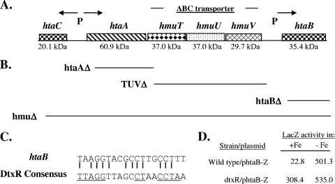
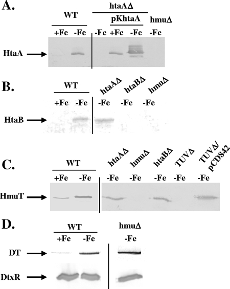
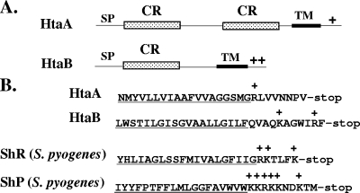
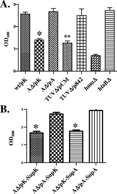
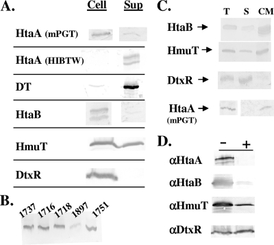
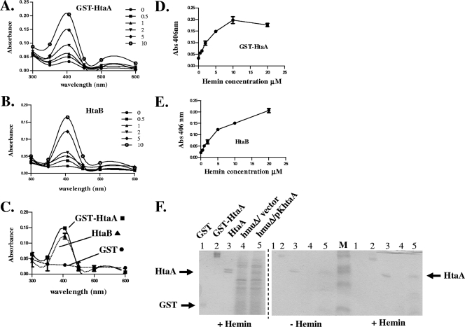
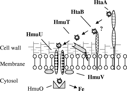
Many human pathogens, including Corynebacterium diphtheriae, the causative agent of diphtheria, use host compounds such as heme and hemoglobin as essential iron sources. In this study, we examined the Corynebacterium hmu hemin transport region, a genetic cluster that contains the hmuTUV genes encoding a previously described ABC-type hemin transporter and three additional genes, which we have designated htaA, htaB, and htaC. The hmu gene cluster is composed of three distinct transcriptional units. The htaA gene appears to be part of an iron- and DtxR-regulated operon that includes hmuTUV, while htaB and htaC are transcribed from unique DtxR-regulated promoters. Nonpolar deletion of either htaA or the hmuTUV genes resulted in a reduced ability to use hemin as an iron source, while deletion of htaB had no effect on hemin iron utilization in C. diphtheriae. A comparison of the predicted amino acid sequences of HtaA and HtaB showed that they share some sequence similarity, and both proteins contain leader sequences and putative C-terminal transmembrane regions. Protein localization studies with C. diphtheriae showed that HtaA is associated predominantly with the cell envelope when the organism is grown in minimal medium but is secreted during growth in nutrient-rich broth. HtaB and HmuT were detected primarily in the cytoplasmic membrane fraction regardless of the growth medium. Hemin binding studies demonstrated that HtaA and HtaB are able to bind hemin, suggesting that these proteins may function as cell surface hemin receptors in C. diphtheriae.


Figures

FIG. 1.
FIG. 1.
(A) Genetic map of the hmu locus in C. diphtheriae. The predicted sizes of the various gene products are indicated below the map. P indicates the presence of a DtxR-regulated promoter, and the arrows indicate the direction of transcription. (B) Regions deleted in the various C. diphtheriae 1737 mutants constructed. The deleted regions are aligned with the genetic map shown in panel A. (C) Alignment of the 19-bp putative DtxR binding site upstream of the C. diphtheriae htaB gene with the consensus DtxR binding site. The underlined sequences are the most highly conserved residues (14). (D) Assessment of the promoter activity (LacZ activity) of an htaB-lacZ transcriptional fusion construct, phtaB-Z, in high-iron HIBTW medium (+Fe) and in iron-depleted conditions (HIBTW medium containing 12 μg/ml EDDA) (−Fe). The values are the means of three experiments. Each result varied less than 15% from the mean. The difference between high- and low-iron conditions for the dtxR mutant was statistically significant (P < 0.05), and this suggests that the point mutant (R47H) maintains some low-level Fe-dependent repressor activity. See Materials and Methods for experimental details.
FIG. 2.
FIG. 2.
Western blot analysis of proteins produced by C. diphtheriae wild-type strain 1737 (WT) and various 1737 deletion mutants. Strains were grown in 1 ml of iron-replete HIBTW medium (+Fe) or iron-depleted medium (HIBTW medium containing 12 μg/ml EDDA) (−Fe), and proteins present either in culture supernatants (HtaA and diphtheria toxin [DT] in panels A and D, respectively) or in whole-cell extracts (HtaB, HmuT, and DtxR in panels B, C, and D, respectively) were detected by Western blot analysis using antiserum raised against the proteins indicated on the left. Samples were normalized using OD600 before SDS gels were loaded. htaAΔ, 1737htaAΔ; TUVΔ, 1737TUVΔ; hmuΔ, 1737hmuΔ; htaBΔ, 1737htaBΔ.
FIG. 3.
FIG. 3.
(A) Comparison of various structural characteristics of the HtaA and HtaB proteins. HtaA and HtaB contain a region of sequence similarity consisting of approximately 200 amino acids designated the conserved region (CR). SP, signal peptide; TM, transmembrane region; +, positively charged residues. (B) Comparison of the amino acid sequences in the C-terminal tail region for HtaA, HtaB, and the surface-anchored hemin binding proteins ShR and ShP from S. pyogenes. The putative membrane-spanning region is underlined, and positively charged residues are indicated by plus signs.
FIG. 4.
FIG. 4.
Utilization of hemoglobin as an iron source by C. diphtheriae wild-type strain 1737 and mutant strains. (A) OD600s of cultures grown for 20 to 22 h in mPGT medium containing 10 μM EDDA and 25 μg/ml hemoglobin (human). The plasmids carried by the various strains included pKhtaA containing the cloned htaA gene (pA), vector pKN2.6Z (pK), vector pCM2.6 (pCM), and pCD842 containing the cloned hmuTUV genes (p842). wt, wild-type strain 1737; AΔ, 1737htaAΔ; TUVΔ, 1737TUVΔ; hmuΔ, 1737hmuΔ; htaBΔ, 1737htaBΔ. (B) Results of experiments performed like the experiments described for panel A, except that cultures of 1737htaAΔ were supplemented with 50 μl of concentrated culture supernatant from 1737hmuΔ that contained either the vector pKN2.6Z (SupK) or the cloned htaA gene on pKhtaA (SupA). The presence of HtaA in culture supernatants in 1737hmuΔ/pKhtaA was confirmed by SDS-PAGE analysis (not shown). The results are the averages and standard deviations of three independent experiments. The values for 1737htaAΔ/pKN2.6Z are significantly different from the values for 1737/pKN2.6Z and 1737htaAΔ/pKhtaA (*, P < 0.05), and the values for 1737TUVΔ/pCM2.6 are significantly different from the values for 1737/pKN2.6Z and 1737TUVΔ/pCD842 (**, P < 0.05).
FIG. 5.
FIG. 5.
(A) Western blot analysis to measure secretion or cell association of proteins expressed from C. diphtheriae wild-type strain 1737. One-milliliter cultures were grown for 20 to 22 h at 37°C in iron-depleted HIBTW medium or in mPGT medium (only results for HtaA are shown for growth in mPGT medium). Polyclonal antiserum (specific to the proteins indicated on the left) was used for detection of proteins associated with the cell pellet (Cell) and the supernatant fraction (Sup). Doublet bands for HtaA and HtaB indicate breakdown products, which exhibited some variability between protein preparations. See Materials and Methods for a description of the method used for sample preparation. DT, diphtheria toxin. (B) Western blot detection of HtaA in culture medium from various C. diphtheriae clinical isolates after growth in iron-depleted HIBTW medium. (C) Western blot analysis to identify the localization of proteins after cell fractionation of C. diphtheriae wild-type strain 1737. Cultures were grown in HIBTW medium or in mPGT medium (only results for HtaA are shown for mPGT medium), and polyclonal antiserum (specific to proteins indicated on the left) was used for detection of total cellular proteins. T, membrane and cytosolic fractions; S, soluble cytosolic proteins; CM, cytoplasmic membrane proteins. The total cellular fraction for HtaA was collected from the whole-cell lysate when the cells were harvested. Samples were loaded using equivalent protein levels. (D) Western blotting to detect protein levels after proteinase K treatment of C. diphtheriae 1737htaΔ/pKhtaA grown in low-iron mPGT medium. The antisera used for detection are indicated on the left (αHtaA, anti-HtaA; αHtaB, anti-HtaB; αHmuT, anti-HmuT; αDtxR, anti-DtxR), and the presence (+) or absence (−) of proteinase K (50 μg/ml) is indicated at the top. See Materials and Methods for a description of experimental details.
FIG. 6.
FIG. 6.
HtaA and HtaB are hemin binding proteins. (A and B) UV-visible spectroscopy was used to examine GST-HtaA (A) or HtaB (B) at a concentration of 3.5 μM in the presence of various hemin concentrations. Absorption scans were done at 300 to 600 nm. (C) Absorption scans for GST-HtaA, HtaB, and GST in the presence of 5 μM hemin. (D and E) Absorption at 406 nm for GST-HtaA (D) and HtaB (E) at various hemin concentrations. Different concentrations of hemin (0 to 20 μM) were incubated with protein (3.5 μM) for at least 15 min at 20 to 25°C prior to measurement of absorbance. The values are the means and standard errors of three independent experiments. (F) Protein preparations were incubated with 0.625 μM hemin (+ Hemin) or with control buffer (− Hemin) prior to separation by SDS-PAGE and staining either with Coomassie blue (left panel) or with TMBZ to detect heme-dependent peroxidase activity as described in Materials and Methods (right panel). Lane 1, purified GST protein; lane 2, purified GST-HtaA fusion; lane 3, purified HtaA; lanes 4 and 5, ammonium sulfate-precipitated supernatant of 1737hmuΔ (hmuΔ) carrying either the vector (lane 4) or the cloned htaA gene on plasmid pKhtaA (lane 5); lane M, molecular weight marker. The + Hemin gel in the right panel (TMBZ stained) is a duplicate of the left panel (Coomassie blue stained).
FIG. 7.
FIG. 7.
Model for hemin transport in C. diphtheriae. It is proposed that hemin initially binds to HtaA, a surface-anchored hemin binding protein that is associated with the cytoplasmic membrane through a C-terminal transmembrane region. Hemin would then be transferred from HtaA to membrane-anchored HtaB (?), or, alternatively, hemin would be passaged directly to the HmuT lipoprotein. Hemin that is bound to HmuT would be transferred to HmuU, which is a membrane-bound permease component of an ABC transporter, and in conjunction with HmuV, an ATPase, would facilitate the movement of hemin into the bacterial cytosol. HmuO is proposed to degrade the intracellular heme and release the iron for cellular metabolism.

Similar articles

Cited by

References

    1. Altschul, S. F., G. Warren, W. Miller, E. W. Meyers, and D. J. Lipman. 1990. Basic local alignment search tool. J. Mol. Biol. 215403-410. - PubMed
    1. Bardsdale, W. L., and A. M. Pappenheimer, Jr. 1954. Phage-host relationships in nontoxigenic and toxigenic diphtheria bacilli. J. Bacteriol. 67220-232. - PMC - PubMed
    1. Bates, C. S., G. E. Montanez, C. R. Woods, R. M. Vincent, and Z. Eichenbaum. 2003. Identification and characterization of a Streptococcus pyogenes operon involved in binding of hemoproteins and acquisition of iron. Infect. Immun. 711042-1055. - PMC - PubMed
    1. Boyd, J. M., O. N. Manish, and J. R. Murphy. 1990. Molecular cloning and DNA sequence analysis of a diphtheria tox iron-dependent regulatory element (dtxR) from Corynebacterium diphtheriae. Proc. Natl. Acad. Sci. USA 875968-5972. - PMC - PubMed
    1. Braun, V. 2005. Bacterial iron transport related to virulence. Contrib. Microbiol. 12210-233. - PubMed

MeSH terms