. 2009 Apr;296(4):L674-83.
doi: 10.1152/ajplung.90585.2008. Epub 2009 Feb 6.

Ozone modulates IL-6 secretion in human airway epithelial and smooth muscle cells

Affiliations

Ozone modulates IL-6 secretion in human airway epithelial and smooth muscle cells

Gautam Damera et al. Am J Physiol Lung Cell Mol Physiol. 2009 Apr.

Abstract

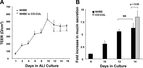
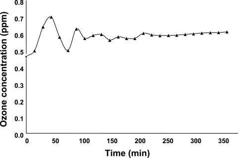
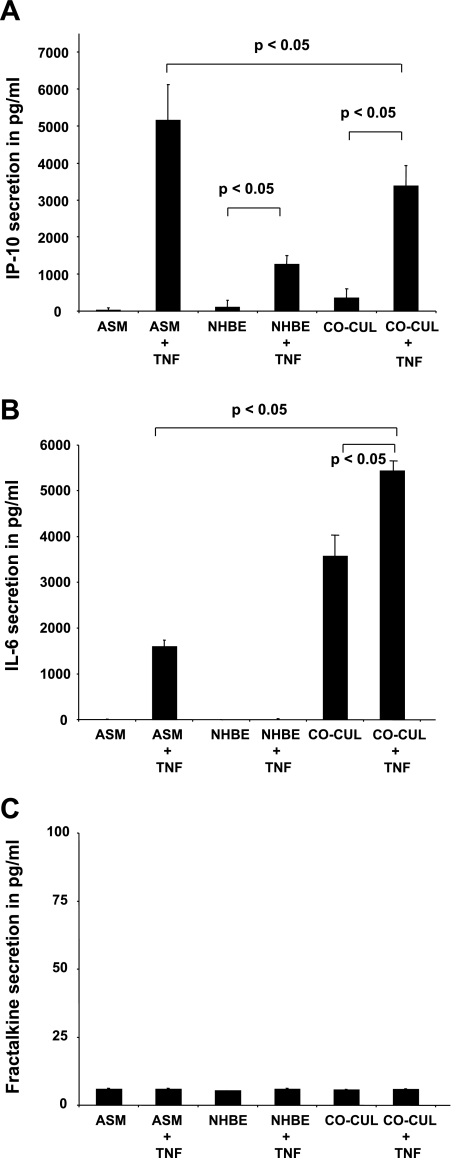
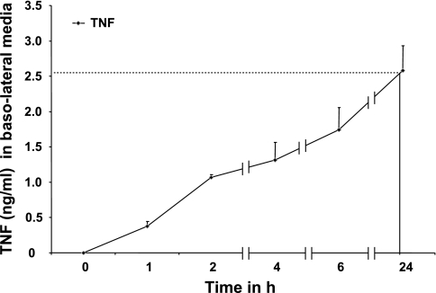
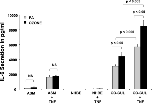
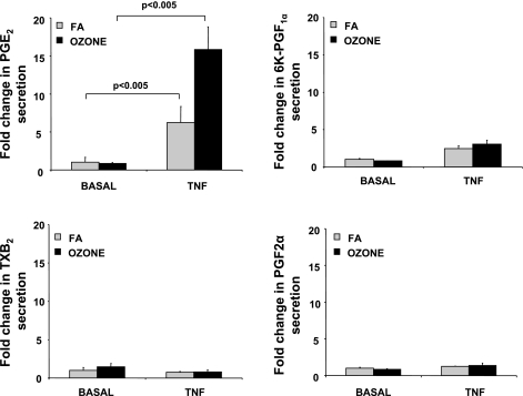
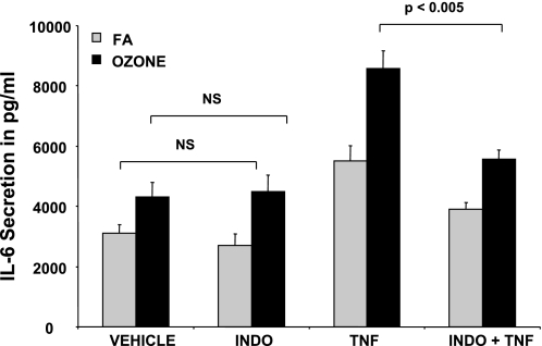
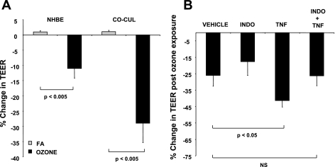
Although ozone enhances leukocyte function and recruitment in airways, the direct effect of ozone in modulating structural cell-derived inflammatory mediators remains unknown. Using a coculture model comprised of differentiated human airway epithelial cells (NHBE) and smooth muscle cells (ASM), we postulate that ozone regulates IL-6 secretion in basal and cytokine-primed structural cells. Air-liquid interface (ALI) cultures of NHBE cells underwent differentiation as determined by mucin secretion, transepithelial electrical resistance (TEER), and ultrastructure parameters. Whereas TNF enhanced basal secretion of IL-6 (57 +/- 3%), ozone exposure at 0.6 ppm for 6 h augmented IL-6 levels in basal (41 +/- 3%) and TNF- (50 +/- 5%) primed cocultures compared with that derived from NHBE or ASM monolayers alone. Levels of PGE(2), 6-keto-PGF(1alpha), PGF(2alpha), and thromboxane B(2) (TxB(2)) levels in basal and TNF-primed cocultures revealed that ozone selectively enhanced PGE(2) production in TNF- (6 +/- 3-fold) primed cocultures, with little effect (P > 0.05) on diluent-treated cultures. In accordance with ozone-induced increases in PGE(2) levels, cyclooxygenase inhibition with indomethacin partially abolished IL-6 secretion. Surprisingly, indomethacin had little effect on constitutive secretion of IL-6 in cocultures, whereas indomethacin completely restored ozone-mediated TEER reduction in TNF-primed cocultures. Collectively, our data for the first time suggest a dual role of ozone in modulating IL-6 secretion and TEER outcomes in a PGE(2)-dependent (in presence of TNF stimulus) and -independent manner (in absence of cytokine stimulus).


Figures

Fig. 1.
Fig. 1.
Normal human bronchial epithelial (NHBE) cell differentiation parameters in air-liquid interface (ALI) cultures. A: transepithelial electrical resistance (TEER) measurements across NHBE monolayers at ALIs for 18 days (n = 6). Solid lines indicate TEER values across NHBE monolayers in singular cultures of NHBE cells alone. Dotted lines indicate TEER values across NHBE monolayers in NHBE-airway smooth muscle (ASM) cell coculture setup (CO-CUL). B: assessment of NHBE monolayer apical mucin secretion by ELISA. Data represent means ± SE from 3 separate experiments done in triplicate. Statistical evaluation was performed by ANOVA. P > 0.05, significant difference. NS, no significant change.
Fig. 2.
Fig. 2.
Evaluation of ozone concentrations in experiments (parts per million) as a function of time (minutes).
Fig. 3.
Fig. 3.
Synthetic responses in basal and TNF-stimulated NHBE-ASM cocultures and in NHBE and ASM cells cultured alone. ELISAs were performed for inducible protein 10 (IP-10) secretion (A), IL-6 secretion (B), and fractalkine secretion (C) in ASM, NHBE, and NHBE-ASM cocultures from basolateral media following apical instillation of vehicle or TNF (10 ng/ml) for 24 h. Summarization of 3 separate experiments performed in triplicate. Data represent means ± SE from 3 separate experiments done in triplicate. Statistical evaluation was performed by ANOVA. P > 0.05, significant difference.
Fig. 4.
Fig. 4.
Time course of TNF permeability across NHBE monolayers at ALIs. TNF (10 ng/ml) was added at the apical surface of NHBE monolayers, and passage of TNF concentrations was determined in the basolateral media at various time points by ELISA. Data represent means ± SE from 3 separate experiments done in triplicate. Statistical evaluation was performed by ANOVA.
Fig. 5.
Fig. 5.
Ozone effects on IL-6 secretion in basal and TNF-stimulated NHBE-ASM cocultures and in NHBE and ASM cells cultured alone. ASM, NHBE, and NHBE-ASM cocultures were treated with TNF (10 ng/ml) or vehicle alone (gray bars) and post-exposed to ozone at 6 ppm (black bars) for 6 h. Cultures were reincubated for an additional 18 h in a 5% CO2-rich incubator and assayed for IL-6 secretion. Data represent means ± SE from 3 separate experiments done in triplicate. Statistical evaluation was performed by ANOVA. P > 0.05, significant difference. FA, forced air.
Fig. 6.
Fig. 6.
Ozone modulates PGE2 levels in basal and TNF-stimulated NHBE-ASM cultures. NHBE-ASM cultures treated with TNF (10 ng/ml) or vehicle alone were exposed to ozone (black bars) or FA (gray bars) for 6 h. Immediately after this period, basolateral media was assayed by liquid chromatography-mass spectrometry (LC/MS) methodology as described in methods. For each prostanoid, data are expressed as fold changes over ozone-unexposed cultures. Data represent means ± SE from 3 separate experiments done in triplicate. Statistical evaluation was performed by ANOVA. P > 0.05, significant difference. 6K-PGF, 6-keto-PGF; TxB2, thromboxane B2.
Fig. 7.
Fig. 7.
Indomethacin (INDO) abrogates IL-6 secretion in ozone-exposed cocultures. NHBE-ASM cultures were pretreated with indomethacin (10 μM) or DMSO in the presence or absence of TNF (10 ng/ml) and postexposed to ozone (black bars) or ambient air (gray bars) for 6 h. IL-6 levels were measured by ELISA 18 h post-ozone exposure. Data represent means ± SE from 3 separate experiments performed in triplicate. Statistical evaluation was performed by ANOVA. P > 0.05, significant difference.
Fig. 8.
Fig. 8.
A: ozone diminishes TEER in NHBE mono- and NHBE-ASM cocultures. TEER values were determined in mono- or cocultures of NHBE cells after exposing them to ozone (0.6 ppm) or FA for 6 h. Data are represented as % changes in TEER. B: indomethacin prevents an ozone-induced decrease in TEER within NHBE monolayers. TEER values were estimated pre- and post-ozone exposure in NHBE-ASM cocultures that were treated with indomethacin (10 μM) or DMSO in the presence or absence of TNF (10 ng/ml). Data show % change in TEER from NHBE monolayers pre-and post-ozone exposure. All data are a summarization of 3 separate experiments performed in triplicate. Statistical evaluation was performed by ANOVA. P > 0.05, significant difference.

Similar articles

Cited by

References

    1. Alpert SE, Walenga RW. Ozone exposure of human tracheal epithelial cells inactivates cyclooxygenase and increases 15-HETE production. Am J Physiol Lung Cell Mol Physiol 269: L734–L743, 1995. - PubMed
    1. Amrani Y, Krymskaya V, Maki C, Panettieri RA Jr. Mechanisms underlying TNFα effects on agonist-mediated calcium homeostasis in human airway smooth muscle cells. Am J Physiol Lung Cell Mol Physiol 273: L1020–L1028, 1997. - PubMed
    1. Amrani Y, Tliba O, Deshpande DA, Walseth TF, Kannan MS, Panettieri RA Jr. Bronchial hyperresponsiveness: insights into new signaling molecules. Curr Opin Pharmacol 4: 230–234, 2004. - PubMed
    1. Anderson JM, Van Itallie CM. Tight junctions and the molecular basis for regulation of paracellular permeability. Am J Physiol Gastrointest Liver Physiol 269: G467–G475, 1995. - PubMed
    1. Appleby SB, Ristimaki A, Neilson K, Narko K, Hla T. Structure of the human cyclo-oxygenase-2 gene. Biochem J 302: 723–727, 1994. - PMC - PubMed

Publication types

MeSH terms