. 2009 Feb 15;182(4):1929-39.
doi: 10.4049/jimmunol.0802703.

The expression of heparin-binding epidermal growth factor-like growth factor by regulatory macrophages

Affiliations

The expression of heparin-binding epidermal growth factor-like growth factor by regulatory macrophages

Justin P Edwards et al. J Immunol. .

Abstract

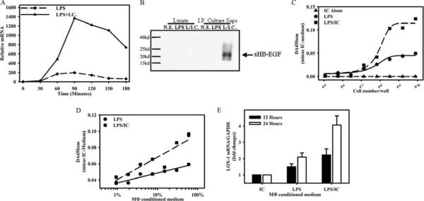
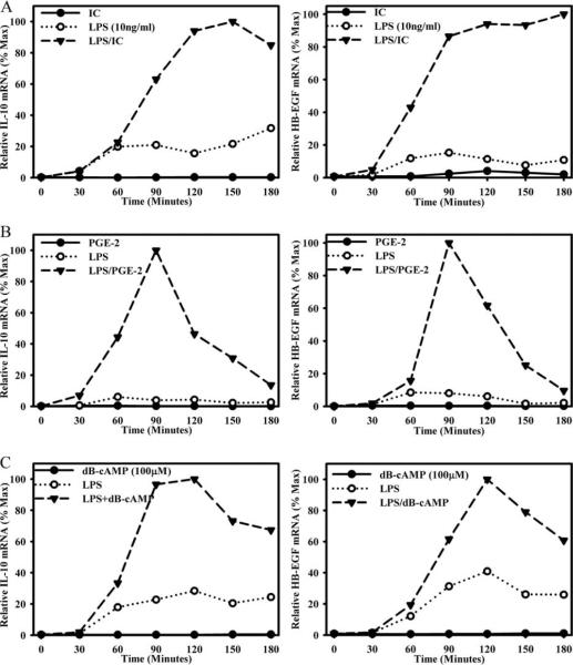
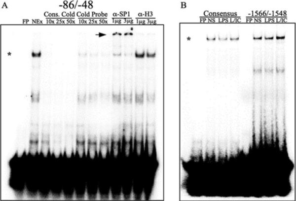
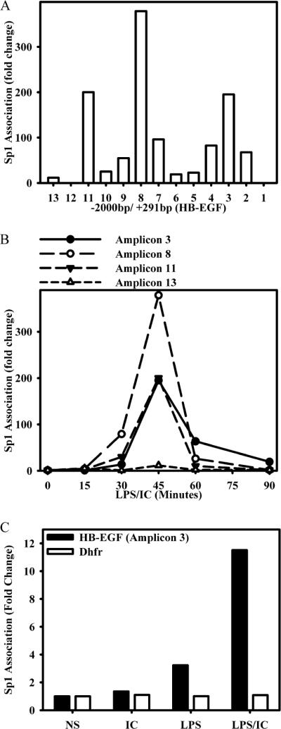
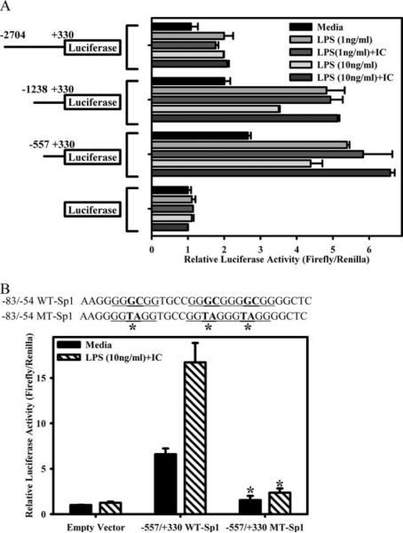
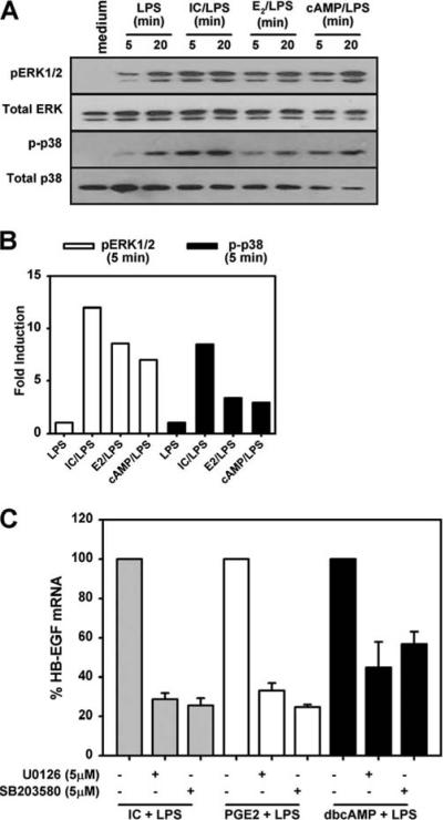
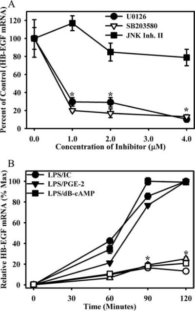
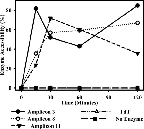
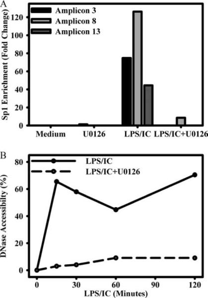
We previously described a population of regulatory macrophages that produced high levels of IL-10 and low levels of IL-12/23. We now describe and characterize the expression of heparin-binding epidermal growth factor (EGF)-like growth factor (HB-EGF) by these macrophages. HB-EGF has previously been associated with a number of physiological and pathological conditions, including tumor growth and angiogenesis. The induction of HB-EGF in regulatory macrophages is due to new transcription and not to increased mRNA stability. The transcription factor Sp1 is a major factor in HB-EGF production, and knockdown of Sp1 substantially diminishes HB-EGF production. Sp1 was recruited to three sites within the first 2 kb of the HB-EGF promoter following stimulation, and the site located at -83/-54 was required for HB-EGF promoter activity. These regions of the promoter become more accessible to endonuclease activity following macrophage activation, and this accessibility was contingent on activation of the MAPK, ERK. We show that several experimental manipulations that give rise to regulatory macrophages also result in HB-EGF production. These observations indicate that in addition to the secretion of the anti-inflammatory cytokine IL-10, another novel characteristic of regulatory macrophages is the production of angiogenic HB-EGF.


Figures

FIGURE 1
FIGURE 1
HB-EGF is induced in BMMϕs in response to LPS and ICs. A, QRT-PCR of HB-EGF expression in BMMϕs after stimulation with LPS (10 ng/ml) alone or LPS plus IgG-OVA ICs. QRT-PCR for HB-EGF mRNA is expressed as a fold change over unstimulated conditions (0 min). B, Western blot for HB-EGF in cell lysates or immunoprecipitated (I.P.) from cell culture supernatants (Sups.) of unstimulated (NS) macrophages (M–) or macrophages stimulated with LPS (10 ng/ml) or LPS plus IC for 16 h. Values are representative of at least three independent experiments. Conditioned medium from IC plus LPS-stimulated macrophages potentiates growth and LOX-1 expression on vascular SMCs. C-E, Macrophage-conditioned medium was generated as described in Materials and Methods from macrophages treated with ICs, LPS, or a combination of LPS plus IC (LPS/IC). C, Murine aortic SMCs (MOVAS) were added to wells of a 96-well plate in DMEM-F-12 medium containing 60% conditioned medium as indicated. Cells were allowed to grow for 48 h, and CellTiter 96 AQueous One Solution Reagents (Promega) were added for 1 h at 37°C. The OD450 was recorded and subtracted from the OD of IC-treated cells. D,1 × 105 MOVAS cells were plated in 96-well plates in DMEM-F-12 medium containing different concentration of IC-, or LPS-, or IC/LPS-conditioned medium. Cells were allowed to grow for 48 h and CellTiter 96 AQueous One Solution Reagents were added as above. E,2 × 106 MOVAS cells were plated in six-well plates in DMEM-F-12 medium containing 60% conditioned medium. Total RNA was isolated after cell culture for 12 h or 24 h, and LOX-1 gene expression was analyzed as described in Materials and Methods. LOX-1 mRNA expression of each sample was normalized to GAPDH mRNA. The normalized LOX-1 mRNA expression of IC-conditioned medium-treated MOVAS cells was arbitrarily set as 1. Each data point represents the mean ± SD of two sets of experiments.
FIGURE 2
FIGURE 2
Regulatory macrophages express HB-EGF. BMMϕs were stimulated with ICs (A), 10−8M PGE2 (B), or 100 μM dbcAMP (C) in the presence or absence of LPS (10 ng/ml). IL-10 (left) and HB-EGF (right) mRNA was measured at 0, 30, 60, 90, 120, 150, and 180 min by QRT-PCR and expressed relative to the maximum level of HB-EGF mRNA during the stimulation conditions. Values are representative of at least three independent experiments.
FIGURE 3
FIGURE 3
Sp1 binds to portions the HB-EGF promoter in vitro. A, 32P-labeled double-stranded oligomers corresponding to a −86/−48 fragment of the HB-EGF promoter were incubated on ice for 1 h with nuclear extracts (NEx) from LPS plus IC-stimulated (45 min) RAW264.7 cells. Binding was competed with increasing concentrations (10–50×) of cold unlabeled probe or cold consensus probe. DNA-Sp1 protein complexes were supershifted by incubating nuclear extracts with 1 or 3 μg of Ab against Sp1 or histone H3 for 30 min on ice before incubation with labeled probe. *, Sp1-DNA complexes; arrowheads, supershifted Sp1-DNA complexes. B, Labeled double-stranded oligomers corresponding to −1566/−1548 of the HB-EGF promoter were incubated with nuclear extracts from unstimulated (NS), LPS, or LPS plus IC-stimulated (L/IC) RAW264.7 cells. FP, Free probe; α-, anti-.
FIGURE 4
FIGURE 4
Sp1 is recruited to the HB-EGF promoter in situ by BMMϕs after stimulation with LPS plus IC. A, BMMϕs were stimulated with ICs and 10 ng/ml LPS (LPS/IC) for 0 and 45 min. Cross-linked chromatin fragments were immunoprecipitated with anti-Sp1 Ab. Immunoprecipitated DNA was purified and examined for the presence of HB-EGF promoter sequences corresponding to each of the 13 amplicons with the corresponding primers listed in Table I as measured by QRT-PCR. The data were normalized to inputs at each time point and plotted as fold changes relative to the data at 0 min. B, BMMϕ were stimulated with immune complexes (IC) and 10 ng/ml LPS (IC/LPS) for 0, 15, 30, 45, 60, and 90 min and processed for ChIP as in A. DNA was purified and examined for the presence of HB-EGF promoter sequences corresponding to amplicons 3, 8, 11, and 13 by QRT-PCR. C, BMMϕs were stimulated with ICs, LPS, or LPS/IC for 45 min or left unstimulated (NS) and processed for ChIP as in A. DNA was purified and examined for the presence of the HB-EGF promoter sequence corresponding to amplicon 3 and the Sp1 site within the promoter of the housekeeping gene dihydrofolate reductase (Dhfr). Values are representative of at least two independent experiments.
FIGURE 5
FIGURE 5
Sp1 is required for full expression of HB-EGF. Day 6 or 7 primary macrophages were transfected with Sp1 siRNA (final concentration, 10 or 100 nM) or scrambled sequence dsRNA by Amaxa Nucleofection for 48 h. Sp1 expression levels were assessed without stimulation (A), and expression levels for HB-EGF were assessed without stimulation and after stimulation with LPS plus IC for 2 h (B) by QRT-PCR. Bars, mean ± SD. *, p <0.001. Values are representative of three independent experiments.
FIGURE 6
FIGURE 6
Luciferase activity of RAW264.7 cells transfected with wild-type (WT) and mutant HB-EGF reporter vectors. A, pGL4.19 luciferase reporter constructs containing −2704/+330, −1238/+330, −557/+330, or none of the HB-EGF promoter were transfected into RAW264.7 cells overnight and then stimulated with LPS (1 or 10 ng/ml) or LPS plus IC for 8 h. Each sample was measured for firefly luciferase activity and normalized to Renilla luciferase activity (transfected at a ratio of 40:1). Values are represented as fold changes relative to unstimulated cells transfected with the empty vector. B, Schematic of the mutations made in the Sp1 site contained in the −557/+330 vector. The central GC of the three conserved core GGGCGG sequences was mutated to TA by site-directed mutagenesis. RAW264.7 cells were transfected with the empty vector or the −557/+330 HB-EGF reporter vector containing the wild-type or mutant Sp1-binding site overnight and then stimulated with LPS (10 ng/ml) plus IC for 8 h. Samples were analyzed as described in A and represented as fold changes from unstimulated cells transfected with the empty vector. Bars, mean ± SD. *, p < 0.001 comparing medium or LPS plus IC conditions from wild-type to mutant −557/+330-transfected cells. Values are representative of at least three independent experiments.
FIGURE 7
FIGURE 7
MAPKactivation following stimulation with LPS plus IC. A, Macrophages were stimulated with 10 ng/ml LPS alone or LPS plus IC, PGE2, or dbcAMP (cAMP). After 5 and 20 min, macrophage lysates were prepared for Western blotting analysis using mAbs to phosphorylated ERK or p38. Blots were normalized with Ab to total ERK and p38. B, ERK and p38 phosphorylation at 5 min poststimulation was quantitated using ImageQuant software. The phosphorylation of MAPKs following LPS stimulation was arbitrarily set as 1, and the data were expressed as fold induction. C, Quantitative PCR measurements of HB-EGF mRNA in macrophages stimulated with LPS plus IC, PGE2, or dbcAMP for 2 h in the presence or absence of MAPK inhibitors U0126 or SB203580. All real-time PCR results are normalized to GAPDH and are expressed as percent HB-EGF mRNA.
FIGURE 8
FIGURE 8
HB-EGF induction requires the activity of ERK1/2 and p38. A, BMMϕs were pretreated with increasing concentrations of the inhibitors of the ERK1/2 pathway (U0126), p38 (SB203580), or JNK (JNK Inhibitor (Inh.) II). BMMϕs were then stimulated with LPS (10 ng/ml) + IC for 2 h. HB-EGF mRNA was measured by QRT-PCR and expressed relative to vehicle controls. Values are representative of at least three independent experiments. Symbols, mean ± SD. B, BMMϕs were pretreated with 5 μM U0126 (open symbols) or vehicle (closed symbols) for 1 h and then stimulated with LPS plus IC, PGE2, or dbcAMP for 60, 90, or 120 min. HB-EGF mRNA was measured by QRT-PCR and expressed relative to the maximum level of HB-EGF mRNA during the stimulation conditions. *, p < 0.01 as compared with vehicle control.
FIGURE 9
FIGURE 9
Changes in DNase I accessibility at the HB-EGF promoter. BMMϕs were stimulated with LPS plus IC for 0, 15, 30, 60, or 120 min and then fixed with paraformaldehyde. Nuclei were isolated and treated with DNase for 1 h on ice. DNA was then purified and subject to QRT-PCR. Data are presented as a percent of DNase accessibility to amplicons 3, 8, and 11 as normalized to undigested DNA and then to unstimulated control. The lymphocyte-specific TdT gene is present as a negative control for changes in DNase sensitivity. Values are representative of at least three independent experiments.
FIGURE 10
FIGURE 10
ERK-dependent alterations at the HB-EGF promoter. A, BMMϕs were treated with medium alone or 5 μM U0126 for 1 h and then left unstimulated or stimulated with LPS (10 ng/ml) plus IC for 45 min. Sp1 association was measured as in Fig. 4 and is expressed as fold enrichment from unstimulated vehicle control for amplicons 3, 8, and 13, each of which contain Sp1-binding sites. B, BMMϕs were pretreated with U0126 or vehicle control as in A, then stimulated for 0, 15, 30, 60, or 120 min, then measured for changes in DNase accessibility as in Fig. 9. DNase accessibility is presented as a percentage of digested DNA as normalized to unstimulated and undigested controls. Values are representative of at least two independent experiments.

Similar articles

Cited by

References

    1. Sutterwala FS, Noel GJ, Salgame P, Mosser DM. Reversal of proinflammatory responses by ligating the macrophage Fcγ receptor type I. J. Exp. Med. 1998;188:217–222. - PMC - PubMed
    1. Anderson CF, Mosser DM. Cutting edge: biasing immune responses by directing antigen to macrophage Fcγ receptors. J. Immunol. 2002;168:3697–3701. - PubMed
    1. Edwards JP, Zhang X, Frauwirth KA, Mosser DM. Biochemical and functional characterization of three activated macrophage populations. J. Leukocyte Biol. 2006;80:1298–1307. - PMC - PubMed
    1. Mosser DM. The many faces of macrophage activation. J. Leukocyte Biol. 2003;73:209–212. - PubMed
    1. Miles SA, Conrad SM, Alves RG, Jeronimo SM, Mosser DM. A role for IgG immune complexes during infection with the intracellular pathogen Leishmania. J. Exp. Med. 2005;201:747–754. - PMC - PubMed

Publication types

MeSH terms

Substances