. 2009 Feb 15;182(4):1940-53.
doi: 10.4049/jimmunol.0800443.

Naive mouse macrophages become activated following recognition of L5178Y lymphoma cells via concurrent ligation of CD40, NKG2D, and CD18 molecules

Affiliations

Naive mouse macrophages become activated following recognition of L5178Y lymphoma cells via concurrent ligation of CD40, NKG2D, and CD18 molecules

Ilia N Buhtoiarov et al. J Immunol. .

Abstract

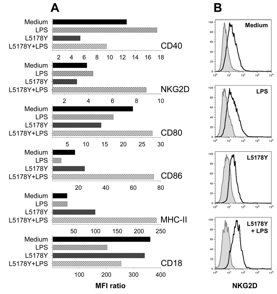
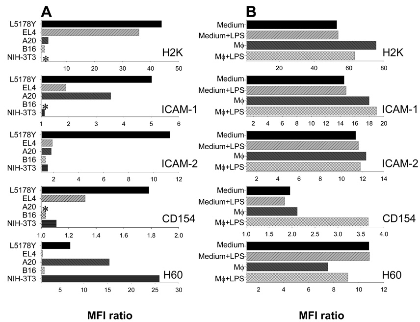
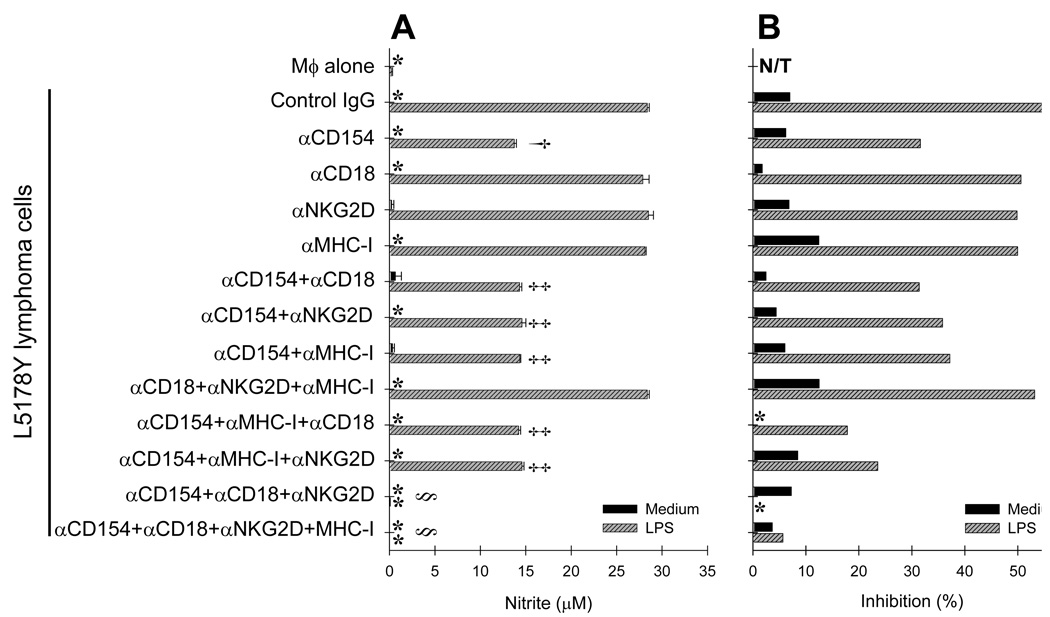
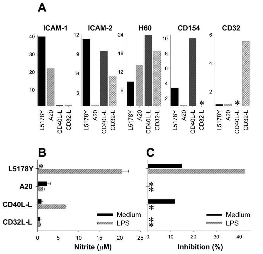
Under different circumstances, tumors can inhibit or activate macrophage (Mphi) effector functions. We studied the mechanisms of tumor-Mphi interactions leading to Mphi activation. The results show that L5178Y mouse T cell lymphoma cells can prime naive mouse Mphi to subsequent LPS stimulation, resulting in increased NO production and antilymphoma effects in vitro. L5178Y cells, but not naive splenocytes, primed Mphi to ligation of TLR4 but not TLR9. L5178Y-primed Mphi incubated with LPS showed down-regulation of CD40 and up-regulation of NKG2D expression. Although L5178Y T cell lymphoma cells primed naive mouse Mphi, several other mouse and human cells lines failed to prime mouse Mphi. Neither L5178Y-conditioned supernatants nor coculture of Mphi and L5178Y cells in Transwells resulted in priming, indicating that direct L5178Y cell-Mphi contact was needed. Several receptor-ligand pairs are reciprocally expressed on Mphi and L5178Y cell membranes and can be potentially involved in Mphi priming. Of these, the CD40-CD154 pair played the most important role, as blocking the interaction of these molecules substantially reduced in vitro Mphi priming. Furthermore, simultaneous blocking of interactions between CD40-CD154, NKG2D-H60, and CD18-ICAM-1/2 led to complete abrogation of Mphi-mediated NO secretion and complete inhibition of Mphi-mediated tumor cell cytostasis. The priming of Mphi to LPS with L5178Y cells was also observed in vivo. These results suggest that contact with certain tumor cells via CD40, NKG2D, and CD18 molecules on the Mphi may facilitate Mphi-mediated antitumor immune surveillance.


Figures

Figure 1
Figure 1. L5178Y lymphoma cells prime naïve mouse Mϕ to LPS, resulting in NO production and anti-proliferative effects in vitro
Flow cytometric profile of PC (A). PC obtained from naïve C57BL/6 mice contained ~35% of CD11b+ cells (Ai). Following 90 min of adhesion, non-adherent PC were removed from cultures by repeated pipetting. The resultant adherent population consisted of 99% CD11b+ Mϕ (Aii,). These adherent cells were comprised of 96% F4/80+ Gr1 Mϕ (Aiii). Naïve adherent C57BL/6 Mϕ were cultured for 24 hr with different mouse and human tumor cell lines in medium with or without LPS. Results are presented as concentration of NO metabolites (µM) in the supernatants (B) or % of inhibition of 3H-TdR incorporation into tumor cells (C). In this experiment, the proliferation of L5178Y cells in medium alone at 24 hrs was 16.5 ± 0.23 × 10³ counts of 3H-TdR; in other experiments where cultures were assayed at 48 hrs, counts for L5178Y cells in medium were 70–110 × 103. * -negligible value.
Figure 2
Figure 2. L5178Y cells induce dose-dependent priming of naïve Mϕ to LPS but not CpG
Adherent C57BL/6 Mϕ were cultured for 48 hr alone or with 2.5×10⁴ well of L5178Y lymphoma cells or autologous splenocytes (A, B), or various numbers of L1578Y cells (as shown in the legend box) in a separate experiment (E, F), in medium with or without LPS (10 ng/ml, as well as 0.1 and 1000 ng/ml in E and F) or CpG (5 µg/ml). Alternatively (C, D), C57BL/6 Mϕ were cultured with 2.5×10⁴ well of L5178Y lymphoma cells or splenocytes from DBA/2 mice for 24 hr, followed by thorough removal of L5178Y cells and splenocytes and placement of 2.5×10⁴/well of new L5178Y cells or B16 melanoma cells, with or without 10 ng/ml LPS, for another 24 hr,. At 42 hr, cell culture supernatants were harvested for the Griess nitrite test, and 1 µCi 3H-TdR was added for 6 hr to the cultures to measure L5178Y cell or splenocyte proliferation. Results are presented as concentration of NO metabolites (µM) in the supernatants (A, C) or % of inhibition of 3H-TdR incorporation into splenocytes (B) or L5178Y lymphoma cells (B, D). *-negligible values. The symbols (in C and D) correspond to the p values for each bar as compared to the previous bar to its left: for C †p=0.0032; ‡p=0.0114; §p=0.0978; ¶p=0.0034; #p=0.0325; @p=0.5572; for D ‡p=0.0221; §p=0.388; #p=0.838; @p=0.958; p values for D were calculated by comparing with Mean ± SEM values of original 3H-TdR incorporation counts. The experiments are representative of 3 separate experiments with similar results.
Figure 3
Figure 3. Contact of Mϕ with L5178Y lymphoma cells must precede or occur simultaneously with LPS stimulation to achieve Mϕ activation
Adherent C57BL/6 Mϕ were cultured in vitro for 48 hr alone or in the presence of various numbers (as shown in the legend box) of L5178Y lymphoma cells. L5178Y cells were added to Mϕ 1 hr after beginning the experiment (A–C, D–F) or 24 hr after beginning the experiment (G–I). Similarly, LPS was added to Mϕ-L5178Y cell cultures 1 hr after beginning the experiment (A–C, G–I) or 24 hr after beginning the experiment (D–F). Cell culture supernatants were taken at 24 hr (A,D,G) (just before L5178Y cells (G) or LPS (D) were added to Mϕ cultures) or at 42 hr (B,E,H), before 1 µCi 3H-TdR was added to the cell cultures. Results are presented as concentration of NO metabolites (µM) in the supernatants (A,B; D,E; G,H) or % of inhibition of 3H-TdR incorporation into L5178Y lymphoma cells (C,F,I). *-negligible values. The experiments are representative of three separate experiments with similar results.
Figure 4
Figure 4. Priming of Mϕ by L5178Y cells results in alterations of Mϕ immunophenotype
Naïve C57Bl/6 Mϕ were cultured for 24 h with or without L5178Y lymphoma cells in medium with or without LPS; Mϕ were then tested by flow cytometry for expression of different surface or intracytoplasmic molecules. Functional Grade Purified anti-mouse CD16+32 FcR block from eBioscience (clone 93, rat IgG2a) was used to pre-coat cells for 30 min on ice followed by washing of the cells. A. Results are presented as MFI ratios calculated as described in the Materials and Methods section. *-negligible values. The experiments are representative of three separate experiments with similar results. B. Representative histograms showing NKG2D expression on Mϕ. These correspond to the data shown for NKG2D expression in Fig. 4A. Dark grey peak – isotype-matched IgG control staining. Solid light grey line with grey fill – staining with anti-NKG2D mAb. MFI values for Mϕ stained with isotype-matched control IgG vs. anti-NKG2D mAb are 6.67 vs. 25.83 (Medium), 6.25 vs. 27.39 (LPS), 7.44 vs. 22.62 (L5178Y), and 6.99 vs. 55.02 (L5178Y+LPS).
Figure 5
Figure 5. L5178Y lymphoma cells have unique immunophenotype that can be altered by exposure to Mϕ
A, L5178Y and EL4 T-cell lymphoma cell lines, A20 B-cell lymphoma cell line, B16 melanoma cell line, and NIH-3T3 fibroblast cell line, cultured in medium alone, were tested by flow cytometry for expression of different surface antigens. Results are presented as MFI ratios calculated as described in Materials and Methods section. B, L5178Y lymphoma cells were cultured alone or in the presence of naïve C57BL/6 Mϕ in medium, with or without LPS. After 12 hr of co-culture, non-adherent cells were harvested and tested for expression of different antigens on F4/80-negative viable L5178Y lymphoma cells by flow cytometry. The experiments are representative of three separate experiments with similar results.
Figure 6
Figure 6. Priming of Mϕ by L5178Y lymphoma requires direct tumor cell-Mϕ contact
Naïve adherent C57BL/6 Mϕ were placed on the bottom of the wells in the transwell (TW) plates. Viable or paraformaldehyde fixed (PF)-L5178Y lymphoma cells were cultured either together with Mϕ (bottom), or placed in the TW-chamber, in the presence or absence of LPS. In a separate group, Mϕ were placed on the bottom of the wells and L5178Y cells were added both to the bottom of the well and into the TW-chamber. In this group, supernatants for NO detection were collected (A), and L5178Y cells (B) were removed for 3H-TdR incorporation evaluation either from the bottom of the wells (dashed rectangle) or from the TW-Chamber (dotted rectangle). Results are presented as concentration of NO metabolites (µM) in the supernatants (A) or % of inhibition of 3H-TdR incorporation into tumor cells (B). C, Naïve adherent Mϕ were cultured (with or without LPS) either with L5178Y lymphoma cells or in L5178Y cell-conditioned medium, or in L5178Y+LPS-conditioned medium. Results are presented as concentration of NO metabolites (µM) in the supernatants at 24 hr of experiment. *-negligible values. N/T-not tested as the PF-L5178Y cells did not incorporate 3H-TdR. The experiments are representative of three separate experiments with similar results.
Figure 7
Figure 7. L5178Y cell-mediated priming of Mϕ involves engagement of CD40, NKG2D, and CD18 on Mϕ
Naïve Mϕ from syngeneic DBA/2 mice were cultured for 24 hr alone or with L5178Y cells in medium with or without LPS and in the presence of either control isotype-matched IgG or blocking mAbs against CD154, NKG2D, CD18, and MHC-I, used alone or in various combinations. Results are presented as concentration of NO metabolites (µM) in the cell culture supernatants (A) or % of inhibition of 3H-TdR incorporation into L5178Y lymphoma cells (B). *-negligible values. N/T-not tested. The experiments are representative of three separate experiments with similar results. †p=0.00064; ‡p>0.05; §p=0.00031 The symbols † corresponds to p value of αCD40 treatment bar compared to control IgG treatment bar; symbols ‡ correspond to p value of either combined treatment bars compared to αCD40 treatment bar; symbols § correspond to p values of αCD40+αCD18+αNKG2D or αCD40+αCD18+αNKG2D+αMHC-I treatments compared to any other combined treatments.
Figure 8
Figure 8. Role of CD40-–CD40L interaction in priming of Mϕ by tumor cells
A. Peritoneal Mϕ from naïve C57BL/6 mice were cultured for 24 hr with L5178Y, A20, and B16 tumor cells in medium with or without 10 ng/ml LPS and agonistic αCD40 mAb, used at concentrations shown in X axis. Tumor cells and αCD40 mAb were added to Mϕ cultures 6 hr before LPS to enable Mϕ priming. At 24 hr, Mϕ activation was evaluated by measuring the cell culture supernatant NO metabolite content (upper panel) and inhibition of 3H-TdR incorporation into tumor cells (lower panel). The amount of NO secreted by Mϕ stimulated with LPS and αCD40 mAb but without tumor cells was negligible and is omitted from the figure. B–E. Peritoneal Mϕ from CD40+/+ (B,C) and CD40−/− (D,E) C57BL/6 mice were cultured for 24 hr with or without L5178Y cells in medium with LPS, αCD40 mAb and mouse IFN-γ. Tumor cells, αCD40 mAb and IFN-γ were added to Mϕ cultures 6 hr before LPS to enable Mϕ priming. At 24 hr of co-culture, Mϕ activation was evaluated by measuring supernatant NO metabolite content (B,D) and inhibition of 3H-TdR incorporation into the tumor cells (C,E). *-value is below the detection limit. N/T - not tested. Mϕ cultured with L5178Y cells without LPS, with or without αCD40 mAb or IFN-γ, secreted negligible amounts of NO and demonstrated no antitumor effect (not shown).
Figure 9
Figure 9. CD154+ICAM-I/II+H60+ L5178Y lymphoma cells but not CD154+ICAM-II+H60+ L-fibroblasts prime Mϕ
A. Lymphoid L5178Y and A20 and non-lymphoid fibroblast CD40L-L and CD32-L cells lines were tested by flow cytometry for expression of ICAM-1, ICAM-2, H60, CD154, and CD32 surface antigens. Results are presented as MFI ratio values, as described in the Materials and Methods. B,C. Naïve adherent C57BL/6 Mϕ were cultured for 24 hr with L5178Y, A20, CD40L-L, and CD32-L cell lines in medium with or without LPS, and Mϕ activation was measured. Results are presented as concentration of NO metabolites (µM) in the supernatants (B) or % of inhibition of 3H-TdR incorporation into L5178Y, A20, CD40L-L, and CD32-L cells (C). * - negligible value.
Figure 10
Figure 10. Synergistic effect of CD40L-L fibroblasts and L5178Y or A20 lymphoma cells on Mϕ priming
A,B. Peritoneal Mϕ (~3×10⁵ cells/well) from naïve C57BL/6 mice were cultured for 24 hr in medium or in the presence of CD40L-L or CD32-L cells (2.5×10⁴ cells/well), with our without LPS in medium. At 1 hr of experiment, different numbers of L5178Y (A) or A20 (B) cells were added to some Mϕ-L cell cultures. At 24 hr, Mϕ activation was evaluated by measuring concentration of NO metabolites in cell culture supernatants. C,D. Peritoneal Mϕ (~3×10⁵ cells/well) from CD40+/+ (C) and CD40−/− (D) C57BL/6 mice were cultured for 24 hr in medium with or without LPS and in the presence of L5178Y cells or CD40L-L cells (1×10⁴ cells/well), added to Mϕ separately or mixed together. At 24 hr, Mϕ activation was evaluated by measuring concentration of NO metabolites in cell culture supernatants.
Figure 11
Figure 11. L5178Y cells and LPS synergize in activation of Mϕ in vivo
A. DBA/2 mice (n=3/group) were injected i.p. with L5178Y cells (5×10⁵/ml) or PBS (control). Twenty-four hrs later, the mice were treated i.v. with 0.2 ml PBS with or without 20 ng LPS. At 48 hr of experiment, mice were euthanized, resident PC collected from each mouse of the experimental group and pooled, and F4/80+ Mϕ tested by flow cytometry for production of IL4, IL10, IFN-γ, TNF-α and IL12 or expression of NKG2D, CD80, CD86, and MHC-II. On histograms: grey filled peaks – staining with isotype-matched control IgG, open black peak – staining with specific mAb. Numbers are MFI ratios calculated as described in Materials and Methods. B. Proliferation of CFSE-labeled L5178Y cells is suppressed in DBA/2 mice after systemic administration of low amount of LPS. CFSE-labeled L5178Y cells (5×10⁵/ml) were injected i.p. into DBA/2 mice (n=3/group). Twenty-four hours after tumor cell implantation the mice were treated i.v. with 0.2 ml PBS with or without 20 ng LPS. After 48 hr, mice were euthanized, resident peritoneal cells + tumor cells collected from each mouse of the group and pooled, and F4/80CFSE+L5178Y were tested by flow cytometry for CFSE fluorescence. In parallel, CFSE-labeled L5178Y cells (5×10⁵/ml, 0.05 ml/well) were cultured in vitro in triplicates with naïve DBA/2 Mϕ as described in Fig. 3D–F. After 48 hr, cell cultures were collected by pipetting and pooled into one analysis sample for each experimental group, and F4/80CFSE+L5178Y were tested by flow cytometry for CFSE fluorescence. As a proliferation suppression control, L5178Y cells were γ-irradiated. Results are presented as CFSE Mean Fluorescence Intensity.

Similar articles

Cited by

References

    1. Blattman JN, Greenberg PD. Cancer immunotherapy: a treatment for the masses. Science. 2004;305:200–205. - PubMed
    1. Chang CC, Ferrone S. NK cell activating ligands on human malignant cells: molecular and functional defects and potential clinical relevance. Semin. Cancer. Biol. 2006;16:383–392. - PubMed
    1. Lamagna C, Aurrand-Lions M, Imhof BA. Dual role of macrophages in tumor growth and angiogenesis. J. Leukoc. Biol. 2006;80:705–713. - PubMed
    1. Greeneltch KM, Schneider M, Steinberg SM, Liewehr DJ, Stewart TJ, Liu K, Abrams SI. Host immunosurveillance controls tumor growth via IFN regulatory factor-8 dependent mechanisms. Cancer. Res. 2007;67:10406–10416. - PubMed
    1. Bottino C, Castriconi R, Moretta L, Moretta A. Cellular ligands of activating NK receptors. Trends Immunol. 2005;26:221–226. - PubMed

Publication types