. 2009 Feb 15;182(4):2002-10.
doi: 10.4049/jimmunol.0803280.

Allergic airway hyperresponsiveness-enhancing gammadelta T cells develop in normal untreated mice and fail to produce IL-4/13, unlike Th2 and NKT cells

Affiliations

Allergic airway hyperresponsiveness-enhancing gammadelta T cells develop in normal untreated mice and fail to produce IL-4/13, unlike Th2 and NKT cells

Niyun Jin et al. J Immunol. .

Abstract

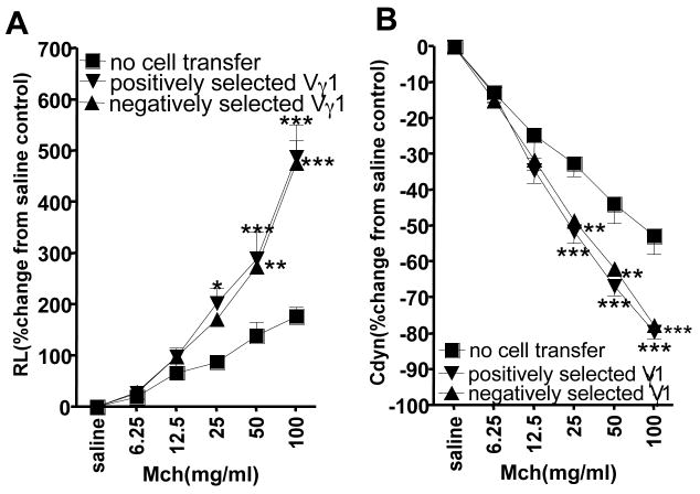
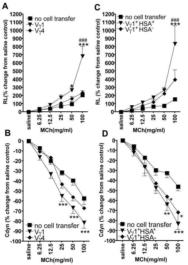
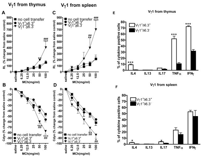
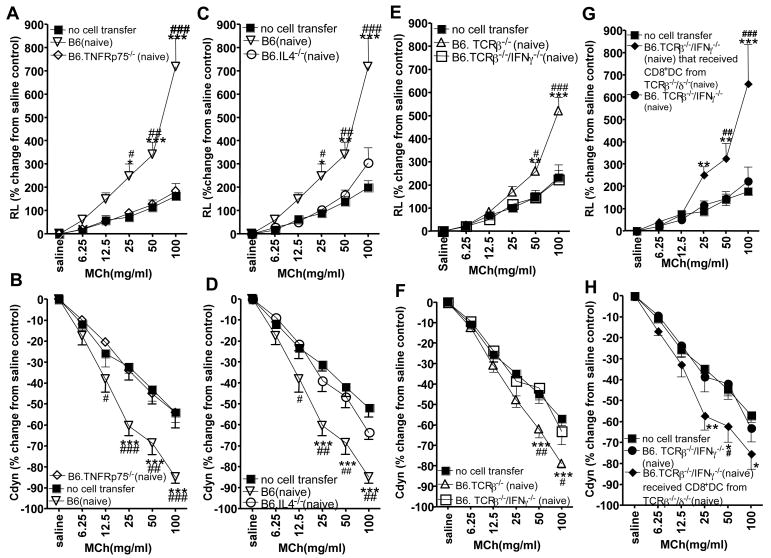
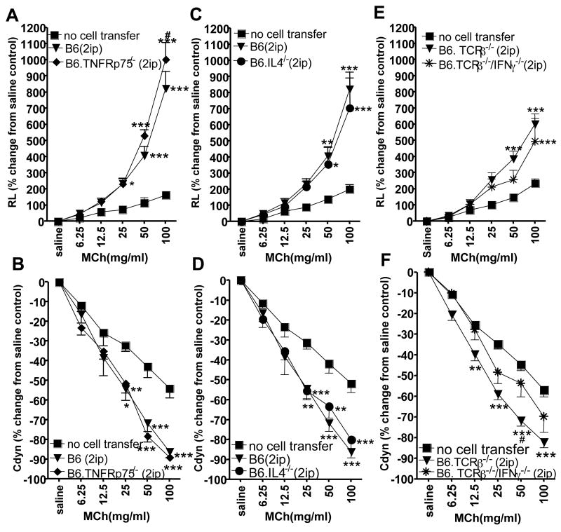
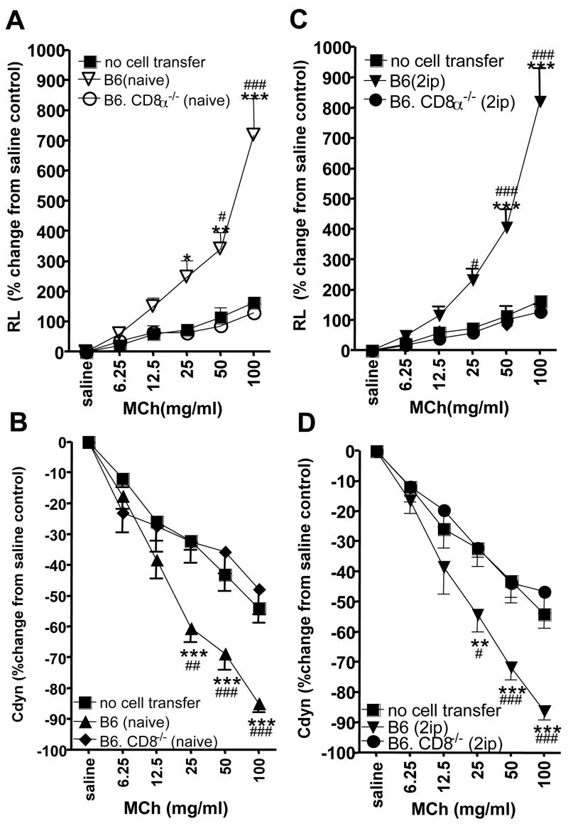
Allergic airway hyperresponsiveness (AHR) in OVA-sensitized and challenged mice, mediated by allergen-specific Th2 cells and Th2-like invariant NKT (iNKT) cells, develops under the influence of enhancing and inhibitory gammadelta T cells. The AHR-enhancing cells belong to the Vgamma1(+) gammadelta T cell subset, cells that are capable of increasing IL-5 and IL-13 levels in the airways in a manner like Th2 cells. They also synergize with iNKT cells in mediating AHR. However, unlike Th2 cells, the AHR enhancers arise in untreated mice, and we show here that they exhibit their functional bias already as thymocytes, at an HSA(high) maturational stage. In further contrast to Th2 cells and also unlike iNKT cells, they could not be stimulated to produce IL-4 and IL-13, consistent with their synergistic dependence on iNKT cells in mediating AHR. Mice deficient in IFN-gamma, TNFRp75, or IL-4 did not produce these AHR-enhancing gammadelta T cells, but in the absence of IFN-gamma, spontaneous development of these cells was restored by adoptive transfer of IFN-gamma-competent dendritic cells from untreated donors. The i.p. injection of OVA/aluminum hydroxide restored development of the AHR enhancers in all of the mutant strains, indicating that the enhancers still can be induced when they fail to develop spontaneously, and that they themselves need not express TNFRp75, IFN-gamma, or IL-4 to exert their function. We conclude that both the development and the cytokine potential of the AHR-enhancing gammadelta T cells differs critically from that of Th2 cells and NKT cells, despite similar influences of these cell populations on AHR.


Figures

Figure 1
Figure 1
Vγ1+ γδ T cells from naïve donors mediate AHR regardless of whether they were purified by positive or negative selection. AHR was monitored by measuring lung resistance (A) and dynamic compliance (B). OVA-sensitized B6.TCR-δ−/− mice received 1 ×10⁴ splenic Vγ1+ γδ T cells from untreated B6.TCR-β−/− donors, prior to airway challenge. The Vγ1+ cells were enriched by positive or negative selection, as described in the Methods. Recipients that were sensitized and challenged but did not receive cells (no cells transferred) are also shown. Results for each group are presented as means +/− SEM (n=7–8). Significant differences between “no cell transfer” and “selected Vγ1” cells transferred groups are indicated: *P<0.05, **P<0.01, ***P<0.001.
Figure 2
Figure 2
Vγ1+ thymocytes mediate AHR. AHR was monitored by measuring lung resistance (A, C) and dynamic compliance (B, D). OVA-sensitized B6.TCR-δ−/− mice received 1×10⁴ splenic Vγ1+ thymocytes from untreated B6. TCR-β−/− donors, prior to airway challenge. The thymocytes were selected for Vγ1 or Vγ4 expression, and Vγ1+ thymocytes were further divided into HSAhi and HSAlo cells, as described in the Methods. Recipients that were sensitized and challenged but did not receive cells (no cell transfer), are also shown. Results for each group are presented as means +/− SEM (n=4–7). Significant differences between “no cell transfer” and “Vγ1+ cells” transferred groups are indicated: *P<0.05, **P<0.01, ***P<0.001. Significant differences between Vγ1+ and Vγ4+ cells transferred groups, or Vγ1/HSA+ and Vγ1/HSA cells transferred groups are also indicated: ###P<0.001.
Figure 3
Figure 3
Vγ1+/Vδ6.3 γδ T cells mediate AHR but fail to express Th2 cytokines. AHR was monitored by measuring lung resistance (A, C) and dynamic compliance (B, D). Cytokine-producing cells were enumerated by intra-cellular staining and cytofluorometry (E, F), and activated cells were enumerated by cell-surface staining and cytofluorometry (G, H). To determine cellular effects on AHR, OVA-sensitized B6.TCR-δ−/− mice received 1×10⁴ Vγ1+ thymocytes or splenic Vγ1+ cells from untreated B6.TCR-β−/− donors, prior to airway challenge. The Vγ1+ cells were further selected into Vδ6.3+ and Vδ6.3 fractions, as described in the Methods. Recipients that were sensitized and challenged but did not receive cells (no cell transfer) are also shown. Results for each group are presented as means +/− SEM (n= 4–6). Significant differences between “no cell transfer” and “Vγ1+ cells” transferred groups are indicated: *P<0.05, **P<0.01, ***P<0.001. Significant differences between “Vγ1/Vδ6.3+” and “Vγ1/Vδ6.3“ cells transferred groups are also indicated: ##P<0.01, ###P<0.001. To determine frequencies of cells with potential for cytokine production, Vγ1 cells from B6.TCR-β−/− thymus and spleen were activated in vitro with PMA/ionophore in the presence of Brefeldin A, stained for TCR-expression and intra-cellular cytokines. Percent fractions of cytokine producing cells among Vγ1+/Vδ6.3+ and Vγ1+/Vδ6.3 subsets are shown (E, F). To determine frequencies of cells expression activation/memory markers, freshly isolated cells were stained directly for TCR expression and cell-surface markers. Percent fractions of marker-positive cells among Vγ1+/Vδ6.3+ and Vγ1+/Vδ6.3 subsets are shown (G, H). Results for each group are presented as mean±SEM (n=8–11). Significant differences between Vγ1+/Vδ6.3 and spleen Vγ1+/V6.3+ groups are indicated: *P<0.05, ** P<0.01, ***P<0.001.
Figure 4
Figure 4
Vγ1+ γδ T cells from naïve donors deficient in TNFRp75, IL-4 and IFN-γ fail to mediate AHR, and transferred CD8+ DC restore this ability in Vγ1+ cells from IFN-γ deficient mice. AHR was monitored by measuring lung resistance (A, C, E, G) and dynamic compliance (B, D, F, H). OVA-sensitized B6.TCR-δ−/− mice received 1×10⁴ splenic Vγ1+ γδ T cells from untreated donors, or from donors that received 1×10⁵ CD8+ splenic DC (G, H, see Methods), prior to airway challenge. In addition to the cells from the cytokine/cytokine-receptor-deficient mice, cells from matched normal controls (C57BL6 or B6.TCR-β−/− mice) were also transferred. Responses of recipients that were sensitized and challenged but did not receive cells (no cell transfer), are also shown. Results for each group are presented as means +/− SEM (A, B n=6; C, D n=8; E, F n=6). Significant differences between “no cells transfer” and “Vγ1+” cells transferred groups are indicated: *P<0.05, **P<0.01, ***P<0.001. Significant differences between wild-type or mutant-derived Vγ1 cells transferred, or between Vγ1+ cells derived from IFN-γ deficient or from IFN-γ-deficient DC-reconstituted mice, are also indicated: #P<0.05, ##P<0.01, ###P<0.001.
Figure 5
Figure 5
Vγ1+ γδ T cells from 2ip-sensitized donors deficient in TNFRp75, IL-4 and IFN-γ mediate AHR. AHR was monitored by measuring lung resistance (A, C, E) and dynamic compliance (B, D, F). OVA-sensitized B6.TCR-δ−/− mice received 1×10⁴ splenic Vγ1+ γδ T cells from 2ip-sensitized donors, prior to airway challenge. In addition to the cells from the cytokine/cytokine-receptor-deficient mice, cells from 2ip-sensitized normal controls (C57BL6 or B6.TCR-β−/− mice) were also transferred. Responses of recipients that were sensitized and challenged but did not receive cells (no cell transfer), are also shown. Results for each group are presented as means +/− SEM (A, B n=6; C, D n=8; E, F n=5). Significant differences between “no cell transfer” and “Vγ1+” cells transferred groups are indicated: *P<0.05, **P<0.01, ***P<0.001. Significant differences between wild-type and mutant-derived “Vγ1+ cells transferred (F), are also indicated: #P<0.01.
Figure 6
Figure 6
Vγ1+ γδ T cells from CD8α-deficient donors fail to mediate AHR even when the donors are 2ip-sensitized. AHR was monitored by measuring lung resistance (A, C) and dynamic compliance (B, D). OVA-sensitized B6.TCR-δ−/− mice received 1 ×10⁴ splenic Vγ1+ γδ T cells from untreated (A, B) or 2ip-sensitized donors, prior to airway challenge. Recipients that were sensitized and challenged but did not receive cells (no cell transfer), are also shown. Results for each group are presented as means +/− SEM (A, B n=3; C, D n=5). Significant differences between “no cell transfer” and “Vγ1+” cells transferred groups are indicated: *P<0.05, **P<0.01, ***P<0.001. Significant differences between wild-type and mutant-derived Vγ1 cells are also indicated: #P<0.05, ##P<0.001, ###P<0.0001.
Figure 7
Figure 7
Vγ1+ thymocytes encounter CD8α+ cells in the thymus. Frozen sections of adult B6.TCR-β−/− thymus were stained with antibodies specific for TCR-δ (red) and CD8α (blue) (A) or Vγ1 (red) and CD8α (blue) (B). Autofluorescence (green) shows thymus tissue. The digital image in panel B is 7.4× enlarged relative to panel A. The arrow in panel B points to the area of contact between the γδ T cell (red) and the CD8+ cell (blue).

Similar articles

Cited by

  • The role of the γ δ T cell in allergic diseases.
    Zheng R, Yang Q. Zheng R, et al. J Immunol Res. 2014;2014:963484. doi: 10.1155/2014/963484. Epub 2014 Jun 3. J Immunol Res. 2014. PMID: 24995350 Free PMC article. Review.
  • Diversity of γδ T-cell antigens.
    Born WK, Kemal Aydintug M, O'Brien RL. Born WK, et al. Cell Mol Immunol. 2013 Jan;10(1):13-20. doi: 10.1038/cmi.2012.45. Epub 2012 Oct 22. Cell Mol Immunol. 2013. PMID: 23085946 Free PMC article. Review.
  • A dual nature of γδ T cell immune memory responses.
    Suen TK, Al B, Scarpa A, Dorhoi A, Netea MG, Placek K. Suen TK, et al. Elife. 2025 Jul 23;14:e104887. doi: 10.7554/eLife.104887. Elife. 2025. PMID: 40700036 Free PMC article. Review.
  • γδ T Lymphocytes in Asthma: a Complicated Picture.
    Zarobkiewicz MK, Wawryk-Gawda E, Kowalska W, Janiszewska M, Bojarska-Junak A. Zarobkiewicz MK, et al. Arch Immunol Ther Exp (Warsz). 2021 Mar 4;69(1):4. doi: 10.1007/s00005-021-00608-7. Arch Immunol Ther Exp (Warsz). 2021. PMID: 33661375 Free PMC article. Review.
  • Role of γδ T Cells in Lung Inflammation.
    Born WK, Roark CL, Jin N, Wands JM, Kemal Aydintug M, Huang Y, Chain JL, Hahn YS, Simonian PL, Fontenot AP, O'Brien RL. Born WK, et al. Open Immunol J. 2009;2:143-150. doi: 10.2174/1874226200902010143. Epub 2009 Oct 23. Open Immunol J. 2009. PMID: 26550059 Free PMC article.

References

    1. Steinman L. A brief history of TH17, the first major revision in the TH1/TH2 hypothesis of T cell-mediated tissue damage. Nature Medicine. 2007;13:139–145. - PubMed
    1. Else KJ, Finkelman FD. Intestinal nematode parasites, cytokines and effector mechanisms. Int J Parasitol. 1998;28:145–1158. - PubMed
    1. Ramalingam TR, Reiman RM, Wynn TA. Exploiting worm and allergy models to understand Th2 cytokine biology. Current Opinion in Allergy and Clinical Immunology. 2005;5:392–398. - PubMed
    1. Li L, Lee HH, Bell JJ, Gregg RK, Ellis JS, Gessner A, Zaghouani H. IL-4 utilizes an alternative receptor to drive apoptosis of Th1 cells and skews neonatal immunity toward Th2. Immunity. 2004;20:429–440. - PubMed
    1. Kaiko GE, Phipps S, Hickey DK, Lam CE, Hansbro PM, Foster PS, Beagley KW. Chlamydia muridarum infection subverts dendritic cell function to promote Th2 immunity and airways hyperreactivity. J Immunol. 2008;180:2225–2232. - PubMed

Publication types

MeSH terms