. 2009 Feb 15;182(4):2131-40.
doi: 10.4049/jimmunol.0802755.

The sequestosome 1/p62 attenuates cytokine gene expression in activated macrophages by inhibiting IFN regulatory factor 8 and TNF receptor-associated factor 6/NF-kappaB activity

Affiliations

The sequestosome 1/p62 attenuates cytokine gene expression in activated macrophages by inhibiting IFN regulatory factor 8 and TNF receptor-associated factor 6/NF-kappaB activity

Ji Young Kim et al. J Immunol. .

Abstract

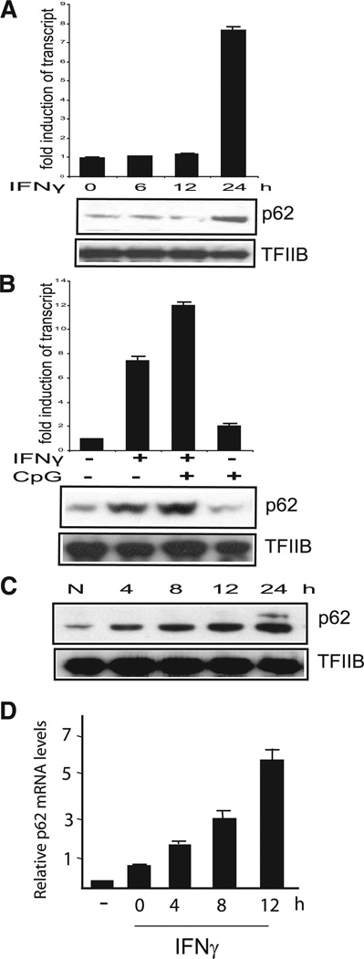
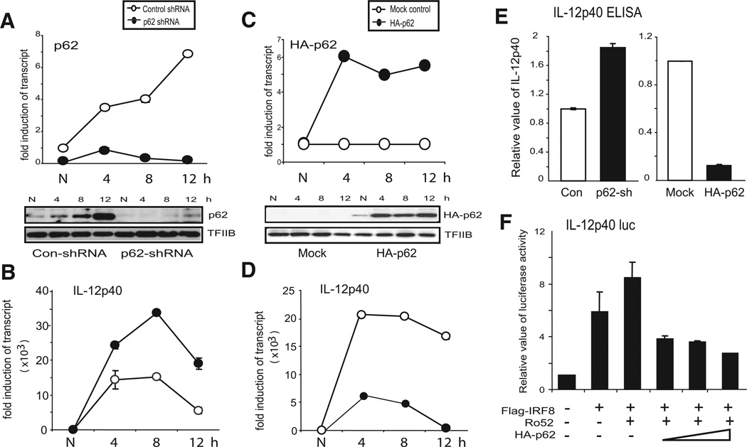
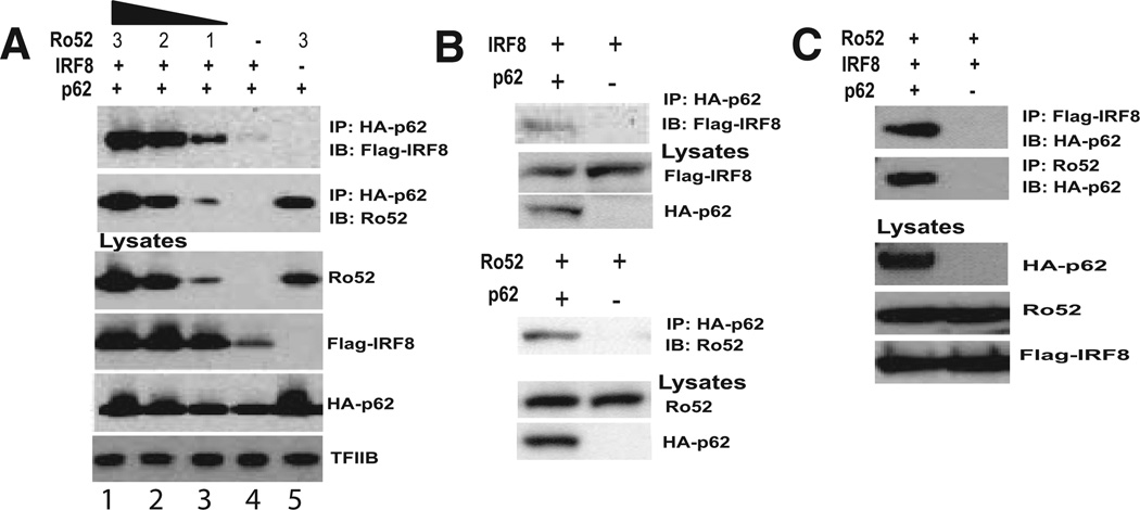
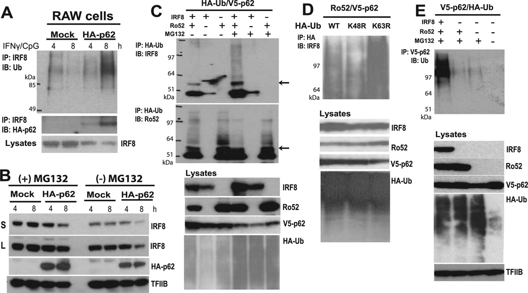
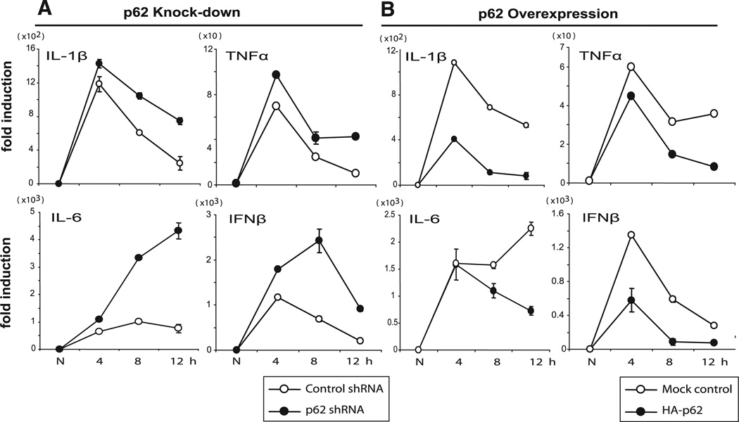
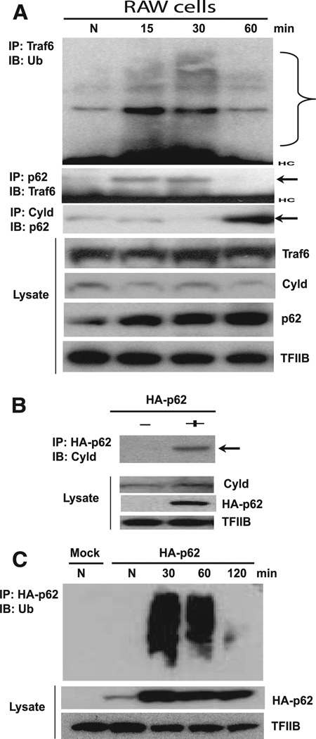
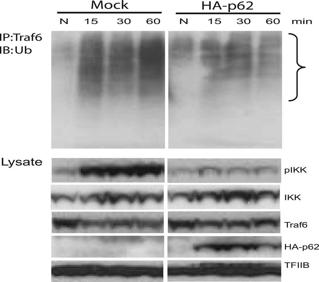
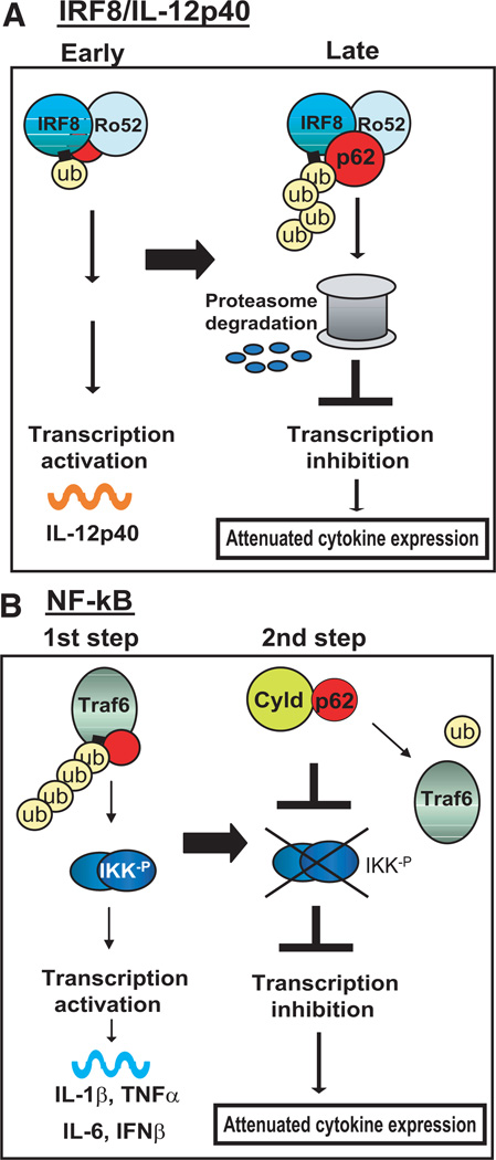
Sequestosome 1/p62 (p62) is a scaffold/adaptor protein with multiple functions implicated for neuronal and bone diseases. It carries a ubiquitin binding domain through which it mediates proteasome-dependent proteolysis. In addition, p62 is reported to regulate NF-kappaB activity in some cells. To date, however, the role of p62 in innate immunity has not been fully elucidated. In this study, we report that IFN-gamma plus TLR signaling stimulates late expression of p62 in murine macrophages. Overexpression of p62 inhibited expression of multiple cytokines, IL-12p40, TNF-alpha, IL-1beta, IL-6, and IFN-beta, whereas p62 underexpression by small hairpin RNA markedly elevated their expression, indicating that p62 is a broad negative regulator of cytokine expression in stimulated macrophages. We show that p62 interacts with IFN regulatory factor 8 and Ro52, the transcription factor and ubiquitin E3 ligase that are important for IL-12p40 expression. This interaction, detectable at a late stage in stimulated macrophages, led to increased polyubiquitination and destabilization of IFN regulatory factor 8. We also show that upon macrophage stimulation, p62 binds to TNFR-associated factor 6, another E3 ligase important for NF-kappaB activation, but later this interaction was replaced by the recruitment of the deubiquitinating enzyme, cylindromatosis, an inhibitor of NF-kappaB activity. Recruitment of cylindromatosis coincided with reduced TNFR-associated factor 6 autoubiquitination and lower NF-kappaB activation. Our results indicate that p62 orchestrates orderly regulation of ubiquitin modification processes in macrophages to ensure attenuation of cytokine transcription postactivation. Together, p62 may provide a mechanism by which to control excessive inflammatory responses after macrophage activation.


Conflict of interest statement

Disclosures

The authors have no financial conflict of interest.

Figures

FIGURE 1
FIGURE 1
Induction of p62 by IFN-γ and CpG stimulation in macrophages. A, RAW cells were stimulated with IFN-γ (150 U/ml) for indicated times. p62 transcripts and p62 protein levels were measured by qRT-PCR and immunoblot, respectively. Values represent the average of three determinations ± SD. B, Cells were pretreated with IFN-γ for 20 h and/or stimulated with CpG (150 ng/ml) for 4 h, and p62 expression was detected as above. C, Cells were pretreated with IFN-γ overnight and then stimulated with CpG (150 ng/ml) for indicated times, and p62 protein expression was tested by immunoblot analysis. N, Indicates neither IFN-γ nor CpG treatment. D, Bone marrow-derived macrophages were treated with IFN-γ (100 U/ml) overnight and then stimulated with CpG (150 ng/ml) for indicated times, and p62 transcript levels were measured as above. –, Denotes no IFN-γ treatment.
FIGURE 2
FIGURE 2
p62 inhibits IFN-γ/CgG-stimulated IL-12p40 expression. A, Knockdown of p62 by shRNA. RAW cells were transduced with a retroviral vector for p62 shRNA or control shRNA and stimulated with IFN-γ overnight, followed by CpG for indicated period of time. Upper panel, Represents the levels of p62 transcripts; lower panel, shows p62 protein expression. B, Enhanced expression of IL-12p40 in p62 knockdown cells. Cells with p62 shRNA (●) or control shRNA (○) were stimulated with IFN-γ/CpG, as in A, and IL-12p40 mRNA levels were quantified by qRT-PCR. The values represent the average of three determinations ± SD. C, Overexpression of HA-tagged p62. RAW cells were stably transfected with HA-p62, and a selected clone was tested for expression of HA-p62 transcripts and proteins, as above. D, Reduced expression of IL-12p40 transcripts by p62 overexpression. Cells expressing HA-p62 or empty vector (Mock) were stimulated with IFN-γ/CpG, and IL-12p40 mRNA levels were measured, as in B. E, Cells with p62 knockdown cells, cells expressing HA-p62, and respective control cells were stimulated with IFN-γ/CpG for 24 h, and IL-12p40 in supernatants was measured by ELISA. The amounts of IL-12p40 were normalized to respective control cells, which produced 350–425 pg/ml proteins. F, Inhibition of IL-12p40 reporter activity by ectopic HA-p62 expression. Cells were transiently transfected with IL-12p40 luciferase reporter along with expression vectors for IRF8, Ro52, and p62 (150, 300, and 550 ng) for 30 h. pcDNA-HA was added to adjust the total amount of plasmids to be 850 ng in all samples. Reporter activity was normalized by Renilla luciferase activity. Values represent the average of three assays ± SD.
FIGURE 3
FIGURE 3
Ro52-mediated interaction of p62 with IRF8. A, 293T cells were transfected with expression vectors for Ro52 (3, 2, and 1 µg) along with HA-p62 (3 µg) and Flag-IRF8 (3 µg). Lysates were precipitated with anti-HA Ab, and the eluted materials were blotted against anti-Flag Ab (IRF8, upper panel) and anti-Ro52 Ab (lower panel). The bottom four panels represent immunoblot data to confirm the expression of transfected proteins. B, Cells were transfected with Flag-IRF8 (upper panel) or Ro52 (lower panel) along with HA-p62 or empty vector. Lysates were precipitated with anti-HA Ab and blotted for Flag-IRF8 or Ro52. C, Cells were transfected with vectors for Flag-IRF8, Ro52, along with HA-p62 or empty vector. Lysates were precipitated with anti-Flag (upper panel) or anti-Ro52 Ab and blotted for HA-p62.
FIGURE 4
FIGURE 4
p62 binds to polyubiquitinated IRF8. A, RAW cells transfected with empty vector (Mock) or HA-p62 were stimulated with IFN-γ/CpG for indicated periods. MG132 was added to the culture for the final 4 h. Lysates were precipitated by anti-IRF8 Ab and tested for for ubiquitin (Ub, upper panel) and p62 (lower panel) by immunoblot analysis. B, RAW cells transfected with empty vector (Mock) or HA-p62 were stimulated with IRFγ/CpG for 4 and 8 h in the presence or absence of MG132 for the final 4 h. Lysates were immunoblotted for indicated proteins. IRF8 levels were tested after a short (S) or a long (L) exposure. C, 293T cells were transfected with expression plasmids for HA-ubiquitin, V5-p62, along with Flag-IRF8 and Ro52 in the presence or absence of MG132. Lysates were precipitated with anti-HA Ab and blotted for IRF8 (upper panel) or Ro52 (lower panel). Middle panel, Total lysates were immunoblotted for indicated proteins to confirm proper transfection. Bottom panel, Indicates total HA-ubiquitin-conjugated proteins. D, 293T cells were transfected with Flag-IRF8, Ro52, V5-p62, and HA wild-type ubiquitin (Ub, WT) or HA-tagged ubiquitin mutants in which lysine (K) residue at 48 or 63 was replaced by arginine (R) (K48R, K63R). Lysates were precipitated by anti-HA Ab and blotted for IRF8. MG132 was added for the final 6. E, Cells were transfected with V5-p62 and HA-ubiquitin along with Flag-IRF8 and Ro52 in the presence or absence of MG132 for the final 6 h. Lysates were precipitated with anti-V5 Ab and blotted for HA-ubiquitin by HA Ab. Middle panels, Lysates were immunoblotted for indicated proteins to monitor expression of transfected proteins and total ubiquitin-conjugated proteins.
FIGURE 5
FIGURE 5
The opposite regulation of proinflammatory cytokine gene expression by p62 knockdown and p62 overexpression. A, RAW cells expressing control or p62 shRNA were stimulated with IFN-γ/CpG for indicated times (h), and transcripts for the indicated cytokine were measured by qRT-PCR. Values represent the average of three assays ± SD. B, RAW cells overexpressing HA-p62 were tested for expression of the cytokines, as above. Three other clones expressing HA-p62 tested showed similar inhibition.
FIGURE 6
FIGURE 6
Interaction of p62 with TRAF6 and CYLD in stimulated macrophages. A, Parental RAW cells were stimulated with IFN-γ/CpG for indicated times. Lysates were precipitated by anti-TRAF6 Ab and blotted for ubiquitin by immunoblot (upper panel). The range of ubiquitinated proteins is bracketed. Lower panels, Lysates were precipitated with Ab against p62 or CYLD, and blotted for TRAF6 or p62 (marked by arrows). Bottom panels, Total lysates were tested for the expression of indicated proteins. B, RAW cells transfected with HA-p62 were stimulated without (−) or with (+) IFN-γ/CpG, and HA-p62 immune precipitates were blotted for the endogenous CYLD. Bottom panels, Indicate the expression of indicated proteins in total lysates. C, RAW cells transfected with empty vector (Mock) or HA-p62 vector were stimulated with IFN-γ/CpG for indicated periods, and HA-p62 immune precipitates were tested for reactivity with ubiquitin (Ub).
FIGURE 7
FIGURE 7
Reduced autoubiquitination of TRAF6 by p62 overexpression. Cells transfected with empty vector (Mock) or HA-62 were stimulated with IFN-γ/CpG for indicted times, and TRAF6 immune complexes were blotted for ubiquitin (upper panel). Total lysates were tested for phosphorylated IKK, total IKK, and indicated proteins (lower panel).
FIGURE 8
FIGURE 8
A model for a dual mode of p62 action. A, IRF8 and Ro52, transcription factors important for IL-12p40 expression, are induced by IFN-γ/TLR early, whereas p62 is induced later in macrophages. In an early stage, Ro52-mediated ubiquitination of IRF8 enhances IL-12p40 transcription. In a later stage as the p62 expression increases, the p62-Ro52 interaction increases, facilitating polyubiquitination of IRF8 and the subsequent degradation. B, p62 interacts with another E3 ubiquitin ligase, TRAF6, upon IFN-γ/TLR stimulation, causing TRAF6 autoubiquitination and NF-κB activation, leading to the induction of proinflammatory cytokines (the first step). Subsequently, the interaction of p62 with TRAF6 is replaced by that with CYLD to reduce TRAF6 autoubiquitination, weakening NF-κB activation (the second step). In this model, p62 orchestrates timely attenuation of cytokine expression by coordinating ubiquitin modification of signaling molecules and transcription factors.

Similar articles

Cited by

References

    1. Kawai T, Akira S. TLR signaling. Semin. Immunol. 2007;19:24–32. - PubMed
    1. Boehm U, Klamp T, Groot M, Howard JC. Cellular responses to interferon-γ. Annu. Rev. Immunol. 1997;15:749–795. - PubMed
    1. O’Shea JJ, Murray PJ. Cytokine signaling modules in inflammatory responses. Immunity. 2008;28:477–487. - PMC - PubMed
    1. Saccani S, Pantano S, Natoli G. Two waves of nuclear factor κBrecruitment to target promoters. J. Exp. Med. 2001;193:1351–1359. - PMC - PubMed
    1. Hayden MS, Ghosh S. Shared principles in NF-κB signaling. Cell. 2008;132:344–362. - PubMed

Publication types

MeSH terms