. 2009 Feb 15;182(4):2439-48.
doi: 10.4049/jimmunol.0803357.

TNF-like weak inducer of apoptosis (TWEAK) activates proinflammatory signaling pathways and gene expression through the activation of TGF-beta-activated kinase 1

Affiliations

TNF-like weak inducer of apoptosis (TWEAK) activates proinflammatory signaling pathways and gene expression through the activation of TGF-beta-activated kinase 1

Mukesh Kumar et al. J Immunol. .

Abstract

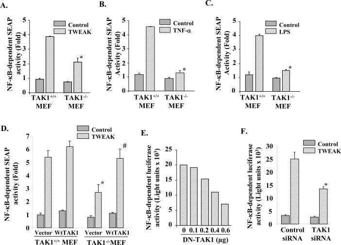
TWEAK, TNF-like weak inducer of apoptosis, is a relatively recently identified proinflammatory cytokine that functions through binding to Fn14 receptor in target cells. Although TWEAK has been shown to modulate several biological responses, the TWEAK-induced signaling pathways remain poorly understood. In this study, we tested the hypothesis that TAK1 (TGF-beta-activated kinase 1) is involved in TWEAK-induced activation of NF-kappaB and MAPK and expression of proinflammatory protein. TWEAK increased the phosphorylation and kinase activity of TAK1 in cultured myoblast and fibroblast cells. The activation of NF-kappaB was significantly inhibited in TAK1-deficient (TAK1(-/-)) mouse embryonic fibroblasts (MEF) compared with wild-type MEF. Deficiency of TAK1 also inhibited the TWEAK-induced activation of IkappaB kinase and the phosphorylation and degradation of IkappaBalpha protein. However, there was no difference in the levels of p100 protein in TWEAK-treated wild-type and TAK1(-/-) MEF. Furthermore, TWEAK-induced transcriptional activation of NF-kappaB was significantly reduced in TAK1(-/-) MEF and in C2C12 myoblasts transfected with a dominant-negative TAK1 or TAK1 short interfering RNA. TAK1 was also required for the activation of AP-1 in response to TWEAK. Activation of JNK1 and p38 MAPK, but not ERK1/2 or Akt kinase, was significantly inhibited in TAK1(-/-) MEF compared with wild-type MEF upon treatment with TWEAK. TWEAK-induced expression of proinflammatory genes such as MMP-9, CCL-2, and VCAM-1 was also reduced in TAK1(-/-) MEF compared with wild-type MEF. Furthermore, the activation of NF-kappaB and the expression of MMP-9 in response to TWEAK involved the upstream activation of Akt kinase. Collectively, our study demonstrates that TAK1 and Akt are the important components of TWEAK-induced proinflammatory signaling and gene expression.


Figures

Figure 1
Figure 1. Effect of TWEAK on the activation of TAK
A). C2C12 myoblasts were treated with TWEAK (100 ng/ml) for indicated time interval, cell extracts made were used for western blotting or immunoprecipitated with TAK1 antibody for in vitro kinase assay. Representative gel pictures presented here show that TWEAK augments the phosphorylation of TAK1 at Ser412 (top panel) and Thr184/187 (second from top) residues and increased its enzymatic activity (third from top). Treatment of cells with TWEAK did not affect the cellular level of TAK1, TRAF-6, TAB1, and TAB2 protein. B). TWEAK treatment also increased the phosphorylation of TAK1 in murine embryonic fibroblasts (MEF) without affecting the total level of TAK1 protein. KA, kinase assay.
Figure 2
Figure 2. Role of TAK1 in TWEAK-induced activation of NF-κB pathways
A). TAK1+/+ and TAK1−/− MEF were treated with recombinant TWEAK protein (100 ng/ml) for indicated time intervals and the activation of NF-κB was measured by EMSA. A representative EMSA gel presented here show that compared to TAK1+/+ MEF, the activation of NF-κB was significantly inhibited in TAK1−/− MEF in response to TWEAK-treatment. B). Cell extracts prepared from TWEAK-treated TAK1+/+ and TAK1−/− MEF were subjected to western blotting to measure the levels of phosphorylated and total IκBα protein. Representative immunoblots presented here show that TWEAK-induced phosphorylation (upper panel) and degradation (middle panel) of IκBα protein was significantly inhibited in TAK1−/− cells compared to TAK1+/+ MEF. The level of an unrelated protein β-actin did not change due to TWEAK treatment. C). TAK1+/+ and TAK1−/− MEF were treated with 100 ng/ml TWEAK protein for indicated time intervals and the activation of IKK was measured. Data presented here show that the activation of IKK was significantly inhibited in TAK1−/− MEF as compared to TAK1+/+ MEF (top panel). There was no difference in the total level of IKKα/β proteins in TWEAK-treated TAK1+/+ and TAK1−/− cells (middle panel). Western blotting confirmed the deficiency of full length TAK1 protein in TAK1−/− MEF (lower panel). D). TAK1+/+ and TAK1−/− MEF were treated with 100 ng/ml TWEAK protein for indicated time intervals and the level of p100 and p52 protein was measured by western blotting. Data presented here show that TWEAK induces the degradation of p100 in MEF but there was no significant difference in the amount of p100 between TAK1+/+ and TAK1−/− MEF at indicated time points.
Figure 3
Figure 3. Role of TAK1 in transcriptional activation NF-κB
A). TAK1+/+ and TAK1−/− MEF were transiently transfected with pNF-κB-SEAP plasmid for 36h followed by treatment with A) TWEAK (100 ng/ml), B) TNF-α (10 ng/ml), or C) LPS (1 μg/ml) for 18h and measurement of SEAP activity in culture supernatants. Data presented here show the deficiency of TAK1 (i.e. TAK1−/−) in MEF significantly reduced the transcriptional activation of NF-κB in response to TWEAK, TNF-α, or LPS. *p<0.01, values significantly different from TAK1+/+ MEF treated with TWEAK, TNF-α or LPS. D). TAK1+/+ and TAK1−/− MEF were transiently transfected with vector alone or wild-type (Wt)-TAK1 along with pNF-κB-SEAP plasmid (in 1:10 ratio). After 36h of transfection, the cells were treated with TWEAK (100 ng/ml) for 18h and the production of SEAP in culture supernatants was measured. Data presented here demonstrate that overexpression wild-type TAK1 protein restores the TWEAK-induced activation of NF-κB in TAK1−/−MEF. *p <0.05, values significantly different from vector alone transfected and TWEAK-treated TAK1+/+ MEF. #p <0.05, values significantly different from vector alone transfected and TWEAK-treated TAK1−/− MEF. E). C2C12 myoblasts were transiently transfected with indicated amounts DN-TAK1 plasmid or control vector along with pNF-κB-Luc (1:10) for 36h. The cells were then treated with TWEAK for 18h and the expression of luciferase in the cell extracts was measured. Data presented here show that overexpression of DN-TAK1 inhibits the TWEAK-induced transcriptional activation of NF-κB in C2C12 myoblasts. F). C2C12 myoblasts were first transfected with control or mouse TAK1 SMARTpool siRNA duplexes for 24h. The cells were then transfected with pNF-κB-Luc plasmid. After 24h the cells were treated with TWEAK (100ng/ml) and expression of luciferase in myoblasts was assayed. Data presented here show that siRNA-mediated knockdown of TAK1 also inhibits the TWEAK-induced activation of NF-κB in C2C12 myoblasts. *p <0.05, values significantly different from control siRNA-transfected and TWEAK-treated C2C12 myoblasts.
Figure 4
Figure 4. Role of TAK1 in TWEAK-induced activation of AP-1 transcription factor
A). TAK1+/+ and TAK1−/− MEF were treated with recombinant TWEAK protein (100 ng/ml) for indicated time intervals and the activation of AP-1 was measured by EMSA. A representative EMSA gel presented here show that compared to TAK1+/+ MEF, TWEAK-induced activation of AP-1 was inhibited in TAK1−/− MEF. B). C2C12 myoblasts were transiently transfected with indicated amounts DN-TAK1 plasmid or control vector along with AP1-Luc (1:10) plasmid for 36h. The cells were then treated with TWEAK for 18h and the expression of luciferase in cell extracts was measured. Data presented here show that the overexpression of DN-TAK1 blocked the TWEAK-induced transcriptional activation of AP-1 in a dose-dependent manner.
Figure 5
Figure 5. Role of TAK1 in TWEAK-induced activation of ERK1/2, JNK1, p38 MAPK and Akt
TAK1+/+ and TAK1−/− MEF were treated with TWEAK (100 ng/ml) for indicated time intervals and the activation of A) ERK1/2, B) JNK1 C) p38MAPK, and D) Akt kinase was measured. Representative gel pictures presented here show that the TWEAK-induced activation of JNK1 and p38 MAPK but not ERK1/2 or Akt kinase was significantly inhibited in TAK1−/− MEF compared to TAK1+/+ MEF. There was no significant difference in total level of ERK1/2, JNK1, p38MAPK, or Akt kinase between TAK1+/+ and TAK1−/− MEF or after treatment with TWEAK.
Figure 6
Figure 6. Involvement of TAK1 in TWEAK-induced expression of MMP-9, CCL-2, and VCAM-1
TAK1+/+ and TAK1−/− MEF were treated with TWEAK (100 ng/ml) for 12h, the total RNA was extracted and used for measuring the transcript level of MMP-9, CCL-2, and VCAM-1. A). Representative agarose gels pictures of semi-quantitative RT-PCR assay show that TWEAK-induced expression of MMP-9, CCL-2, and VCAM-1 protein was significantly inhibited in TAK1−/− MEF compared to TAK1+/+ MEF. B). Reduced level of MMP-9, CCL-2, or VCAM-1 mRNA in TAK1−/− MEF compared to TAK1+/+ MEF was also confirmed by quantitative real-time PCR. *p < 0.05 values significantly different from corresponding TWEAK-treated TAK1+/+ MEF.
Figure 7
Figure 7. Role of Akt kinase in TWEAK-induced activation of NF-κB and expression of MMP-9
A). C2C12 myotubes were treated with 100 ng/ml TWEAK protein for indicated time intervals and the phosphorylation of Akt was measured by western blotting using antibody that recognizes phosphorylated Akt protein. Data presented here show that TWEAK augments the phosphorylation of Akt without affecting its total cellular levels. B). C2C12 myoblasts were transiently transfected with vector alone or expressing DN-Akt protein along with pNF-κB-SEAP plasmid in 1:10 ratio. The myoblasts were differentiated into myotubes, treated with TWEAK for 18h and the production of SEAP in culture supernatants was measured. Data presented here show that overexpression of DN-Akt inhibits the TWEAK-induced production of SEAP in culture supernatants. *p<0.01, values significantly different from vector alone transfected TWEAK-treated myotubes. C). C2C12 myotubes were preincubated with 50μM LY294002 for 2h followed by treatment with TWEAK (100 ng/ml) for 24h. The production of MMP-9 in culture supernatants was measured by gelatin zymography (upper panel) or western blotting (lower panel). Data presented here demonstrate that LY294002 significantly inhibited the TWEAK-induced MMP-9 production in myotubes. D). C2C12 myoblasts were transiently transfected with vector alone or plasmid expressing DN-Akt protein along with mouse MMP-9 promoter luciferase plasmid in 1:10 ratio. The myoblasts were then differentiated into myotubes, treated with TWEAK for 24h, and the expression of luciferase in cell extracts was measured. Data presented here show that the overexpression of DN-Akt significantly inhibits the TWEAK-induced activation of MMP-9 promoter in myotubes. #p<0.01, values significantly different from vector alone transfected TWEAK-treated myotubes. E). TAK1+/+ and TAK1−/− MEF were transfected with vector alone or DN-Akt plasmid along with pNF-κB-SEAP plasmid in 1:10 ratio for 36h. The cells were then treated with TWEAK (100ng/ml) for additional 18h and the production of SEAP in culture supernatants was measured. Data presented here demonstrate that overexpression of DN-Akt more drastically reduced the TWEAK-induced transcriptional activation of NF-κB in TAK1−/− MEF. p<0.01, values significantly different from vector alone transfected and TWEAK-treated TAK1+/+ MEF. #p<0.01, values significantly different from vector alone transfected and TWEAK-treated TAK1+/+ MEF. @p<0.05, values significantly different from vector alone transfected and TWEAK-treated TAK1−/− MEF.
Figure 8
Figure 8. Proposed signaling pathway involving TAK1 and Akt in TWEAK-induced activation of NF-κB
Binding of TWEAK to Fn14 receptors leads to the activation of TAK1 and Akt through recruitment of TRAF-1, 2, 3, and/or 5 proteins. Activated TAK1 and Akt then phosphorylate and activate IKKβ in IKK complex leading to the activation NF-κB through canonical pathway. Activated TAK1 can also activate JNK1 and p38 MAPK which in their turn activates AP-1. Increased activation of NF-κB and AP-1 leads to the increased expression of proinflammatory genes.

Similar articles

Cited by

References

    1. Aggarwal BB. Signalling pathways of the TNF superfamily: a double-edged sword. Nat Rev Immunol. 2003;3:745–756. - PubMed
    1. Locksley RM, Killeen N, Lenardo MJ. The TNF and TNF receptor superfamilies: integrating mammalian biology. Cell. 2001;104:487–501. - PubMed
    1. Aggarwal BB. Tumour necrosis factors receptor associated signalling molecules and their role in activation of apoptosis, JNK and NF-kappaB. Ann Rheum Dis. 2000;59(Suppl 1):i6–16. - PMC - PubMed
    1. Chicheportiche Y, Bourdon PR, Xu H, Hsu YM, Scott H, Hession C, Garcia I, Browning JL. TWEAK, a new secreted ligand in the tumor necrosis factor family that weakly induces apoptosis. J Biol Chem. 1997;272:32401–32410. - PubMed
    1. Winkles JA. The TWEAK-Fn14 cytokine-receptor axis: discovery, biology and therapeutic targeting. Nat Rev Drug Discov. 2008;7:411–425. - PMC - PubMed

Publication types

MeSH terms