. 2009 Feb 15;182(4):2449-57.
doi: 10.4049/jimmunol.0802770.

Regulation of mature ADAM17 by redox agents for L-selectin shedding

Affiliations

Regulation of mature ADAM17 by redox agents for L-selectin shedding

Yue Wang et al. J Immunol. .

Abstract

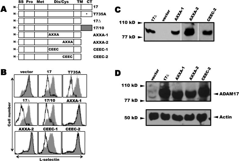
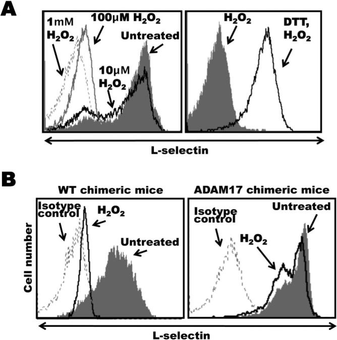
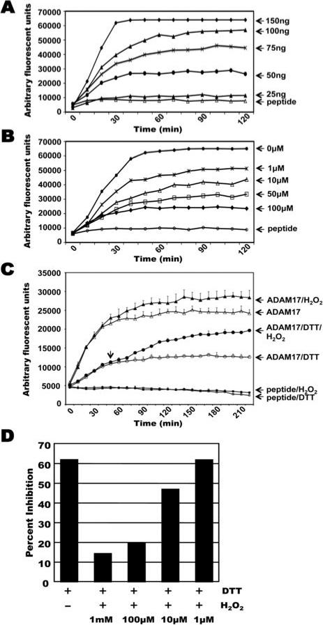
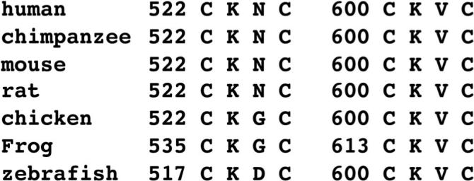
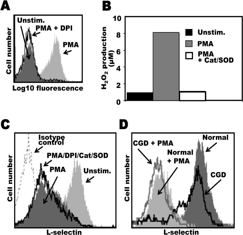
L-selectin is constitutively expressed by neutrophils and plays a key role in directing these cells to sites of inflammation. Upon neutrophil activation, L-selectin is rapidly and efficiently down-regulated from the cell surface by ectodomain shedding. We have directly shown that A disintegrin and metalloprotease 17 (ADAM17) is a primary and nonredundant sheddase of L-selection by activated neutrophils in vivo. Following cell activation, intracellular signals lead to the induction of ADAM17's enzymatic activity; however, the target of this inducer mechanism remains unclear. Our study provides evidence of an activation mechanism that involves the extracellular region of the mature form of cell surface ADAM17 and not its intracellular region. We demonstrate that the catalytic activity of purified ADAM17 lacking a prodomain and its intracellular region is diminished under mild reducing conditions by DTT and enhanced by H(2)O(2) oxidation. Moreover, H(2)O(2) reversed ADAM17 inhibition by DTT. The treatment of neutrophils with H(2)O(2) also induced L-selectin shedding in an ADAM17-dependent manner. These findings suggest that thiol-disulfide conversion occurring in the extracellular region of ADAM17 may be involved in its activation. An analysis of ADAM17 revealed that within its disintegrin/cysteine-rich region are two highly conserved, vicinal cysteine sulfhydryl motifs (cysteine-X-X-cysteine), which are well-characterized targets for thiol-disulfide exchange in various other proteins. Using a cell-based ADAM17 reconstitution assay, we demonstrate that the cysteine-X-X-cysteine motifs are critical for L-selectin cleavage. Taken together, our findings suggest that reduction-oxidation modifications of cysteinyl sulfhydryl groups in mature ADAM17 may serve as a mechanism for regulating the shedding of L-selectin following neutrophil stimulation.


Figures

Figure 1
Figure 1. Effects of ADAM17 reconstitution on L-selectin shedding
A, ADAM17 cDNA constructs. 17, full length ADAM17; T735A, full length ADAM17/T735A; 17Δ, ADAM17 cytoplasmic tail deletion; 17/10, ADAM17 containing an ADAM10 cytoplasmic tail; AXXA-1, C522KNC → A522KNA; AXXA-2, C600KVC → A600KVA; CEEC-1, C522KNC → C522EEC; CEEC-2, C600KVC → C600EEC. All constructs lacking a cytoplasmic tail (CT) contained a C-terminus FLAG epitope. SS, signal sequence; Pro, prodomain; Met, metalloprotease; Dis/Cys, disintegrin/cysteine-rich; TM, transmembrane; B, ADAM17-deficient cells stably expressing wild-type L-selectin were transduced with a bicistronic retroviral vector for proportional expression of an ADAM17 construct and GFP. Each transductant was stained with an L-selectin mAb and then dual analyzed by flow cytometry. GFP expressing cells (open histograms) and non-expressing cells (filled histograms) were each electronically gated and their relative expression of L-selectin was determined, as previously described (54). Cells transfected with an empty vector are indicated (vector). The x-axis = Log10 fluorescence and y-axis = cell number. Results are from representative experiments performed 3−5 times. C and D, The EC2 transductants expressing 17Δ, AXXA-1, AXXA-2, CEEC-2, or empty vector (vector) were sorted to match their levels of EGFP expression, which ranged from 77 to 81%. C, Detergent lysates from equivalent cell numbers of each transductant were treated with wheat germ agglutinin agarose to selectively precipitate glycoproteins, which were then subjected to reducing SDS-PAGE and immunoblotting with an anti-FLAG mAb, as described in the Material and Methods. D, Equivalent cell numbers of the EC2 transductants expressing 17Δ, AXXA-1, AXXA-2, CEEC-2 or empty vector (vector) were biotinylated to label cell surface proteins, then detergent lysed and directly subjected to SDS-PAGE for the detection of actin content to assess cell equivalency among the samples (lower panel), or treated with streptavidin agarose to selectively precipitate the biotinylated proteins, which were subjected to reducing SDS-PAGE and immunoblotting with anti-mouse ADAM17 polyclonal sera (upper panel), as described in the Material and Methods.
Figure 2
Figure 2. ADAM17-dependent L-selectin shedding by H2O2-treated neutrophils
A, Bone marrow leukocytes from C57BL/6 mice were either untreated or treated with H2O2 at the indicated concentrations for 15 min at 37°C (left panel). In addition, some leukocytes were initially incubated with DTT (200 μM) for 15 min at 25°C then H2O2 (100 μM) for 15 min at 37°C, as indicated (right panel). All cells were then double-stained for surface expression of L-selectin and the neutrophil marker Gr-1. B, Radiation chimeric mice reconstituted with hematopoietic cells either lacking functional ADAM17 (ADAM17 chimeric mice) or expressing functional ADAM17 (wild-type chimeric mice) were generated as described in the Materials and Methods. Bone marrow leukocytes from age-matched, ADAM17 or wild-type (WT) chimeric mice were either untreated or treated with H2O2 (4mM) for 15 min at 37 °C, as indicated. All cells were then double-stained for surface expression of L-selectin and the neutrophil marker Gr-1. Relative staining levels were determined by flow cytometry (A and B). For all histogram plots, the dashed line indicates staining by an isotype-matched negative control mAb. The y axis = cell number and the x axis = Log 10 fluorescence. Results are representative of 3 independent experiments.
Figure 3
Figure 3. The activity of purified ADAM17 is modulated by redox conditions
A, Various concentrations of recombinant human ADAM17 lacking a pro-domain and intracellular region was incubated with an internally-quenched fluorogenic peptide substrate. B, ADAM17 (75ng) was incubated with peptide substrate in the absence or presence of various concentrations of the reducing agent DTT, as indicated. C, ADAM17 (75ng) was incubated with peptide substrate in the presence of DTT (50 μM), H2O2 (4 mM), or DTT for 50 min and then H2O2. The fluorescent signal of the peptide substrate alone (peptide), or when in the presence of DTT or H2O2 (peptide/DTT or peptide/H2O2, respectively) is plotted. D, ADAM17 (75ng) was incubated with peptide substrate in the presence of DTT (50 μM) or DTT with varying concentrations of H2O2. Data are expressed as percent inhibition as follows: percent inhibition = (1 – fluorescence intensity of mixture solution of ADAM17 with DTT ± H2O2 / that of ADAM17 without DTT ± H2O2) × 100%. In panels A, B, and D, each reaction was done in triplicate and the average is shown. Representative data from 3 separate experiments are shown. For panel C, each data point represents the mean ± SD of three separate experiments. The variability in the fluorescent signal between panels A-D is due to the use of different lot preparations of the commercially available ADAM17 and fluorogenic peptide. All results shown were consistent between lot preparations.
Figure 4
Figure 4
Alignment of the two CXXC motifs in the disintegrin/cysteine-rich region of ADAM17 from various animal species.
Figure 5
Figure 5. NADPH oxidase-generated ROS is not critical for the induction of L-selectin shedding by human neutrophils
A and B, The efficiency of the NADPH oxidase inhibitor DPI on intracellular ROS production and the ROS scavengers catalase and SOD on extracellular ROS production by human neutrophils were assessed using the indicators DHR123 and Amplex Red, respectively. Neutrophils from normal healthy donors were stimulated with PMA (10 ng/ml) in the absence or presence of DPI or catalase plus SOD, as described in the Material and Methods. Fluorescence emission upon DHR123 conversion to rhodamine was measured by flow cytometry (A), and Amplex Red conversion to resorufin was measured by a fluorescence plate reader (B). C, Neutrophils were either stimulated with PMA (10 ng/ml) for 15 min at 37°C in the absence or presence of DPI/catalase/SOD, as indicated, or they were not stimulated PMA (Unstim.). D, Neutrophils from normal healthy donors or from CGD patients were incubated at 37°C in the presence or absence of PMA for 15 min, as indicated. Neutrophils were then labeled with PE-conjugated LAM1−116 to determine relative L-selectin expression (C and D). Treatment of neutrophils with the appropriate concentrations of carrier alone had no effect on L-selectin expression (data not shown). Non-specific antibody labeling was determined using the appropriate isotype negative control mAb, as indicated. Cells were analyzed by flow cytometry and the mean fluorescence intensity of 10,000 cells was determined for each cell sample. The data in panels A-C are representative of at least three independent experiments using neutrophils isolated from separate donors. The data in panel D is representative of two independent experiments using neutrophils isolated from separate donors.

Similar articles

Cited by

References

    1. Rainer TH. L-selectin in health and disease. Resuscitation. 2002;52:127–141. - PubMed
    1. Bargatze RF, Kurk S, Butcher EC, Jutila MA. Neutrophils roll on adherent neutrophils bound to cytokine- induced endothelial cells via L-selectin on the rolling cells. J. Exp. Med. 1994;180:1785–1792. - PMC - PubMed
    1. Walcheck B, Moore KL, McEver RP, Kishimoto TK. Neutrophil-neutrophil interactions under hydrodynamic shear stress involve L-selectin and PSGL-1. A mechanism that amplifies initial leukocyte accumulation on P-selectin in vitro. J. Clin. Invest. 1996;98:1081–1087. - PMC - PubMed
    1. Sperandio M, Smith ML, Forlow SB, Olson TS, Xia L, McEver RP, Ley K. P-selectin glycoprotein ligand-1 mediates L-selectin-dependent leukocyte rolling in venules. J. Exp. Med. 2003;197:1355–1363. - PMC - PubMed
    1. St Hill CA, Alexander SR, Walcheck B. Indirect capture augments leukocyte accumulation on P-selectin in flowing whole blood. J. Leukoc. Biol. 2003;73:464–471. - PubMed

Publication types

MeSH terms