. 2009 Mar 3;106(9):3443-8.
doi: 10.1073/pnas.0810473106. Epub 2009 Feb 6.

Acute inflammatory proteins constitute the organic matrix of prostatic corpora amylacea and calculi in men with prostate cancer

Affiliations

Acute inflammatory proteins constitute the organic matrix of prostatic corpora amylacea and calculi in men with prostate cancer

Karen S Sfanos et al. Proc Natl Acad Sci U S A. .

Abstract

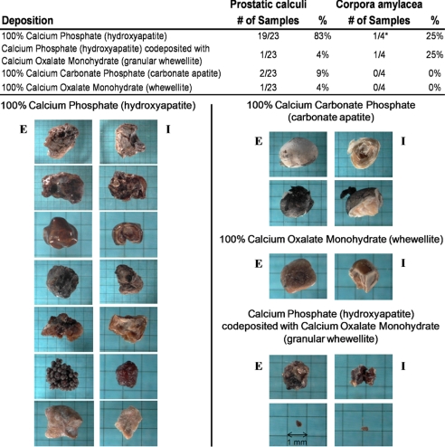
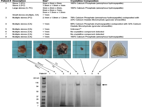
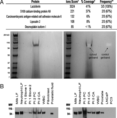
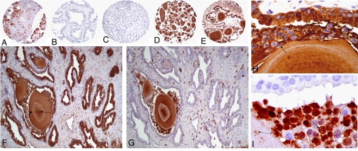
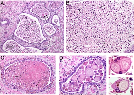
Corpora amylacea (CA) are a frequent microscopic finding in radical prostatectomy specimens from men undergoing treatment for prostate cancer. Although often observed histologically to be associated with inflammation, the contribution of CA to prostatitis-related symptoms of unknown etiology or to prostate carcinogenesis remains unclear. Prostatic calculi (PC), which potentially represent calcified forms of CA, are less common but can cause urological disease including urinary retention and prostatitis. We conducted a comprehensive compositional analysis of CA/PC to gain insight into their biogenesis. Infrared spectroscopy analysis of calculi collected from 23 patients confirmed a prevalence of calcium phosphate in the form of hydroxyapatite. This result sets PC apart from most urinary stones, which largely are composed of calcium oxalate. Tandem mass spectrometry-based proteomic analysis of CA/PC revealed that lactoferrin is the predominant protein component, a result that was confirmed by Western blot analysis. Other proteins identified, including calprotectin, myeloperoxidase, and alpha-defensins, are proteins contained in neutrophil granules. Immunohistochemistry (IHC) suggested the source of lactoferrin to be prostate-infiltrating neutrophils as well as inflamed prostate epithelium; however, IHC for calprotectin suggested prostate-infiltrating neutrophils as a major source of the protein, because it was absent from other prostate compartments. This study represents a definitive analysis of the protein composition of prostatic CA and calculi and suggests that acute inflammation has a role in their biogenesis--an intriguing finding, given the prevalence of CA in prostatectomy specimens and the hypothesized role for inflammation in prostate carcinogenesis.


Conflict of interest statement

The authors declare no conflict of interest.

Figures

Fig. 1.
Fig. 1.
Crystalline composition of prostatic calculi and corpora amylacea. (Top) Results of stone analysis by infrared spectroscopy. *Two CA samples contained no detectable mineral deposition. (Bottom) Variation in size, color, and shape among different crystalline compositions. E = exterior of stone; I = interior of stone.
Fig. 2.
Fig. 2.
Protein extraction from prostatic corpora amylacea and calculi. (Top) Description and crystalline composition of samples subject to protein extraction. The sample set included 2 separate PC from 1 patient (“stone 1” and “stone 2”) and PC versus CA from a separate patient. In all, the protein composition of a total of 10 samples from 8 patients was determined. *Average size if multiple stones. **Sample not available for chemical stone analysis. (Bottom) Examples of SDS/PAGE of extracted proteins on 4%–20% gradient gels visualized by Coomassie blue stain. Samples from separate gels have been grouped together. Arrow indicates predominant protein band observed at ≈100 kDa.
Fig. 3.
Fig. 3.
Identification of predominant protein band as lactoferrin. (A, Top) Results of in-gel peptide analysis by LC/MS/MS. (Bottom) Example of protein band excised from SDS/PAGE gel. *For multiple matches, the highest score observed in a single case is shown. **Peptide hit(s) also were observed in a single case for the following proteins: α-1-antichymotrypsin, TIMP-1, myeloperoxidase, uromodulin, plakophilin 1 isoform 1b, annexin A2, ubiquitin, ARG1, caspase 14 precursor, stratifin, and catalase. (B) Western blot analysis for human lactoferrin. Membranes were probed with rabbit anti-human lactoferrin (Sigma) at a dilution of 1:5000. Milk LF = human milk lactoferrin (0.5 μg), Neutrophil LF = human neutrophil lactoferrin (0.5 μg), WBC = human peripheral blood white blood cell lysate (5 μg), Fibroblast = human skin fibroblast lysate (5 μg), Prostatic fluid (5 μg), Prostate = human prostate tissue lysate (5 μg), LNCaP lysate (5 μg), PC3 lysate (5 μg).
Fig. 4.
Fig. 4.
IHC for lactoferrin and calprotectin in human prostate tissues. Tissue sections were probed with rabbit anti-human lactoferrin at 1:2000 (A–F and H) or mouse anti-human calprotectin (Dako) at 1:300 (G and I). (A) Human salivary gland tissue (lactoferrin positive control). (B) Normal human prostate. (C) Prostate cancer. (D) CA. (E) Inflamed prostate tissue with CA inside atrophic gland. (F) Positive lactoferrin staining of atrophic prostate epithelium, acute inflammatory cells, and CA. (G) Calprotectin staining of section adjacent to (F) showing positive staining of infiltrating leukocytes and CA only. CA stain was positive for both lactoferrin (D–F) and calprotectin (G) with areas of dense staining (dashed arrow in F) and concentric rings that do not stain positive (solid arrow in F). (H) Example of epithelium, neutrophils (arrows, as evidenced by multilobed nuclei), and CA staining positive for lactoferrin. (I) Section adjacent to (H) showing negative epithelium, focal positive calprotectin staining of CA (dashed arrow) and strong calprotectin staining of neutrophils (solid arrow).
Fig. 5.
Fig. 5.
Presence of CA within acutely inflamed prostate glands. (A) Mixed neutrophil and macrophage infiltrates and CA (arrows) within glands and stromal lymphocytes surrounding glands. (B) Dense population of primarily neutrophils and CA (arrows) within prostate gland. (C) Dense population of primarily macrophages and CA (arrows) within prostate gland. (D) Examples of engulfment of CA by macrophages and multinucleated giant cells.

References

    1. Thompson SH. The Diseases of the Prostate: Their Pathology and Treatment. London: J. & A. Churchill; 1883.
    1. Shah SK, Chau MH, Schnepper GD, Lui PD. Open prostatolithotomy for the management of giant prostatic calculi. Urology. 2007;70:1008.e1009–1010. - PubMed
    1. Moore RA. Morphology of prostatic corpora amylacea and calculi. Arch Pathol. 1936;22:24–40.
    1. Izzidien AY. Prostatic calcification in a 4-year-old boy. Arch Dis Child. 1980;55:963–964. - PMC - PubMed
    1. Bedir S, et al. Endoscopic treatment of multiple prostatic calculi causing urinary retention. International Journal of Urology. 2005;12:693–695. - PubMed

Publication types

MeSH terms