

Methylphenidate-induced dendritic spine formation and DeltaFosB expression in nucleus accumbens
- PMID: 19202072
- PMCID: PMC2650365
- DOI: 10.1073/pnas.0813179106
Methylphenidate-induced dendritic spine formation and DeltaFosB expression in nucleus accumbens
Abstract
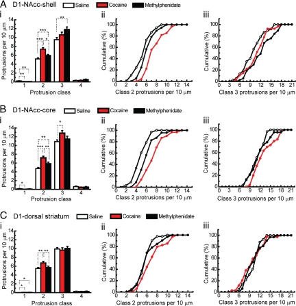
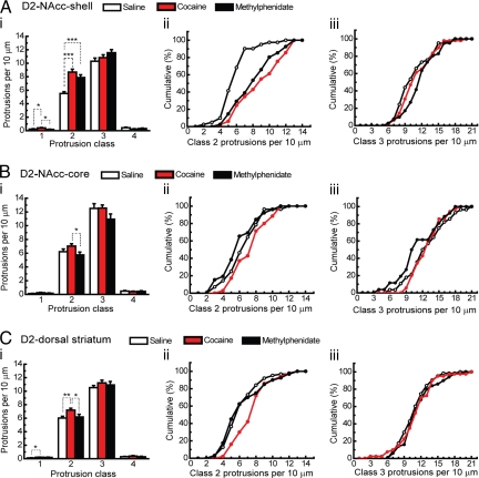
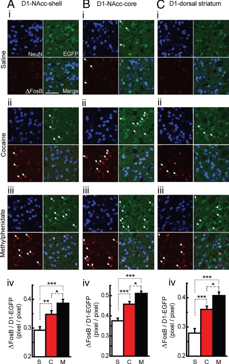
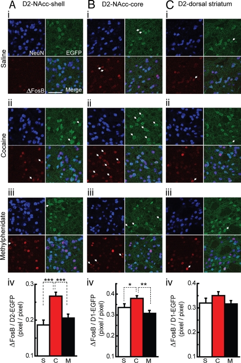
Methylphenidate is the psychostimulant medication most commonly prescribed to treat attention deficit hyperactivity disorder (ADHD). Recent trends in the high usage of methylphenidate for both therapeutic and nontherapeutic purposes prompted us to investigate the long-term effects of exposure to the drug on neuronal adaptation. We compared the effects of chronic methylphenidate or cocaine (15 mg/kg, 14 days for both) exposure in mice on dendritic spine morphology and DeltaFosB expression in medium-sized spiny neurons (MSN) from ventral and dorsal striatum. Chronic methylphenidate increased the density of dendritic spines in MSN-D1 (MSN-expressing dopamine D1 receptors) from the core and shell of nucleus accumbens (NAcc) as well as MSN-D2 (MSN-expressing dopamine D2 receptors) from the shell of NAcc. In contrast, cocaine increased the density of spines in both populations of MSN from all regions of striatum. In general, the effect of methylphenidate on the increase of shorter spines (class 2) was less than that of cocaine. Interestingly, the methylphenidate-induced increase in the density of relatively longer spines (class 3) in the shell of NAcc was bigger than that induced by cocaine. Furthermore, methylphenidate exposure increased expression of DeltaFosB only in MSN-D1 from all areas of striatum, and surprisingly, the increase was greater than that induced by cocaine. Thus, our results show differential effects of methylphenidate and cocaine on neuronal adaptation in specific types of MSN in reward-related brain regions.
Conflict of interest statement
The authors declare no conflict of interest.
Figures
Similar articles
-
Cocaine-induced dendritic spine formation in D1 and D2 dopamine receptor-containing medium spiny neurons in nucleus accumbens.Proc Natl Acad Sci U S A. 2006 Feb 28;103(9):3399-404. doi: 10.1073/pnas.0511244103. Epub 2006 Feb 21. Proc Natl Acad Sci U S A. 2006. PMID: 16492766 Free PMC article.
-
Behavioral and structural responses to chronic cocaine require a feedforward loop involving ΔFosB and calcium/calmodulin-dependent protein kinase II in the nucleus accumbens shell.J Neurosci. 2013 Mar 6;33(10):4295-307. doi: 10.1523/JNEUROSCI.5192-12.2013. J Neurosci. 2013. PMID: 23467346 Free PMC article.
-
Cocaine-Induced Structural Plasticity in Input Regions to Distinct Cell Types in Nucleus Accumbens.Biol Psychiatry. 2018 Dec 15;84(12):893-904. doi: 10.1016/j.biopsych.2018.04.019. Epub 2018 May 9. Biol Psychiatry. 2018. PMID: 29921416 Free PMC article.
-
Cell-Type-Specific Adaptions in Striatal Medium-Sized Spiny Neurons and Their Roles in Behavioral Responses to Drugs of Abuse.Front Synaptic Neurosci. 2021 Dec 14;13:799274. doi: 10.3389/fnsyn.2021.799274. eCollection 2021. Front Synaptic Neurosci. 2021. PMID: 34970134 Free PMC article. Review.
-
Chronic Methylphenidate Effects on Brain Gene Expression: An Exploratory Review.Psychol Res Behav Manag. 2024 Feb 15;17:577-592. doi: 10.2147/PRBM.S445719. eCollection 2024. Psychol Res Behav Manag. 2024. PMID: 38379637 Free PMC article. Review.
Cited by
-
Age differences to methylphenidate-NAc neuronal and behavioral recordings from freely behaving animals.J Neural Transm (Vienna). 2022 Aug;129(8):1061-1076. doi: 10.1007/s00702-022-02526-0. Epub 2022 Jul 16. J Neural Transm (Vienna). 2022. PMID: 35842551
-
Prenatal Exposure to Alcohol Induces Functional and Structural Plasticity in Dopamine D1 Receptor-Expressing Neurons of the Dorsomedial Striatum.Alcohol Clin Exp Res. 2018 Jun 5:10.1111/acer.13806. doi: 10.1111/acer.13806. Online ahead of print. Alcohol Clin Exp Res. 2018. PMID: 29870053 Free PMC article.
-
D1 and D2 specific dopamine antagonist modulate the caudate nucleus neuronal responses to chronic methylphenidate exposure.J Neural Transm (Vienna). 2017 Feb;124(2):159-170. doi: 10.1007/s00702-016-1647-x. Epub 2016 Nov 16. J Neural Transm (Vienna). 2017. PMID: 27853928
-
Early developmental changes in GABAA receptor expression in nucleus accumbens medium spiny neurons.Front Neurosci. 2024 Dec 12;18:1445162. doi: 10.3389/fnins.2024.1445162. eCollection 2024. Front Neurosci. 2024. PMID: 39726828 Free PMC article.
-
MEF-2 regulates activity-dependent spine loss in striatopallidal medium spiny neurons.Mol Cell Neurosci. 2010 May;44(1):94-108. doi: 10.1016/j.mcn.2010.01.012. Epub 2010 Mar 1. Mol Cell Neurosci. 2010. PMID: 20197093 Free PMC article.
References
-
- Biederman J, Faraone SV. Attention-deficit hyperactivity disorder. Lancet. 2005;366:237–248. - PubMed
-
- Robison LM, Sclar DA, Skaer TL, Galin RS. National trends in the prevalence of attention-deficit/hyperactivity disorder and the prescribing of methylphenidate among school-age children: 1990–1995. Clin Pediatr (Phila) 1999;38:209–217. - PubMed
-
- Volkow ND, et al. Dopamine transporter occupancies in the human brain induced by therapeutic doses of oral methylphenidate. Am J Psychiatry. 1998;155:1325–1331. - PubMed
-
- Volkow ND, et al. Is methylphenidate like cocaine? Studies on their pharmacokinetics and distribution in the human brain. Arch Gen Psychiatry. 1995;52:456–463. - PubMed
Publication types
MeSH terms
Substances
Grants and funding
LinkOut - more resources
Full Text Sources
Molecular Biology Databases
Research Materials
Miscellaneous
