Interferometric fluorescent super-resolution microscopy resolves 3D cellular ultrastructure
- PMID: 19202073
- PMCID: PMC2637278
- DOI: 10.1073/pnas.0813131106
Interferometric fluorescent super-resolution microscopy resolves 3D cellular ultrastructure
Abstract
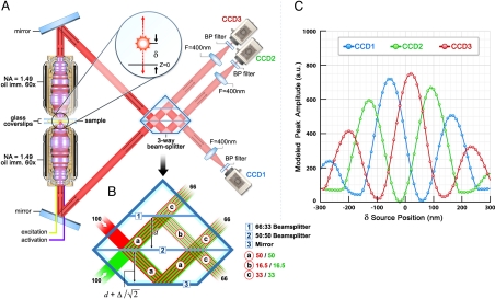
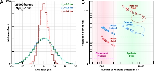
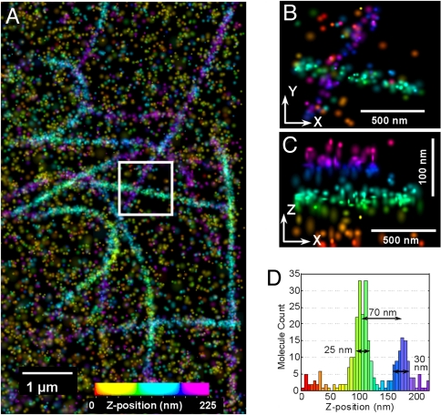
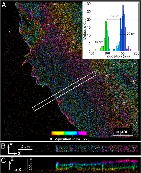
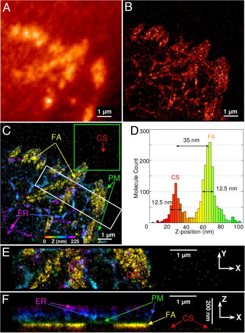
Understanding molecular-scale architecture of cells requires determination of 3D locations of specific proteins with accuracy matching their nanometer-length scale. Existing electron and light microscopy techniques are limited either in molecular specificity or resolution. Here, we introduce interferometric photoactivated localization microscopy (iPALM), the combination of photoactivated localization microscopy with single-photon, simultaneous multiphase interferometry that provides sub-20-nm 3D protein localization with optimal molecular specificity. We demonstrate measurement of the 25-nm microtubule diameter, resolve the dorsal and ventral plasma membranes, and visualize the arrangement of integrin receptors within endoplasmic reticulum and adhesion complexes, 3D protein organization previously resolved only by electron microscopy. iPALM thus closes the gap between electron tomography and light microscopy, enabling both molecular specification and resolution of cellular nanoarchitecture.
Conflict of interest statement
The authors declare no conflict of interest.
Figures


Similar articles
-
Three-dimensional Super Resolution Microscopy of F-actin Filaments by Interferometric PhotoActivated Localization Microscopy (iPALM).J Vis Exp. 2016 Dec 1;(118):54774. doi: 10.3791/54774. J Vis Exp. 2016. PMID: 27929472 Free PMC article.
-
Imaging cellular ultrastructure by PALM, iPALM, and correlative iPALM-EM.Methods Cell Biol. 2014;123:273-94. doi: 10.1016/B978-0-12-420138-5.00015-X. Methods Cell Biol. 2014. PMID: 24974033
-
Three-dimensional super-resolution imaging by stochastic optical reconstruction microscopy.Science. 2008 Feb 8;319(5864):810-3. doi: 10.1126/science.1153529. Epub 2008 Jan 3. Science. 2008. PMID: 18174397 Free PMC article.
-
About samples, giving examples: Optimized Single Molecule Localization Microscopy.Methods. 2020 Mar 1;174:100-114. doi: 10.1016/j.ymeth.2019.05.008. Epub 2019 May 10. Methods. 2020. PMID: 31078795 Review.
-
Super-resolution microscopy demystified.Nat Cell Biol. 2019 Jan;21(1):72-84. doi: 10.1038/s41556-018-0251-8. Epub 2019 Jan 2. Nat Cell Biol. 2019. PMID: 30602772 Review.
Cited by
-
Quantitative multicolor subdiffraction imaging of bacterial protein ultrastructures in three dimensions.Nano Lett. 2013 Mar 13;13(3):987-93. doi: 10.1021/nl304071h. Epub 2013 Feb 25. Nano Lett. 2013. PMID: 23414562 Free PMC article.
-
Resolution improvement by 3D particle averaging in localization microscopy.Methods Appl Fluoresc. 2015 Mar;3(1):014003. doi: 10.1088/2050-6120/3/1/014003. Methods Appl Fluoresc. 2015. PMID: 25866640 Free PMC article.
-
Distinct, but not completely separate spatial transport routes in the nuclear pore complex.Nucleus. 2013 May-Jun;4(3):166-75. doi: 10.4161/nucl.24874. Epub 2013 May 1. Nucleus. 2013. PMID: 23669120 Free PMC article. Review.
-
Bleaching/blinking assisted localization microscopy for superresolution imaging using standard fluorescent molecules.Proc Natl Acad Sci U S A. 2011 Dec 27;108(52):21081-6. doi: 10.1073/pnas.1117430109. Epub 2011 Dec 13. Proc Natl Acad Sci U S A. 2011. PMID: 22167805 Free PMC article.
-
Augmented 3D super-resolution of fluorescence-free nanoparticles using enhanced dark-field illumination based on wavelength-modulation and a least-cubic algorithm.Sci Rep. 2016 Sep 13;6:32863. doi: 10.1038/srep32863. Sci Rep. 2016. PMID: 27619347 Free PMC article.
References
Publication types
MeSH terms
Grants and funding
LinkOut - more resources
Full Text Sources
Other Literature Sources
Research Materials
Miscellaneous