. 2009 Feb 24;106(8):2729-34.
doi: 10.1073/pnas.0813175106. Epub 2009 Feb 6.

Anti-CTLA-4 therapy results in higher CD4+ICOShi T cell frequency and IFN-gamma levels in both nonmalignant and malignant prostate tissues

Affiliations

Anti-CTLA-4 therapy results in higher CD4+ICOShi T cell frequency and IFN-gamma levels in both nonmalignant and malignant prostate tissues

Hong Chen et al. Proc Natl Acad Sci U S A. .

Abstract

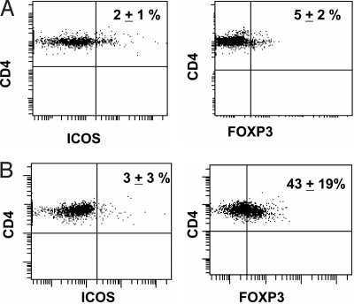
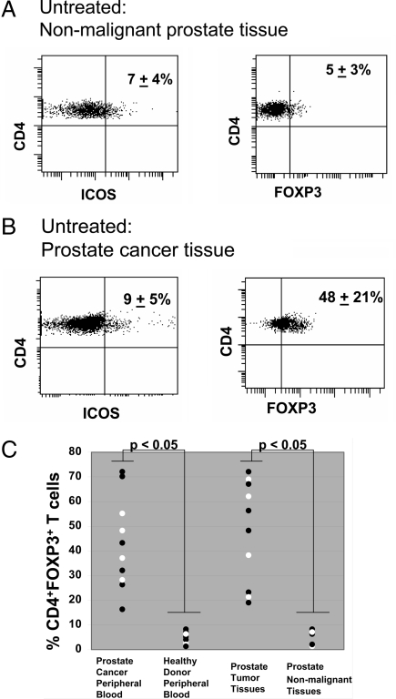
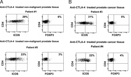
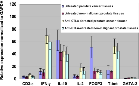
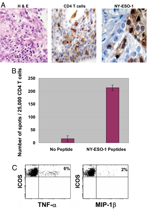
Cytotoxic lymphocyte antigen-4 (CTLA-4) blockade is an active immunotherapeutic strategy that is currently in clinical trials in cancer. There are several ongoing trials of anti-CTLA-4 in the metastatic setting of prostate cancer patients with reported clinical responses consisting of decreases in the prostate specific antigen (PSA) tumor marker for some patients. Immunologic markers that correlate with these clinical responses are necessary to guide further development of anti-CTLA-4 therapy in the treatment of cancer patients. We recently reported that CD4(+) inducible co-stimulator (ICOS)(hi) T cells that produce interferon-gamma (IFN-gamma) are increased in the peripheral blood and tumor tissues of bladder cancer patients treated with anti-CTLA-4 antibody. Here we present data from the same clinical trial in bladder cancer patients demonstrating a higher frequency of CD4(+)ICOS(hi) T cells and IFN-gamma mRNA levels in nonmalignant prostate tissues and incidental prostate tumor tissues removed at the time of radical cystoprostatectomy. Our data suggest immunologic markers that can be used to monitor prostate cancer patients who receive anti-CTLA-4 therapy and indicate that the immunologic impact of anti-CTLA-4 antibody can occur in both tumor and nonmalignant tissues. These data should be taken into consideration for evaluation of efficacy as well as immune-related adverse events associated with anti-CTLA-4 therapy.


Conflict of interest statement

Conflict of interest statement: P.S. has served as a consultant on advisory boards for Bristol-Myers Squibb.

Figures

Fig. 1.
Fig. 1.
Expression of ICOS and FOXP3 by CD4 T cells in peripheral blood of prostate cancer patients. (A) In healthy donors (n = 10), ≈2% of CD4 T cells express ICOS (Left) and 5% of CD4 T cells express FOXP3 (Right). (B) In untreated prostate cancer patients (n = 10), ≈3% of CD4 T cells express ICOS (Left) and 43% of CD4 T cells express FOXP3 (Right). A representative figure from one healthy donor (A) and one untreated prostate cancer patient (B) is shown, with numbers indicating mean percentages (± standard deviations) calculated from samples from 10 healthy donors and 10 patients with untreated prostate cancer.
Fig. 2.
Fig. 2.
Expression of ICOS and FOXP3 by CD4 T cells in normal and malignant prostate tissues of untreated patients. (A) In nonmalignant prostate tissues (n = 5), ≈7% of CD4 T cells express ICOS (Left) and 5% of CD4 T cells express FOXP3 (Right). (B) In prostate cancer tissues (n = 10), ≈9% of CD4 T cells express ICOS (Left) and 48% of CD4 T cells express FOXP3 (Right). A representative figure from one nonmalignant tissue sample (A) and from prostate cancer tissue sample from one untreated patient (B) is shown with numbers indicating mean percentages (± standard deviations) calculated from samples from 5 nonmalignant prostate tissue samples and 10 prostate cancer tissue samples. (C) Graphical representation of significantly (P < 0.05) higher frequency of CD4+FOXP3+ T cells in peripheral blood of prostate cancer patients (n = 10) as compared to peripheral blood of healthy donors, and in prostate tumor tissues (n = 10) as compared to nonmalignant prostate tissues (n = 5).
Fig. 3.
Fig. 3.
Expression of ICOS and FOXP3 by CD4 T cells in normal and malignant prostate tissues from patients treated with anti–CTLA-4. (A) CD4 T cells had a higher frequency of ICOS expression at 29% (patient 1, Left Upper) and 23% (patient 5, Left Lower) in nonmalignant prostate tissues after patients received treatment with anti-CTLA-4 antibody while FOXP3 levels remained similarly low in both patients (Right) as compared to untreated nonmalignant tissues shown in Fig. 2A. (B) CD4 T cells had a higher frequency of ICOS expression at 31% (patient 3, Left Upper) and 22% (patient 4, Left Lower) in prostate cancer tissues after patients received treatment with anti-CTLA-4 antibody, while frequency of FOXP3 expression was lower at 5% (patient 3, Right Upper) and 4% (patient 4, Right Lower) as compared to untreated prostate cancer tissues shown in Fig. 2B.
Fig. 4.
Fig. 4.
Changes in expression of specific gene transcripts in prostate tissues following treatment with anti-CTLA-4. Fold induction of CD3-ε, IFN-γ, IL-10, IL-2, FOXP3, T-bet, and GATA-3 mRNA levels in untreated prostate cancer tissues, untreated nonmalignant prostate tissues, anti-CTLA-4 antibody treated prostate cancer tissues, and anti-CTLA-4 antibody treated nonmalignant prostate tissues as compared to GAPDH mRNA levels as assessed by real-time PCR. Anti-CTLA-4 treatment induced higher IFN-γ and T-bet mRNA levels in both cancer and nonmalignant tissues as well as lower FOXP3 mRNA levels in cancer tissues as compared to untreated cancer tissues.
Fig. 5.
Fig. 5.
NY-ESO-1 is recognized by CD4 tumor-infiltrating T cells from an anti-CTLA-4 treated patient sample. (A) H & E staining of patient 4 prostate tissues (Right), demonstrating CD4 T cell infiltration (Middle) and NY-ESO-1 expression on prostate tumor cells (Left). (B) An ELISPOT assay demonstrating CD4 T cell recognition of antigen-presenting cells (APCs) pulsed with NY-ESO-1 overlapping peptides as compared to unpulsed APCs (no peptides). The assay was done in triplicate and data were plotted as mean ± standard deviation. (C) CD4+ICOShi T cells produce TNF-α (Right) and MIP-1β (Left) in the presence of APCs pulsed with NY-ESO-1 overlapping peptides.

Similar articles

Cited by

References

    1. Walunas TL, et al. CTLA-4 can function as a negative regulator of T cell activation. Immunity. 1994;1:405–413. - PubMed
    1. Krummel MF, Allison JP. CD28 and CTLA-4 have opposing affects on the response of T cells to stimulation. J Exp Med. 1995;182:459–465. - PMC - PubMed
    1. Krummel MF, Allison JP. CTLA-4 engagement inhibits IL-2 accumulation and cell cycle progression upon activation of resting T cells. J Exp Med. 1996;183:2533–2540. - PMC - PubMed
    1. Leach DR, Krummel MF, Allison JP. Enhancement of antitumor immunity by CTLA-4 blockade. Science. 1996;271:1734–1736. - PubMed
    1. van Elas A, Hurwitz AA, Allison JP. Combination immunotherapy of B16 melanoma using anti-cytotoxic T lymphocyte-associated antigen 4 (CTLA-4) and granulocyte/macrophage colony-stimulating factor (GM-CSF)-producing vaccines induces rejection of subcutaneous and metastatic tumors accompanied by autoimmune depigmentation. J Exp Med. 1999;190:355–366. - PMC - PubMed

Publication types