. 2009 Apr 3;284(14):9558-65.
doi: 10.1074/jbc.M808906200. Epub 2009 Feb 7.

Human CtIP mediates cell cycle control of DNA end resection and double strand break repair

Affiliations

Human CtIP mediates cell cycle control of DNA end resection and double strand break repair

Pablo Huertas et al. J Biol Chem. .

Abstract

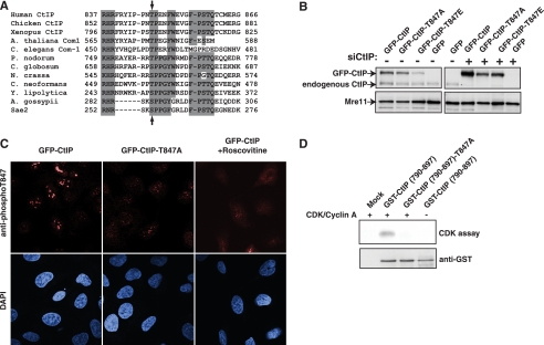
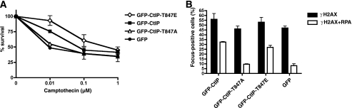
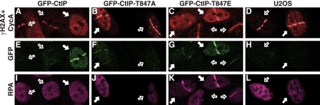
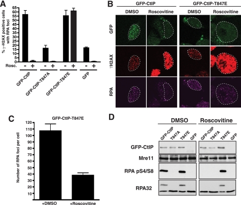
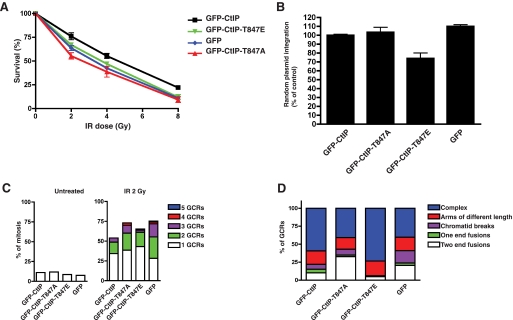
In G(0) and G(1), DNA double strand breaks are repaired by nonhomologous end joining, whereas in S and G(2), they are also repaired by homologous recombination. The human CtIP protein controls double strand break (DSB) resection, an event that occurs effectively only in S/G(2) and that promotes homologous recombination but not non-homologous end joining. Here, we mutate a highly conserved cyclin-dependent kinase (CDK) target motif in CtIP and reveal that mutating Thr-847 to Ala impairs resection, whereas mutating it to Glu to mimic constitutive phosphorylation does not. Moreover, we show that unlike cells expressing wild-type CtIP, cells expressing the Thr-to-Glu mutant resect DSBs even after CDK inhibition. Finally, we establish that Thr-847 mutations to either Ala or Glu affect DSB repair efficiency, cause hypersensitivity toward DSB-generating agents, and affect the frequency and nature of radiation-induced chromosomal rearrangements. These results suggest that CDK-mediated control of resection in human cells operates by mechanisms similar to those recently established in yeast.


Figures

FIGURE 1.
FIGURE 1.
Functional effects of mutating Thr-847 of CtIP. A, alignment of the region conserved among Sae2/CtIP orthologues. Arrows show the position of the conserved CtIP Thr-847 and Sae2 Ser-267. A. thaliana, Arabidopsis thaliana; C. elegans, Caenorhabditis elegans; P. nodorum, Phaeosphaeria nodorum; C. globosum, Chaetomium globosum; N. crassa, Neurospora crassa; C. neoformans, Cryptococcus neoformans; Y. lipolytica, Yarrowia lipolytica; A. gossypii, Ashbya gossypii. B, expression levels of GFP-CtIP derivatives in stably transfected clones before (left) or after (right) siRNA depletion of endogenous CtIP (siCtIP). C, representative confocal microscope images of cells expressing wild-type or T847A CtIP variants after immunostaining with a phospho-specific antibody raised against phosphorylated Thr-847. Cells were incubated in the presence of the CDK inhibitor roscovitine where indicated. D, a GST-fused wild-type or T847A mutant CtIP C-terminal fragment (residues 750-897) was affinity-purified with glutathione-Sepharose 4B and then incubated with [γ-32P]ATP in the presence or absence of recombinant CDK2/cyclin A, separated by 10% SDS-PAGE, and transferred to nitrocellulose membrane. Proteins were detected with an anti-GST antibody (bottom), and phosphorylation was visualized by autoradiography (CDK assay; top).
FIGURE 2.
FIGURE 2.
Effects of CtIP mutations on cellular responses to camptothecin-induced DNA damage. A, cell survival after exposing cells expressing GFP-CtIP fusions to the indicated doses of camptothecin; averages and standard deviations (error bars) of three independent experiments are shown. B, quantification of γH2AX and RPA foci-positive cells and γH2AX focus-positive cells for the indicated CtIP variants after 1 h of treatment with 1 μm camptothecin. Averages and standard deviations (error bars) of four independent experiments are shown. At least 200 cells were counted per experiment.
FIGURE 3.
FIGURE 3.
Effects of CtIP mutations on recruitment of proteins to laser-induced damage. Representative images of cells expressing GFP-CtIP variants after laser damage are shown. Cells were immunostained for RPA (magenta) and γH2AX plus cyclin A (red). Damaged cells not expressing cyclin A (G1) and cells positive for cyclin A (S/G2) are marked with empty and filled arrows, respectively.
FIGURE 4.
FIGURE 4.
CtIP mutations affect DSB processing. A, cells expressing CtIP variants were treated with DMSO (-) or 25 μm roscovitine (Rosc.)(+) and then irradiated with 10 Gy of IR. One h later, cells were immunostained for RPA or γH2AX. Averages and standard deviations (error bars) of three independent experiments are shown. At least 200 cells were counted per experiment. B, representative images of cells treated in A. C, the number of RPA foci per cell in cells expressing the GFP-CtIP-T847E mutant in the presence or absence of the CDK inhibitor roscovitine. Error bars, standard deviations. D, an immunoblot of protein extracts, collected 1 h after irradiation (10 Gy), of cells expressing the indicated GFP-CtIP fusions. Panels to the left and right contain samples derived from cells treated in the absence or presence of roscovitine, respectively.
FIGURE 5.
FIGURE 5.
Effects of CtIP mutations on DNA repair and chromosome integrity. A, the survival of U2OS cells expressing GFP-CtIP fusions after treatment with IR. Averages and standard deviations (error bars) of three independent experiments are shown. B, the effects of CtIP mutations on NHEJ efficiency as measured by random plasmid integration. Frequencies of integration were normalized to the values of wild-type GFP-CtIP, set as 100%. Averages and standard deviations (error bars) of three independent experiments are shown. C, the percentage of mitoses showing 1, 2, 3, 4, or 5 GCRs in cells expressing GFP-CtIP variants either untreated (left) or treated with 2 Gy of IR (right). At least 100 mitoses were analyzed per experiment. Error bars, standard deviations. D, the percentage of each GCR type in the cells analyzed in C.

References

    1. Shrivastav, M., De Haro, L. P., and Nickoloff, J. A. (2008) Cell Res. 18 134-147 - PubMed
    1. Sonoda, E., Hochegger, H., Saberi, A., Taniguchi, Y., and Takeda, S. (2006) DNA Repair 5 1021-1029 - PubMed
    1. Lieber, M. R. (2008) J. Biol. Chem. 283 1-5 - PubMed
    1. West, S. C. (2003) Nat. Rev. Mol. Cell Biol. 4 435-445 - PubMed
    1. Aylon, Y., Liefshitz, B., and Kupiec, M. (2004) EMBO J. 23 4868-4875 - PMC - PubMed

Publication types

MeSH terms