

Mitochondria targeted peptides protect against 1-methyl-4-phenyl-1,2,3,6-tetrahydropyridine neurotoxicity
- PMID: 19203217
- PMCID: PMC2819801
- DOI: 10.1089/ars.2009.2445
Mitochondria targeted peptides protect against 1-methyl-4-phenyl-1,2,3,6-tetrahydropyridine neurotoxicity
Abstract
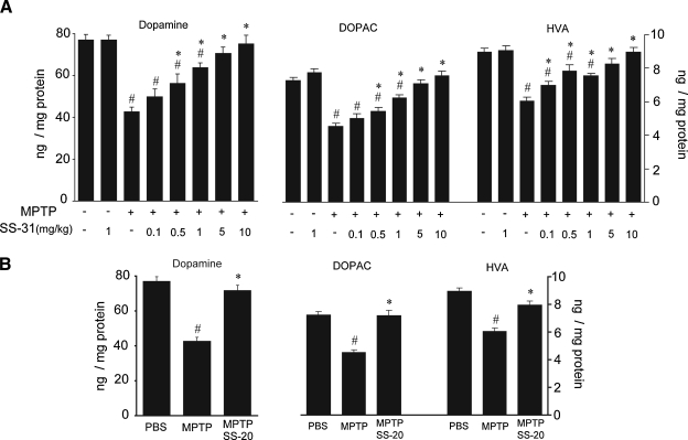
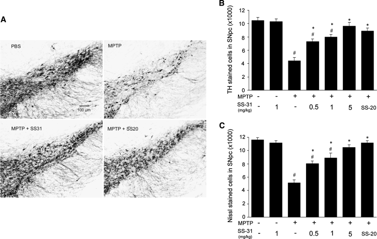
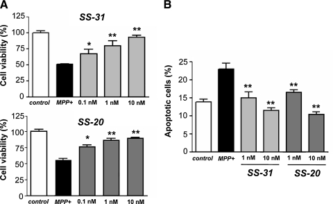
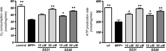
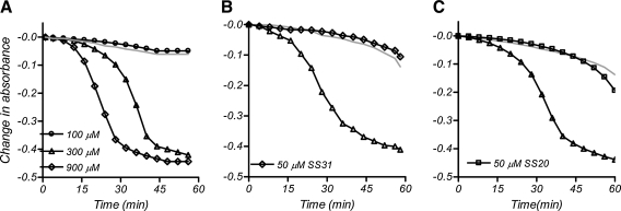
A large body of evidence suggests that mitochondrial dysfunction and oxidative damage play a role in the pathogenesis of Parkinson's disease (PD). A number of antioxidants have been effective in animal models of PD. We have developed a family of mitochondria-targeted peptides that can protect against mitochondrial swelling and apoptosis (SS peptides). In this study, we examined the ability of two peptides, SS-31 and SS-20, to protect against 1-methyl-4-phenyl-1,2,3,6-tetrahydropyridine (MPTP) neurotoxicity in mice. SS-31 produced dose-dependent complete protection against loss of dopamine and its metabolites in striatum, as well as loss of tyrosine hydroxylase immunoreactive neurons in substantia nigra pars compacta. SS-20, which does not possess intrinsic ability in scavenging reactive oxygen species, also demonstrated significant neuroprotective effects on dopaminergic neurons of MPTP-treated mice. Both SS-31 and SS-20 were very potent (nM) in preventing MPP+ (1-methyl-4-phenylpyridinium)-induced cell death in cultured dopamine cells (SN4741). Studies with isolated mitochondria showed that both SS-31 and SS-20 prevented MPP+-induced inhibition of oxygen consumption and ATP production, and mitochondrial swelling. These findings provide strong evidence that these neuroprotective peptides, which target both mitochondrial dysfunction and oxidative damage, are a promising approach for the treatment of PD.
Figures
Similar articles
-
EGb761 protects against nigrostriatal dopaminergic neurotoxicity in 1-methyl-4-phenyl-1,2,3,6-tetrahydropyridine-induced Parkinsonism in mice: role of oxidative stress.Eur J Neurosci. 2008 Jul;28(1):41-50. doi: 10.1111/j.1460-9568.2008.06314.x. Eur J Neurosci. 2008. PMID: 18662333
-
Six weeks of voluntary exercise don't protect C57BL/6 mice against neurotoxicity of MPTP and MPP(+).Neurotox Res. 2014 Feb;25(2):147-52. doi: 10.1007/s12640-013-9412-5. Epub 2013 Jul 20. Neurotox Res. 2014. PMID: 23873578
-
Neuroprotective effects of total flavonoid fraction of the Epimedium koreanum Nakai extract on dopaminergic neurons: In vivo and in vitro.Biomed Pharmacother. 2017 Jul;91:656-663. doi: 10.1016/j.biopha.2017.04.083. Epub 2017 May 8. Biomed Pharmacother. 2017. PMID: 28494419
-
Metallobiology of 1-methyl-4-phenyl-1,2,3,6-tetrahydropyridine neurotoxicity.Metallomics. 2013 Feb;5(2):91-109. doi: 10.1039/c2mt20164j. Metallomics. 2013. PMID: 23322189 Review.
-
SS-31, a Mitochondria-Targeting Peptide, Ameliorates Kidney Disease.Oxid Med Cell Longev. 2022 Jun 6;2022:1295509. doi: 10.1155/2022/1295509. eCollection 2022. Oxid Med Cell Longev. 2022. PMID: 35707274 Free PMC article. Review.
Cited by
-
The entangled ER-mitochondrial axis as a potential therapeutic strategy in neurodegeneration: A tangled duo unchained.Cell Calcium. 2016 Sep;60(3):218-34. doi: 10.1016/j.ceca.2016.04.010. Epub 2016 May 7. Cell Calcium. 2016. PMID: 27212603 Free PMC article. Review.
-
Tenofovir-induced nephrotoxicity: incidence, mechanism, risk factors, prognosis and proposed agents for prevention.Eur J Clin Pharmacol. 2014 Sep;70(9):1029-40. doi: 10.1007/s00228-014-1712-z. Epub 2014 Jun 25. Eur J Clin Pharmacol. 2014. PMID: 24958564 Review.
-
Elamipretide: A Review of Its Structure, Mechanism of Action, and Therapeutic Potential.Int J Mol Sci. 2025 Jan 23;26(3):944. doi: 10.3390/ijms26030944. Int J Mol Sci. 2025. PMID: 39940712 Free PMC article. Review.
-
Interactions of amyloidogenic proteins with mitochondrial protein import machinery in aging-related neurodegenerative diseases.Front Physiol. 2023 Nov 2;14:1263420. doi: 10.3389/fphys.2023.1263420. eCollection 2023. Front Physiol. 2023. PMID: 38028797 Free PMC article. Review.
-
Improving mitochondrial function with SS-31 reverses age-related redox stress and improves exercise tolerance in aged mice.Free Radic Biol Med. 2019 Apr;134:268-281. doi: 10.1016/j.freeradbiomed.2018.12.031. Epub 2018 Dec 28. Free Radic Biol Med. 2019. PMID: 30597195 Free PMC article.
References
-
- Adams JD., Jr. Klaidman LK. Leung AC. MPP+ and MPDP+ induced oxygen radical formation with mitochondrial enzymes. Free Radic Biol Med. 1993;15:181–186. - PubMed
-
- Bates TE. Heales SJ. Davies SE. Boakye P. Clark JB. Effects of 1-methyl-4-phenylpyridinium on isolated rat brain mitochondria: Evidence for a primary involvement of energy depletion. J Neurochem. 1994;63:640–648. - PubMed
-
- Beal MF. Matthews RT. Tieleman A. Shults CW. Coenzyme Q10 attenuates the 1-methyl-4-phenyl-1,2,3,tetrahydropyridine (MPTP) induced loss of striatal dopamine and dopaminergic axons in aged mice. Brain Res. 1998;783:109–114. - PubMed
-
- Betarbet R. Sherer TB. MacKenzie G. Garcia–Osuna M. Panov AV. Greenamyre JT. Chronic systemic pesticide exposure reproduces features of Parkinson's disease. Nat Neurosci. 2000;3:1301–1306. - PubMed
-
- Canet–Aviles RM. Wilson MA. Miller DW. Ahmad R. McLendon C. Bandyopadhyay S. Baptista MJ. Ringe D. Petsko GA. Cookson MR. The Parkinson's disease protein DJ-1 is neuroprotective due to cysteine-sulfinic acid-driven mitochondrial localization. Proc Natl Acad Sci USA. 2004;101:9103–9108. - PMC - PubMed
Publication types
MeSH terms
Substances
Grants and funding
LinkOut - more resources
Full Text Sources
Other Literature Sources
Miscellaneous
