

Embryonic stem cells in cardiac repair and regeneration
- PMID: 19203218
- PMCID: PMC2788053
- DOI: 10.1089/ars.2009.2491
Embryonic stem cells in cardiac repair and regeneration
Abstract
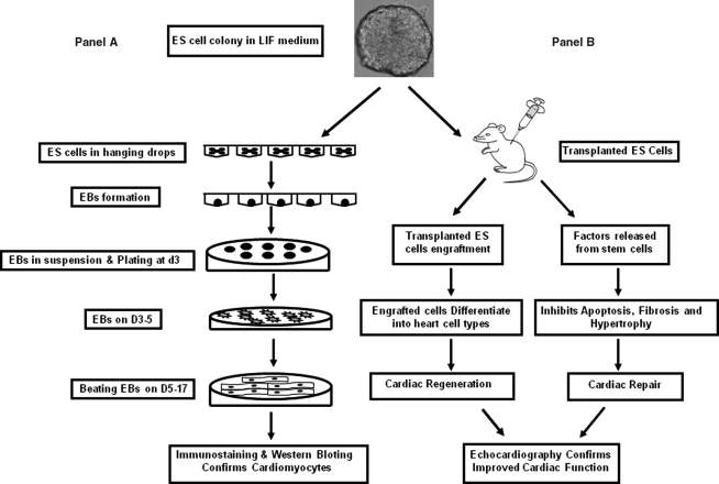
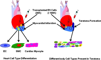
Cell transplantation is a subject of fast-growing research with a potential of a therapeutic approach for the treatment of heart diseases. Clinical applications require preparation of large number of donor cells. Stem cell studies published to date demonstrate that scientists have not reached the general consensus to use an optimal cell type for better cardiac repair and regeneration. We used embryonic stem (ES) cells and their released factors for cardiac repair and regeneration. The major concern of cardiac regeneration with stem cells includes engraftment, differentiation, and teratoma formation after ES cell transplantation. Our current knowledge of ES cell transplantation in the heart is very limited. This review discusses the use of various growth factors to enhance ES cells engraftment and differentiation, as well as the issue of teratoma formation.
Figures
Similar articles
-
Effects of cell number on teratoma formation by human embryonic stem cells.Cell Cycle. 2009 Aug 15;8(16):2608-12. doi: 10.4161/cc.8.16.9353. Epub 2009 Aug 24. Cell Cycle. 2009. PMID: 19597339 Free PMC article.
-
Spatial and temporal kinetics of teratoma formation from murine embryonic stem cell transplantation.Stem Cells Dev. 2007 Dec;16(6):883-91. doi: 10.1089/scd.2007.0160. Stem Cells Dev. 2007. PMID: 17896868
-
Transplantation of undifferentiated murine embryonic stem cells in the heart: teratoma formation and immune response.FASEB J. 2007 May;21(7):1345-57. doi: 10.1096/fj.06-6769com. Epub 2007 Feb 6. FASEB J. 2007. PMID: 17284483
-
Pre-transplantation specification of stem cells to cardiac lineage for regeneration of cardiac tissue.Stem Cell Rev Rep. 2009 Mar;5(1):51-60. doi: 10.1007/s12015-009-9050-8. Epub 2009 Jan 30. Stem Cell Rev Rep. 2009. PMID: 19184567 Free PMC article. Review.
-
Embryonic stem cells: differentiation into cardiomyocytes and potential for heart repair and regeneration.Coron Artery Dis. 2005 Mar;16(2):111-6. doi: 10.1097/00019501-200503000-00006. Coron Artery Dis. 2005. PMID: 15735404 Review.
Cited by
-
The Potential Properties of Natural Compounds in Cardiac Stem Cell Activation: Their Role in Myocardial Regeneration.Nutrients. 2021 Jan 19;13(1):275. doi: 10.3390/nu13010275. Nutrients. 2021. PMID: 33477916 Free PMC article. Review.
-
Reprogramming of skeletal myoblasts for induction of pluripotency for tumor-free cardiomyogenesis in the infarcted heart.Circ Res. 2011 Jun 24;109(1):60-70. doi: 10.1161/CIRCRESAHA.110.240010. Epub 2011 May 12. Circ Res. 2011. PMID: 21566212 Free PMC article.
-
Exogenous Nitric Oxide Protects Human Embryonic Stem Cell-Derived Cardiomyocytes against Ischemia/Reperfusion Injury.Oxid Med Cell Longev. 2016;2016:4298945. doi: 10.1155/2016/4298945. Epub 2016 Jun 15. Oxid Med Cell Longev. 2016. PMID: 27403231 Free PMC article.
-
Stem cells in the infarcted heart.J Cardiovasc Transl Res. 2010 Feb;3(1):73-8. doi: 10.1007/s12265-009-9151-4. Epub 2009 Nov 20. J Cardiovasc Transl Res. 2010. PMID: 20560035 Review.
-
Preconditioning and stem cell survival.J Cardiovasc Transl Res. 2010 Apr;3(2):89-102. doi: 10.1007/s12265-009-9161-2. Epub 2009 Dec 22. J Cardiovasc Transl Res. 2010. PMID: 20560023 Review.
References
-
- Anversa P. Leri A. Kajstura J. Nadal-Ginard B. Myocyte growth and cardiac repair. J Mol Cell Cardiol. 2002;34:91–105. - PubMed
-
- Anversa P. Olivetti G. Leri A. Liu Y. Kajstura J. Myocyte cell death and ventricular remodeling. Curr Opin Nephrol Hypertens. 1997;6:169–176. - PubMed
-
- Behfar A. Zingman LV. Hodgson DM. Rauzier JM. Kane GC. Terzic A. Puceat M. Stem cell differentiation requires a paracrine pathway in the heart. FASEB J. 2002;16:1558–1566. - PubMed
-
- Bel A. Messas E. Agbulut O. Richard P. Samuel JL. Bruneval P. Hagege AA. Menasche P. Transplantation of autologous fresh bone marrow into infarcted myocardium: a word of caution. Circulation. 2003;108(suppl 1):II247–II252. - PubMed
-
- Chen TL. Wang JA. Shi H. Gui C. Luo RH. Xie XJ. Xiang MX. Zhang X. Cao J. Cyclosporin A pre-incubation attenuates hypoxia/reoxygenation-induced apoptosis in mesenchymal stem cells. Scand J Clin Lab Invest. 2008:1–9. Epub ahead of print. - PubMed
Publication types
MeSH terms
Grants and funding
LinkOut - more resources
Full Text Sources
Other Literature Sources
