. 2009 Sep;26(9):1557-66.
doi: 10.1089/neu.2008.0765.

Impaired expression of neuroprotective molecules in the HIF-1alpha pathway following traumatic brain injury in aged mice

Affiliations

Impaired expression of neuroprotective molecules in the HIF-1alpha pathway following traumatic brain injury in aged mice

Joshua Anderson et al. J Neurotrauma. 2009 Sep.

Abstract

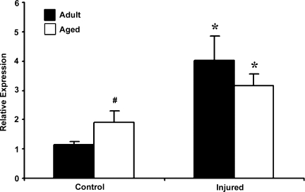
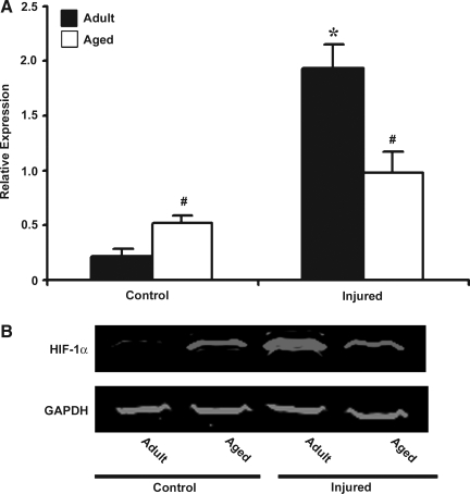
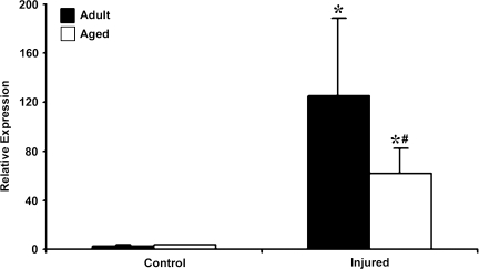
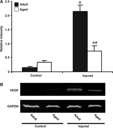
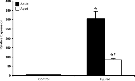
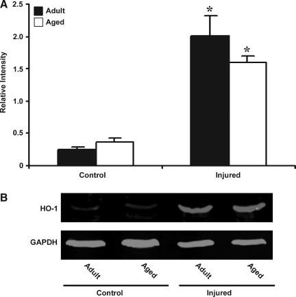
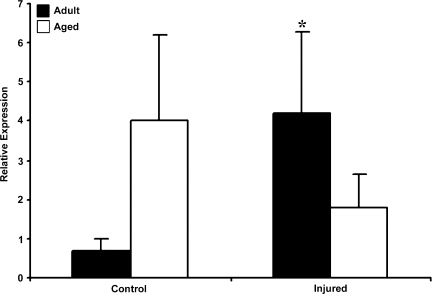
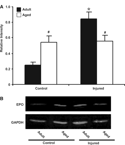
Elderly traumatic brain injury (TBI) patients have higher rates of mortality and worse functional outcome than non-elderly TBI patients. The mechanisms involved in poor outcomes in the elderly are not well understood. Hypoxia-inducible factor-1 alpha (HIF-1alpha) is a basic helix-loop-helix transcription factor that modulates expression of key genes involved in neuroprotection. In this study, we studied the expression of HIF-1alpha and its target survival genes, heme oxygenase-1 (HO-1), vascular endothelial growth factor (VEGF), and erythropoietin (EPO) in the brains of adult versus aged mice following controlled cortical impact (CCI) injury. Adult (5-6 months) and aged (23-24 months) C57Bl/6 mice were injured using a CCI device. At 72 h post-injury, mice were sacrificed and the injured cortex was used for mRNA and protein analysis using real-time reverse transcription--polymerase chain reaction (RT-PCR) and Western blotting protocols. Following injury, HIF-1alpha, HO-1, and VEGF showed upregulation in both the young and aged mice, but in the aged animals the increase in HIF-1alpha and VEGF in response to injury was much lower than in the adult injured animals. EPO was upregulated in the adult injured brain, but not in the aged injured brain. These results support the hypothesis that reduced expression of genes in the HIF-1alpha neuroprotective pathway in aging may contribute to poor prognosis in the elderly following TBI.


Figures

FIG. 1.
FIG. 1.
Expression of HIF-1α. mRNA in cerebral cortex of adult and aged mice 3 days post-injury analyzed by real-time reverse transcription—polymerase chain reaction (RT-PCR). Real-time reactions were performed in duplicate for both the target gene and glyceraldehyde-3-phosphate dehydrogenase (GAPDH), which was used as a housekeeping control. Relative expression was calculated using delta-delta Ct method. Data are presented as mean ± standard error of the mean (SEM; n = 6/group). Statistical significance, Student's t-test: *p < 0.05 injured versus respective control; #p < 0.05 aged versus adult.
FIG. 2.
FIG. 2.
Western blot analysis of HIF-1α in protein extract from cerebral cortex of adult and aged mice 3 days post-injury. (A) Relative levels of protein based on optical density normalized to glyceraldehyde-3-phosphate dehydrogenase (GAPDH) signal. Data are presented as mean ± SEM (n = 3/group). (B) Representative Western blot for HIF-1α, along with GAPDH used as loading control. Statistical significance, Student's t-test: *p < 0.05 injured versus respective control; #p < 0.05 aged versus adult.
FIG. 3.
FIG. 3.
Expression of vascular endothelial growth factor (VEGF) mRNA in cerebral cortex of adult and aged mice 3 days post-injury analyzed by real-time reverse transcription—polymerase chain reaction (RT-PCR). Real-time reactions were performed in duplicate for both the target gene and glyceraldehyde-3-phosphate dehydrogenase (GAPDH), which was used as a housekeeping control. Relative expression was calculated using delta-delta Ct method. Data are presented as mean ± standard error of the mean (SEM; n = 6/group). Statistical significance, Student's t-test: *p < 0.05 injured versus respective control; #p < 0.05 aged versus adult.
FIG. 4.
FIG. 4.
Western blot analysis of vascular endothelial growth factor (VEGF) in protein extract from cerebral cortex of adult and aged mice 3 days post-injury. (A) Relative levels of protein based on optical density normalized to glyceraldehyde-3-phosphate dehydrogenase (GAPDH) signal. Data are presented as mean ± standard error of the mean (SEM; n = 3/group). (B) Representative Western blot for VEGF along with GAPDH used as loading control. Statistical significance, Student's t-test: *p < 0.05 injured versus respective control; #p < 0.05 aged versus adult.
FIG. 5.
FIG. 5.
Expression of heme oxygenase–1 (HO-1) mRNA in cerebral cortex of adult and aged mice 3 days post-injury analyzed by real-time reverse transcription—polymerase chain reaction (RT-PCR). Real-time reactions were performed in duplicate for both the target gene and glyceraldehyde-3-phosphate dehydrogenase (GAPDH), which was used as a housekeeping control. Relative expression was calculated using delta-delta Ct method. Data are presented as mean ± standard error of the mean (SEM; n = 6/group). Statistical significance, Student's t-test: *p < 0.05 injured versus respective control; #p < 0.05 aged versus adult.
FIG. 6.
FIG. 6.
Western blot analysis of heme oxygenase–1 (HO-1) in protein extract from cerebral cortex of adult and aged mice 3 days post-injury. (A) Relative levels of protein based on optical density normalized to glyceraldehyde-3-phosphate dehydrogenase (GAPDH) signal. Data are presented as mean ± standard error of the mean (SEM; n = 3/group). (B) Representative Western blot for HO-1 along with GAPDH used as loading control. Statistical significance, Student's t-test: *p < 0.05 injured versus respective control.
FIG. 7.
FIG. 7.
Expression of erythropoietin (EPO) mRNA in cerebral cortex of adult and aged mice 3 days post-injury analyzed by real-time reverse transcription—polymerase chain reaction (RT-PCR). Real-time reactions were performed in duplicate for both the target gene and glyceraldehyde-3-phosphate dehydrogenase (GAPDH), used as a housekeeping control. The relative expression was calculated using delta-delta Ct method. Data are presented as mean ± standard error of the mean (SEM; n = 6/group). Statistical significance, Student's t-test: *p < 0.05 injured versus respective control.
FIG. 8.
FIG. 8.
Western blot analysis of erythropoietin (EPO) in protein extract from cerebral cortex of adult and aged mice 3 days post-injury. (A) Relative levels of protein based on optical density normalized to glyceraldehyde-3-phosphate dehydrogenase (GAPDH) signal. Data are presented as mean ±standard error of the mean (SEM; n = 3/group). (B) Representative Western blot for EPO along with GAPDH used as loading control. Statistical significance, Student's t-test: *p < 0.05 injured versus control; #p < 0.05 aged versus adult.

References

    1. Adekoya N. Thurman D.J. White D.D. Webb K.W. Surveillance for traumatic brain injury deaths—United States, 1989–1998. MMWR Surveill. Summ. 2002;51:1–14. - PubMed
    1. Alam J. Cook J.L. How many transcription factors does it take to turn on the heme oxygenase–1 gene? Am. J. Respir. Cell Mol. Biol. 2007;36:166–174. - PubMed
    1. Baranova O. Miranda L.F. Pichiule P. Dragatsis I. Johnson R.S. Chavez J.C. Neuron-specific inactivation of the hypoxia inducible factor 1 alpha increases brain injury in a mouse model of transient focal cerebral ischemia. J. Neurosci. 2007;27:6320–6332. - PMC - PubMed
    1. Belayev L. Zhao W. Busto R. Ginsberg M.D. Transient middle cerebral artery occlusion by intraluminal suture: I. Three-dimensional autoradiographic image-analysis of local cerebral glucose metabolism-blood flow interrelationships during ischemia and early recirculation. J. Cereb. Blood Flow Metab. 1997;17:1266–1280. - PubMed
    1. Bergeron M. Yu A.Y. Solway K.E. Semenza G.L. Sharp F.R. Induction of hypoxia-inducible factor–1 (HIF-1) and its target genes following focal ischaemia in rat brain. Eur. J. Neurosci. 1999;11:4159–4170. - PubMed

Publication types

MeSH terms

Substances