. 2009 Feb 9:2:5.
doi: 10.1186/1756-6606-2-5.

Cathepsin D expression level affects alpha-synuclein processing, aggregation, and toxicity in vivo

Affiliations

Cathepsin D expression level affects alpha-synuclein processing, aggregation, and toxicity in vivo

Valerie Cullen et al. Mol Brain. .

Abstract

Background: Elevated SNCA gene expression and intracellular accumulation of the encoded alpha-synuclein (aSyn) protein are associated with the development of Parkinson disease (PD). To date, few enzymes have been examined for their ability to degrade aSyn. Here, we explore the effects of CTSD gene expression, which encodes the lysosomal protease cathepsin D (CathD), on aSyn processing.

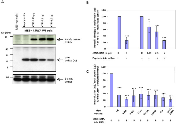
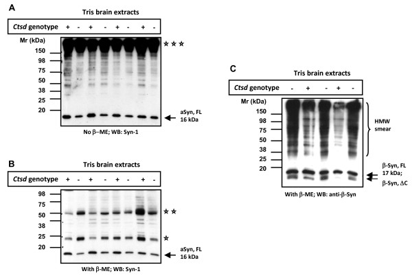
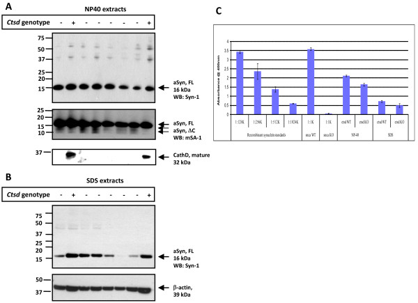
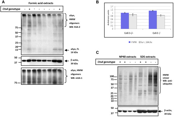
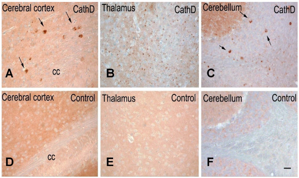
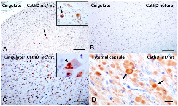
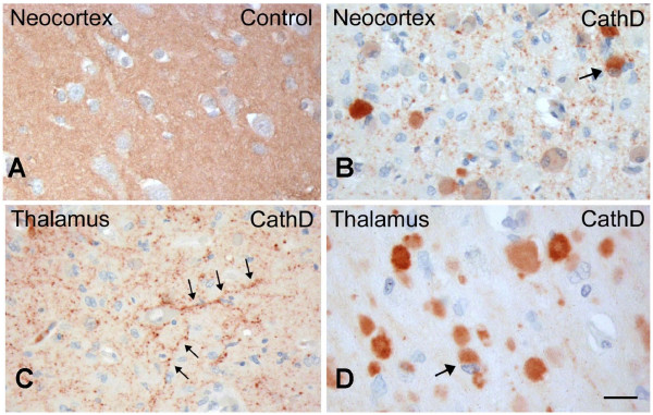
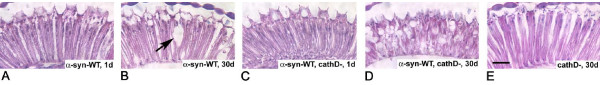
Results: Over-expression of human CTSD cDNA in dopaminergic MES23.5 cell cultures induced the marked proteolysis of exogenously expressed aSyn proteins in a dose-dependent manner. Unexpectedly, brain extractions, Western blotting and ELISA quantification revealed evidence for reduced levels of soluble endogenous aSyn in ctsd knock-out mice. However, these CathD-deficient mice also contained elevated levels of insoluble, oligomeric aSyn species, as detected by formic acid extraction. In accordance, immunohistochemical studies of ctsd-mutant brain from mice, sheep and humans revealed selective synucleinopathy-like changes that varied slightly among the three species. These changes included intracellular aSyn accumulation and formation of ubiquitin-positive inclusions. Furthermore, using an established Drosophila model of human synucleinopathy, we observed markedly enhanced retinal toxicity in ctsd-null flies.

Conclusion: We conclude from these complementary investigations that: one, CathD can effectively degrade excess aSyn in dopaminergic cells; two, ctsd gene mutations result in a lysosomal storage disorder that includes microscopic and biochemical evidence of aSyn misprocessing; and three, CathD deficiency facilitates aSyn toxicity. We therefore postulate that CathD promotes 'synucleinase' activity, and that enhancing its function may lower aSyn concentrations in vivo.


Figures

Figure 1
Figure 1
Over-expression of Cathepsin D lowers α-synuclein in dopaminergic cells. (A) Western blot analyses under reducing conditions of MES23.5 cells [38] expressing untagged, wild-type human SNCA (hSNCA WT) or vector (vec) plasmid DNA following transfection with increasing amounts of CTSD cDNA. Lysates collected 24 hours after transfection show an inverse decrease in the level of full-length (FL) α-synuclein (aSyn), as shown with polyclonal antibodies (Ab), 7071AP [46], hSA-2 [39,40], and monoclonal Ab Syn-1 (not shown); anti-β-actin was used as loading control. Note, no low molecular fragments of aSyn are detected under these conditions. (B) ELISA [hSA2/Syn1-B] and statistical analyses of MES-aSyn cells transfected with increasing amounts of CTSD cDNA, demonstrating the quantitation of the aSyn-lowering effect by ectopic Cathepsin D (**, P < 0.01; ***, P < 0.001). Note, the addition of Pepstatin A to the cell lysis buffer did not change the observed reduction of the intracellular aSyn concentration (up to < 80 per cent). (C) ELISA [hSA2/Syn1-B] and statistical analyses of MES23.5 cells demonstrating the quantitation of ectopic Cathepsin D expression (5 μg per dish) on mutant aSyn proteins and wild-type rat aSyn (see text for details). Data in (B) and (C) are expressed as mean [± standard deviation] of the intracellular concentration of aSyn (in pg/per μg total protein) in MES23.5 cell lysates from each dish and graphically displayed in per cent numbers of vector control without ectopic CTSD expression. Graphs reflect six independent experiments.
Figure 2
Figure 2
Mice lacking ctsd gene expression show reduced levels of endogenous α-synuclein in Tris-soluble brain extracts. Western blot analyses of Tris brain extracts from 24-day old, wild-type (+) and ctsd-null mice (-) carried out under non-reducing (A) and reducing (B) conditions. Probing with monoclonal aSyn Ab, Syn-1 (and rabbit anti-mouse IgG secondary Ab) demonstrates a subtle reduction in the full-length, monomeric aSyn protein (aSyn, FL) at the 16 kDa position in ctsd-null mice. Representative examples of 24 mouse brains are shown (n = 4 per genetic group under non-reducing conditions; n = 16 per genotype under reducing conditions). The same blots reveal a broad, high molecular weight (HMW) band above 150 kDa (triple asterisks), and reactive bands at 50 kDa (double asterisks) and 25 (asterisk) kDa positions under non-reducing (A) and reducing (B) conditions, respectively. (C) Immunoblotting with anti-β-synuclein Ab under reducing conditions reveals no difference at the 17 kDa monomeric (β-Syn, FL) protein position, but indicates elevation of lower molecular weight fragment(s) at ~13 kDa (β-Syn, ΔC) and a HMW-immunoreactive smear in cathD-deficient mice. (Note, no additional anti-β-synuclein Ab is available for corroboration).
Figure 3
Figure 3
CathD-deficient mice show reduced α-synuclein levels in detergent soluble brain extracts. Western blot analyses of fractionated whole brain homogenates from 24-day old, wild-type (+) and ctsd-/- mice (-) carried out under reducing conditions showing NP-40 (A) and SDS extracts (B). Immunoblots were probed with anti-aSyn Abs (Syn-1; mSA-1), anti-β-actin and anti-CathD Abs. Primary Ab detection was carried out with sheep anti-mouse and donkey anti-rabbit Ab, respectively. Under these immunoblotting conditions, variable amounts of the 16 kDa aSyn monomer (aSyn, FL) and traces of 12.5 kDa and 10 kDa (aSyn, ΔC) aSyn-reactive proteins are detectable; no reproducible > 150, 50 and 25 kDa aSyn-positive bands are seen.(C) ELISA [hSA2/Syn1-B] quantification of NP-40 and SDS extracts from ctsd wild-type (WT) and mutant ctsd-/- (KO) mouse brains and of snca WT and snca-/- (KO) lysates serving as positive and negative controls. Ctsd-genotyped brain extracts were loaded in triplicates at three different dilutions each (1:500 to 1:2,000) onto a 384-well plate and read at 405 nm, as described [39,40]. Note, the absence of OD405 nm signals in snca KO mouse tissue. Bar graphs represent the mean group values (± SD) of mice that were individually analyzed (numbers as in A-C). The concentrations of total aSyn (in ng/μl) present in NP-40 extracts measured: ctsd WT, 1.34 ± 0.25; ctsd KO, 1.09 ± 0.26; and in the SDS fractions: ctsd WT, 0.60 ± 0.25; and ctsd KO, 0.48 ± 0.28.
Figure 4
Figure 4
CathD-deficient brains show increased HMW α-synuclein-positive oligomers in formic acid extracts. (A) Western blot analyses carried out under reducing conditions of formic acid extracts obtained from previously generated SDS pellets of wild-type (+) and ctsd-/- (-) mouse brains (see Fig. 3 above). Immunoblots were probed with high sensitivity, polyclonal aSyn Abs, hSA-2 and mSA-1, and anti-β-actin Ab, as indicated. Under these conditions, HMW aSyn-positive smears and trace amounts of monomeric, 16 kDa aSyn (aSyn, FL) are detectable in these formic acid extracts.(B) Quantification of aSyn-reactive species by densitometry examining the ratio of HMW smears (blue bars) and 16 kDa monomer (empty bars) versus the signal of β-actin-positive bands. Note, an ~15 per cent increase in HMW oligomers of aSyn is detected in formic acid extracts of ctsd-/- brain by independent analysis of both mSA-1 (B) and hSA-2 immunoblots. (C) NP-40 and SDS extracts probed with anti-ubiquitin consistently reveal HMW immunoreactive smears in SDS brain extracts of ctsd-/- mice. Representative examples of twelve mouse brains are shown (n = 4, ctsd+/+; n = 8, ctsd-/-).
Figure 5
Figure 5
Mice lacking ctsd gene expression exhibit intracellular α-synuclein accumulation in several regions of the brain. Serial, paraffin-embedded sections from 24-day old cathepsin D knock-out mice (CathD) (A-C) and age-matched, wild-type control (D-F) mice (n = 5) were probed by immunohistochemistry with affinity-purified, polyclonal aSyn Ab, hSA-2. (A) In the frontal cortex of cathepsin D knock-out mice, scattered neurons with prominent intracellular aSyn-reactivity are detectable (arrows). (B) In the thalamus of CathD-deficient mice, the neuropil staining is reduced when compared with control animals, and small grain-like aggregates of aSyn are visible. (C) In the cerebellum of CathD deficient mice, abnormal aSyn-positive accumulations of varying size can be observed in the granular cell layer and deep nuclei; those that appear cytoplasmic are identified by arrows. (D-F) As expected, aSyn-positive aggregates are not found in age-matched, wild-type mice processed in parallel. CC denotes corpus callosum. Scale bar, 25 μm.
Figure 6
Figure 6
Sheep expressing a homozygous mutant of Cathepsin D exhibit accumulation of phosphorylated α-synuclein in cingulate cortex. (A) Select cortical neurons (arrow) of cingulate gyrus in homozygous mutants (CathD mt/mt) show cytoplasmic immunostaining with Ser129-phosphorylated aSyn Ab. (B) Absence of Ser129-phosphorylated aSyn-reactivity in the cingulate gyrus of age-matched, heterozygous CTSD (CathD hetero) sheep brain. (C) Anti-ubiquitin antibody staining reveals extensive, often multiple, intra-cellular inclusion formation in neurons (arowhead) of affected sheep throughout the cortex including cingulate cortex. (D) Immunostaining for aSyn with polyclonal Ab, hSA-2, reveals abundant aSyn deposits and axonal swelling (arrows) in the internal capsule of homozygous mutant sheep but not wild-type (not shown) or heterozygous sheep (not shown). Scale bars, 100 μm in A, B, C, and 15 μm in D.
Figure 7
Figure 7
Human neuronal ceroid lipofuscinosis due to Cathepsin D deficiency leads to intracellular accumulation of α-synuclein. (A) Paraffin-embedded brain sections of a neurologically normal infant (Control) stained with polyclonal aSyn Ab, hSA-2, show strong reactivity of the neuropil in the neocortex. (B) Immunohistochemistry of brain specimens from CathD-deficient infants (CathD; n = 3), carried out in parallel, demonstrates marked reduction of neuropil staining. These sections also display scattered, cytoplasmic aSyn aggregates within neurons (identified by arrow). (C) Sections of the thalamus from the same individual as in B also reveal neuritic pathology (arrows), as well as (D) intracellular accumulation of aSyn, sometimes seen as a juxtanuclear inclusions (arrow). Scale bar, 15 μm.
Figure 8
Figure 8
Cathepsin D deficiency enhances α-synuclein toxicity in the retina of fly brain. (A) Normal retina in one-day old, human SNCA cDNA-transgenic flies (α-Syn-WT; Feany and Bender, 2000). (B) At day 30 these flies show mild degeneration; arrow indicates vacuole. (C) Normal morphology of retina at day 1 in flies expressing human SNCA cDNA in the absence of endogenous fly cathD (α-Syn-WT, cathD-). (D) Marked degeneration of retinal neurons occurs by day 30 in flies ectopically expressing human SNCA cDNA in the absence of fly cathD. Note the appearance of numerous vacuoles, and the thinning and loss of retinal architecture. (E) Deletion of fly ctsd in the absence of aSyn expression (cathD-)does not cause retinal degeneration at day 30. Scale bar, 20 μm.

Similar articles

Cited by

References

    1. Polymeropoulos MH, Lavedan C, Leroy E, Ide SE, Dehejia A, Dutra A, Pike B, Root H, Rubenstein J, Boyer R, et al. Mutation in the alpha-synuclein gene identified in families with Parkinson's disease. Science. 1997;276:2045–2047. doi: 10.1126/science.276.5321.2045. - DOI - PubMed
    1. Kruger R, Kuhn W, Muller T, Woitalla D, Graeber M, Kosel S, Przuntek H, Epplen JT, Schols L, Riess O. Ala30Pro mutation in the gene encoding alpha-synuclein in Parkinson's disease. Nat Genet. 1998;18:106–108. doi: 10.1038/ng0298-106. - DOI - PubMed
    1. Singleton AB, Farrer M, Johnson J, Singleton A, Hague S, Kachergus J, Hulihan M, Peuralinna T, Dutra A, Nussbaum R, et al. alpha-Synuclein locus triplication causes Parkinson's disease. Science. 2003;302:841. doi: 10.1126/science.1090278. - DOI - PubMed
    1. Trojanowski JQ, Lee VM. Aggregation of neurofilament and alpha-synuclein proteins in Lewy bodies: implications for the pathogenesis of Parkinson disease and Lewy body dementia. Arch Neurol. 1998;55:151–152. doi: 10.1001/archneur.55.2.151. - DOI - PubMed
    1. Goedert M. Parkinson's disease and other alpha-synucleinopathies. Clin Chem Lab Med. 2001;39:308–312. doi: 10.1515/CCLM.2001.047. - DOI - PubMed

Publication types

MeSH terms