Review
. 2009 Apr;60(1):2-23.
doi: 10.1016/j.brainresrev.2008.12.018. Epub 2008 Dec 31.

Roles of transient receptor potential channels in pain

Affiliations
Review

Roles of transient receptor potential channels in pain

Cheryl L Stucky et al. Brain Res Rev. 2009 Apr.

Abstract

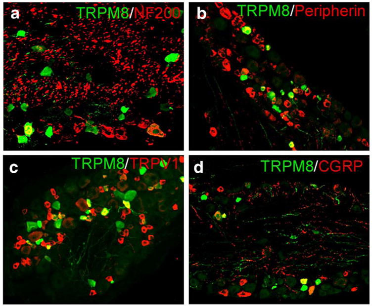
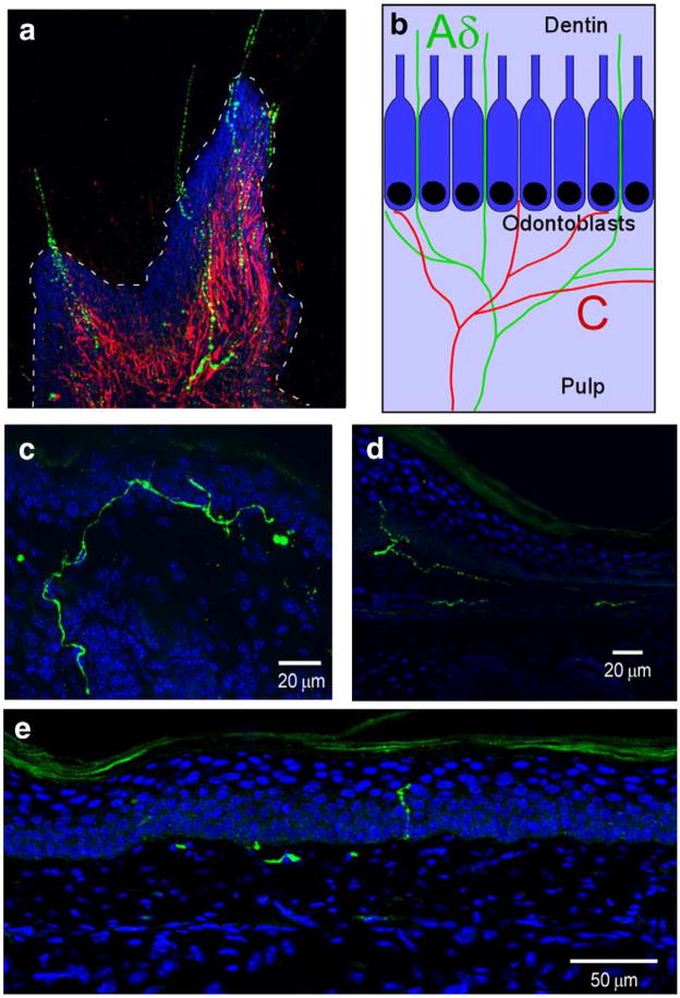
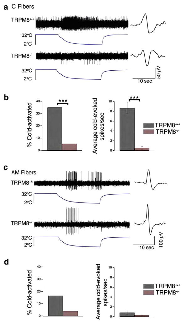
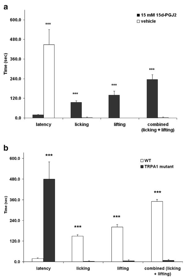
Pain perception begins with the activation of primary sensory nociceptors. Over the past decade, flourishing research has revealed that members of the Transient Receptor Potential (TRP) ion channel family are fundamental molecules that detect noxious stimuli and transduce a diverse range of physical and chemical energy into action potentials in somatosensory nociceptors. Here we highlight the roles of TRP vanilloid 1 (TRPV1), TRP melastatin 8 (TRPM8) and TRP ankyrin 1 (TRPA1) in the activation of nociceptors by heat and cold environmental stimuli, mechanical force, and by chemicals including exogenous plant and environmental compounds as well as endogenous inflammatory molecules. The contribution of these channels to pain and somatosensation is discussed at levels ranging from whole animal behavior to molecular modulation by intracellular signaling proteins. An emerging theme is that TRP channels are not simple ion channel transducers of one or two stimuli, but instead serve multidimensional roles in signaling sensory stimuli that are exceptionally diverse in modality and in their environmental milieu.


Figures

Fig. 1
Fig. 1
TRPM8 co-localizes with a diverse array of afferent markers in somatosensory neurons. TRPM8 neurons in both TG and DRG were immunoreactive for the intermediate filaments NF200 (a; TG: 25.5±2.6%, n=329; DRG: 13.9±3.7%, n=129) and peripherin (b; TG: 28.0±3.8%, n=239; DRG: 24.8±2.8%, n=272), as well as TRPV1 (c; TG: 38.8±2.2%, n=719; DRG: 23.7±4.6%, n=221) and CGRP (d; TG: 32.1±3.1%, n=511; DRG: 19.9±2.4%, n=288; see Takashima et al., 2007).
Fig. 2
Fig. 2
Peripheral terminals of TRPM8 axons are found in functionally distinct regions in the tooth and skin. (a) In a longitudinal section of a de-calcified molar, TRPM8 fibers (green) cross the odontoblast layer located at the pulp–dentin border (dashed line) and extend into the dentin, while peripherin+ axons (red) are restricted to the pulp. Cell nuclei were stained with DAPI (blue). (b) Schematic diagram of mouse molars indicating the termination zones of cold-sensitive C (red) and Aδ fibers (green) localized in the pulp and dentin, respectively. Cell rich odontoblast layer that separates the pulp and dentin is shown in blue (blue box). (c–e) In mouse skin, TRPM8 axons (green) terminate in a heterogeneous manner throughout the skin dermis and epidermis. Cell nuclei were stained with DAPI (blue).
Fig. 3
Fig. 3
TRPM8-deficient cutaneous primary afferent fibers show a significant loss of cold sensitivity. (a) Example of a typical response of a wild type (top) and TRPM8-deficient (bottom) cutaneous C fiber to a cold ramp (32 to 2 °C, over 20 s). The action potential waveform is shown to right of trace. (b) Percentage of C fibers responding to cold ramp in wild type (black; n=60) versus TRPM8−/− (red; n=56) mice (left). Average cold-evoked action potential firing rate in C fibers of wild type (n=21) versus TRPM8−/− (n=3) mice (right; Mean±S.E.M.). (c) Example of a typical response of wild type (top) and TRPM8-deficient (bottom) cutaneous AM fiber to a cold ramp (32 to 2 °C over 20 s). The action potential waveform is shown to right of trace. (d) Percentage of AM fibers responding to cold ramp in wild-type (black; n=24) versus TRPM8−/− (red; n=26) mice (left). Average cold-evoked action potential firing rate in AM fibers of wild type (n=4; Mean±S.E.M.) versus TRPM8−/− (n=1) mice (right). ***p<0.001, Students t-test or Fisher’s exact test (See Bautista et al., 2007).
Fig. 4
Fig. 4
Intraplantar injection of 15d-PGJ2 causes acute nociceptive responses via TRPA1. (a) 10 μL of vehicle (10% DMSO in saline) or 15 mM 15d-PGJ2 was injected into the hind paw of C57BL/6J mice (n =5 per group) and nociceptive behaviors (licking and lifting of the paw) for 10 min. 15d-PGJ2 caused significant nociceptive responses compared to vehicle. (b) 15d-PGJ2-induced nociceptive behaviors are absent in TRPA1 knockout mice (n=5 per group; ***p<0.001).
Fig. 5
Fig. 5
Formaldehyde and the endogenous inflammatory mediator 4-hydroxynonenal activate recombinant TRPA1. a,b) Increased intracellular calcium was observed at concentrations below 1 mM formaldehyde (a) and 4-hydroxynonenal (b) in CHO cells expressing TRPA1 (red) but not naïve CHO cells (black). CHO cells expressing TRPV1 (blue) were not responsive at low concentrations of these electrophiles. (c) Formaldehyde (1 mM, ~0.003%) increased mTRPA1 single channel activity by 1 min (middle panel) and activity remained high after 15 min of washout. Channel activity was monitored via the inside-out configuration using voltage ramps from −50 to +120 mV, rate of 2.8 mV/s every 5 s.
Fig. 6
Fig. 6
Formalin elicits inflammatory pain behaviors through TRPA1. (a) Ratiometric Fura-2 calcium imaging illustrates that formaldehyde (1 mM) evokes increase in Fura-2 ratios in DRG neurons from wild type mice, specifically in neurons also activated by mustard oil and capsaicin (top) but formaldehyde has no effect on neurons from TRPA1-null mice (bottom). (b) Formalin (10 μl of 0.2%) was injected intraplantar (i.pl.) into one hind paw of a mouse. The duration of formalin-induced nocifensive behaviors during the first phase (0–10 min) and second phase (10–30 min) after injection was scored for wild type and TRPA1−/− littermates. TRPA1-deficient mice showed a severe deficit in nociceptive responses during both phases of formalin-induced pain behavior. Responses represent the time mice spent licking, flicking, or lifting the injected hind paw (n>10 mice).
Fig. 7
Fig. 7
AP18 is a potent, selective and reversible antagonist for TRPA1. (a, b) AP18 inhibits increases in intracellular calcium evoked by cinnamaldehyde (Cinn; a; FLIPR technique) and iodoacetamide (IA; b; Fura-2 ratiometric calcium imaging) in CHO cells expressing TRPA1. (c) AP18 (20 μM) strongly inhibited the Cinn-activated TRPA1 currents in inside-out macropatches derived from Xenopus oocytes. The selectivity profile of AP18 is described in the box inset.
Fig. 8
Fig. 8
AP18 partially reverses inflammation-induced mechanical hyperalgesia. CFA injection induces marked mechanical hyperalgesia in both wild type (red) and TRPA1−/− mice (green). AP18 was injected (i.pl.) after the 24 h measurement time point. AP18 significantly reversed the mechanical hyperalgesia in wild type mice but not TRPA1 null littermates (n=12, females). Mice were injected with 10 μg CFA in 10 μL; AP18 (1 mM in 10 μL) was injected in a PBS based vehicle containing 0.5% Tween80 and 1% DMSO. Statistical significance was determined using a two-tailed Student’s t-test.
Fig. 9
Fig. 9
AP18 partially reverses cold hyperalgesia in rats with CFA-induced inflammation. Sprague Dawley rats (n=8) were injected with 50 μg CFA in 100 μL (1:1 emulsion of mineral oil and saline). AP18 (1 mM in 10 μL) was injected in a PBS based vehicle containing 0.5% Tween80 and 1% DMSO. Statistical significance was determined using a two-tailed Student’s t-test.
Fig. 10
Fig. 10
Growth factors are upregulated during inflammation. PCR was used to measure changes in growth factor levels following a single injection of CFA into hindpaw glabrous skin. Artemin mRNA levels increased 10-fold within 24 h.
Fig. 11
Fig. 11
Growth factors inhibit TRPV1 tachyphylaxis. Fura-2 calcium imaging of primary afferent responses to capsaicin (Cap) before and after 10 min exposure to growth factor. In the absence of growth factor, repeated capsaicin responses normally exhibit tachyphylaxis. All four growth factors block tachyphylaxis and produce potentiation (neurturin effect is delayed). Percentage (%) indicates the capsaicin-responsive cells potentiated by each growth factor.
Fig. 12
Fig. 12
Growth factors induce hyperalgesia in vivo. Latency to thermal nociceptive response measured with a Hargreaves apparatus before and after injection of 0.2 μg/20 μl growth factor (or saline control) into the hindpaw of wildtype mice. In wildtype animals, all growth factors tested caused significant thermal hyperalgesia during the first 4 h. Co-injection of NGF/ART resulted in persistent thermal hyperalgesia lasting 6d. *p<0.05.
Fig. 13
Fig. 13
TRPA1 shows prominent tachyphylaxis. Fura-2 calcium imaging was used to record responses to repeated application of 100 μM mustard oil (yellow squares) at 10 min intervals. Cells were first tested with a depolarizing stimulus (50 mM K+) to evaluate cell health and then tested 4 times with mustard oil.
Fig. 14
Fig. 14
TRPA1 activity is modulated in sensory neurons. TRPA1 activation is sensitized by intracellular Ca++ and kinases PKC and PKA. TRPA1 is inhibited by PIP2, and polyphosphates such as IP3 are required for TRPA1 function. Bradykinin, acting through the bradykinin receptor BK2, activates PLC, PKC and PKA to sensitize TRPA1; proteases trypsin and tryptase, acting through PAR2, sensitize TRPA1 through PLC. The effects of these pathways are largely additive.
Fig. 15
Fig. 15
Illustration of hypothetical AKAP150 association with TRPV1 at the plasma membrane. PKA phosphorylation sites, as listed and referenced in the text, are identified by red spheres.

Similar articles

Cited by

References

    1. Ahern GP, Premkumar LS. Voltage-dependent priming of rat vanilloid receptor: effects of agonist and protein kinase C activation. J Physiol. 2002;545:441–451. - PMC - PubMed
    1. Akopian AN, Ruparel NB, Jeske NA, Hargreaves KM. Transient receptor potential TRPA1 channel desensitization in sensory neurons is agonist dependent and regulated by TRPV1-directed internalization. J Physiol. 2007;583:175–193. - PMC - PubMed
    1. Allchorne AJ, Broom DC, Woolf CJ. Detection of cold pain, cold allodynia and cold hyperalgesia in freely behaving rats. Mol Pain. 2005;1:36–41. - PMC - PubMed
    1. Andersson DA, Gentry C, Moss S, Bevan S. Transient receptor potential A1 is a sensory receptor for multiple products of oxidative stress. J Neurosci. 2008;28:2485–2494. - PMC - PubMed
    1. Bandell M, Story GM, Hwang SW, Viswanath V, Eid SR, Petrus MJ, Earley TJ, Patapoutian A. Noxious cold ion channel TRPA1 is activated by pungent compounds and bradykinin. Neuron. 2004;41:849–857. - PubMed

Publication types

MeSH terms