. 2009 Apr 10;284(15):9908-16.
doi: 10.1074/jbc.M806210200. Epub 2009 Feb 9.

Epidermal growth factor-activated aryl hydrocarbon receptor nuclear translocator/HIF-1{beta} signal pathway up-regulates cyclooxygenase-2 gene expression associated with squamous cell carcinoma

Affiliations

Epidermal growth factor-activated aryl hydrocarbon receptor nuclear translocator/HIF-1{beta} signal pathway up-regulates cyclooxygenase-2 gene expression associated with squamous cell carcinoma

Kwang-Yu Chang et al. J Biol Chem. .

Abstract

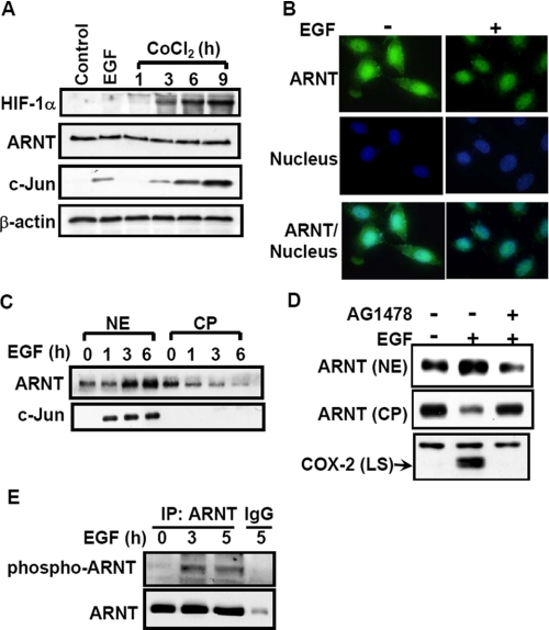
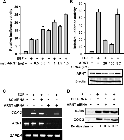
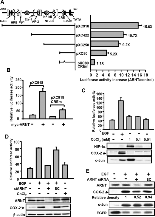
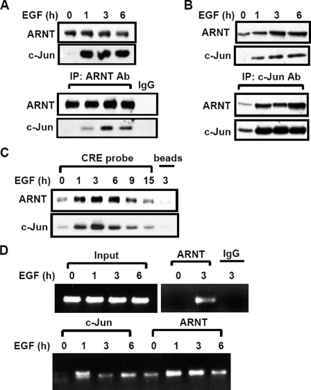
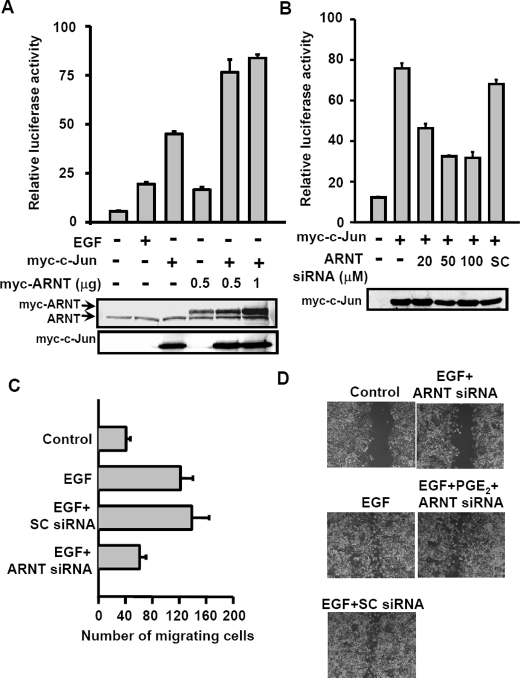
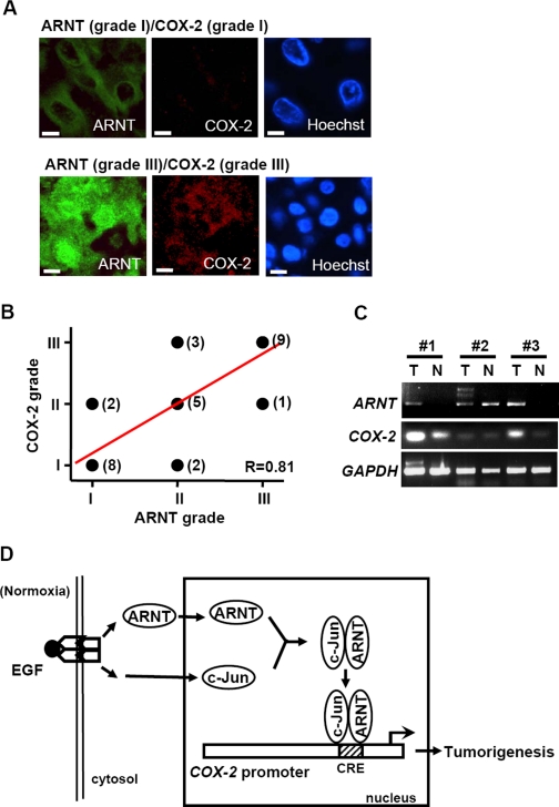
Hypoxia-inducible factor (HIF) accumulates when tumors grow under hypoxic conditions. The genesis of tumors, however, usually involves normoxic conditions. In this study, we were interested in examining the potential role of aryl hydrocarbon receptor nuclear translocator (ARNT)/HIF-1beta in tumor growth under normoxic conditions, specifically when cells are treated with epidermal growth factor (EGF), which is known to affect the gene expression of tumor growth-related protein COX-2 (cyclooxygenase-2). The results showed that EGF receptor inhibitor, AG1478, abolished EGF-induced nuclear accumulation of ARNT as well as the expression of COX-2. ARNT small interfering RNA inhibited the promoter activity, mRNA level, and protein expression of COX-2 in cells treated with EGF. In contrast, CoCl(2)-induced HIF-1alpha exhibited no effect on COX-2 expression. EGF also stimulated the formation of the ARNT.c-Jun complex as well as the complex binding to the COX-2 promoter. ARNT small interfering RNAs blocked EGF-activated cell migration. Moreover, COX-2 and ARNT were cohorts present distinctively in clinical specimens of human cervical squamous cell carcinoma and were almost nondetectable in adjacent normal or noncancerous cervical tissues. Our results revealed that ARNT plays an important role in EGF-regulated COX-2 gene expression and may thus be related to either a cause or a consequence of tumorigenesis in cervical cancer.


Figures

FIGURE 1.
FIGURE 1.
Effect of EGF on HIF expression and translocation. A, cells were starved for 18 h in serum-free culture medium and then treated with 50 ng/ml EGF for 4 h or with 1 mm CoCl2 for various time periods (as indicated) in the culture medium without serum. Lysates of cells were prepared and subjected to SDS-PAGE and analyzed by Western blotting with antibodies against HIF-1α, ARNT, c-Jun, and β-actin. B, cells were treated with 50 ng/ml EGF for 4 h, fixed by using 4% paraformaldehyde, labeled with rabbit anti-ARNT antibodies, and then stained with secondary antibodies conjugated with fluorescein isothiocyanate. DNA was stained with 4,6-diamidino-2-phenylindole. Finally, the cells were examined using a microscope. C, cells were starved and then treated with 50 ng/ml EGF for various time periods (as indicated) in the culture medium without serum. Cytoplasmic fraction (CP) and nuclear extracts (NE) of cells were prepared in the same volume of buffer, and an aliquot of each fraction was subjected to Western blot analysis using antibodies against ARNT and c-Jun. D, cells were starved and then treated with 5 nm AG1478 for 30 min, followed by EGF treatment for 4 h. Nuclear extracts, cytoplasmic fraction, and lysates of cells (LS) were prepared and subjected to Western blot analysis using antibodies specific for ARNT and COX-2. E, cellular lysates of cells were prepared and subjected to immunoprecipitation (IP) with antibodies against ARNT bound to protein A-agarose. The proteins were subjected to SDS-PAGE and analyzed by Western blotting with anti-ARNT and anti-phosphothreonine antibodies.
FIGURE 2.
FIGURE 2.
Effect of ARNT on EGF-induced gene expression of COX-2. A, luciferase vector bearing the COX-2 gene promoter was constructed (pXC918) (25). Cells were transfected with pXC918 and ARNT expression vector by lipofection. Cells were treated with 50 ng/ml EGF and further cultured in fresh medium up to 6 h. The luciferase activities and protein concentrations were then determined and normalized. Values represent means ± S.E. of three determinations. B, cells were transfected with pXC918 and various amounts of ARNT siRNA oligonucleotides by lipofection. After EGF treatment for 6 h, the luciferase activities and protein concentrations were determined and normalized. Values represent means ± S.E. of three determinations. Expressions of ARNT and β-actin proteins were analyzed by Western blot analysis using anti-ARNT and anti-β-actin antibodies, respectively. C, cells were transfected with 50 nm ARNT siRNA or scramble oligonucleotides by lipofection. After EGF treatment for 6 h, total RNA was extracted for reverse transcription PCR with COX-2, ARNT, and glyceraldehyde-3-phosphate dehydrogenase primers. D, expressions of COX-2 and c-Jun proteins were analyzed by Western blot analysis using anti-COX-2 and anti-c-Jun antibodies, respectively. The relative density of COX-2 protein was quantified as indicated.
FIGURE 3.
FIGURE 3.
Analysis of ARNT-responsive regions in the 5′-flanking region of the COX-2 gene. A, luciferase vectors bearing various lengths of the COX-2 gene promoter were constructed as indicated. Site-directed mutagenesis of CRE consensus sequences in the 5′-flanking region ranging from –57 to –53 bp of the COX-2 gene was performed. The potential consensus sequences in the 5′-flanking region are indicated. Plasmid transfection was performed as described under “Experimental Procedures.” The luciferase activities and protein concentrations were then determined and normalized. The expression ratios of ARNT (1 μg)-treated cells to control cells are indicated. Results are expressed as means ± S.E. of three independent experiments in triplicate wells for each construct. B, cells were transfected with pXC918, pXC918 CREm, and ARNT expression vector by lipofection. The luciferase activities and protein concentrations were then determined and normalized. Values represent means ± S.E. of three determinations. C, cells were transfected with pXC918 by lipofection. After EGF or CoCl2 treatment for 6 h, the luciferase activities and protein concentrations were determined and normalized. Values represent means ± S.E. of three determinations. Expressions of HIF-1α, COX-2, and c-Jun proteins were analyzed by Western blot analysis using antibodies against HIF-1α, COX-2, and c-Jun, respectively. D, SiHa cells were transfected with pXC918 and ARNT siRNA oligonucleotides by lipofection. After EGF or CoCl2 treatment for 6 h, the luciferase activities and protein concentrations were determined and normalized. Values represent means ± S.E. of three determinations. Expressions of ARNT, COX-2, and β-actin proteins were analyzed by Western blot analysis using antibodies against ARNT, COX-2, and β-actin, respectively. E, OEC-M1 cells were transfected with ARNT siRNA oligonucleotides by lipofection. After EGF treatment for 6 h, the expression of ARNT, COX-2, c-Jun, EGFR, and β-actin was analyzed by Western blot analysis. The relative density of COX-2 protein was quantified as indicated.
FIGURE 4.
FIGURE 4.
EGF induces the complex formation and the binding of c-Jun·ARNT to the COX-2 gene promoter. A–C, cells were starved for 18 h in serum-free culture medium and then treated with EGF for various time periods (as indicated) in the culture medium without serum. Cellular lysates (A) and nuclear extracts (B) of cells were prepared and subjected to Western blot or immunoprecipitated (IP) with antibodies against c-Jun and ARNT bound to protein A-agarose. The proteins were subjected to SDS-PAGE and analyzed by Western blotting with anti-c-Jun and anti-ARNT antibodies. C, confluent cells were starved for 18 h in serum-free culture medium and then treated with EGF for various time periods (as indicated) in the culture medium without serum. Nuclear extracts were prepared, and DNA affinity precipitation assay was performed, as described under “Experimental Procedures.” Binding of c-Jun and ARNT proteins to CRE probes was analyzed by Western blot. The streptavidin-agarose beads were used to serve as a nonspecific binding control. D, cross-linked chromatin derived from EGF-treated cells was immunoprecipitated with c-Jun and ARNT antibodies and analyzed by PCR with specific primers for the region from –186 to +49 bp of the COX-2 promoter. Input, nonimmunoprecipitated cross-linked chromatin.
FIGURE 5.
FIGURE 5.
Cooperation between ARNT and c-Jun in promoter activation of the COX-2 gene results in cell migration. A, cells were transfected with pXC-918, Myc-c-Jun, and Myc-ARNT expression vector by lipofection. Cells were treated with or without EGF for 6 h. The luciferase activities and protein concentrations were then determined and normalized. Values represent means ± S.E. of three determinations. Expressions of ARNT and Myc-c-Jun proteins were analyzed by Western blot analysis using anti-ARNT and anti-Myc antibodies, respectively. B, cells were transfected with pXC-918, Myc-c-Jun, and ARNT siRNA by lipofection. The luciferase activities and protein concentrations were then determined and normalized. Values represent means ± S.E. of three determinations. Expressions of Myc-c-Jun proteins were analyzed by Western blot analysis using anti-Myc antibodies. C, cells were transfected with 50 nm ARNT siRNA or scramble oligonucleotides by lipofection. Migration was assessed after 15 h in the presence or absence of EGF. The histogram displays the mean number of migrated cells obtained by counting four separate fields in three independent experiments. Error bars represent means ± S.D. D, cells were transfected with 50 nm ARNT siRNA or scramble oligonucleotides by lipofection. After a wound was made with a pipette tip, cells were treated with 50 ng/ml EGF and 10μm PGE2 for 60 h. The extent of wound closure was observed by using a phase-contrast microscope camera (model DMI 4000 B; Leica).
FIGURE 6.
FIGURE 6.
Expression patterns of ARNT and COX-2 in surgical specimens of cervical carcinoma. A, representative pictures of cervical cancer samples at different ARNT/COX-2 grades. The triple-immunofluorescent stain technique was used to identify ARNT (green), COX-2 (red), and nucleus (blue) in cervical cancer tissues. Scale bar, 5 μm. B, a linear correlation (R = 0.81) of ARNT and COX-2 expression in the same surgical specimen of cervical cancer tissues. The case number is indicated in parentheses beside each point. C, total RNA from tumor and normal tissue of different human cervical cancer patients (#1 to #3) was extracted for reverse transcription PCR with COX-2, ARNT, and glyceraldehyde-3-phosphate dehydrogenase primers. D, schematic pathway of COX-2 promoter regulation by EGF stimulation. Activation of EGFR signaling increases nuclear accumulation of ARNT under normoxic conditions. ARNT then interacts with transcriptional factor c-Jun and binds to the CRE site, which results in an increase of COX-2 expression and tumorigenesis.

Similar articles

Cited by

References

    1. Kewley, R. J., Whitelaw, M. L., and Chapman-Smith, A. (2004) Int. J. Biochem. Cell Biol. 36 189–204 - PubMed
    1. Giaccia, A., Siim, B. G., and Johnson, R. S. (2003) Nat. Rev. Drug Discov. 2 803–811 - PubMed
    1. Kozak, K. R., Abbott, B., and Hankinson, O. (1997) Dev. Biol. 191 297–305 - PubMed
    1. Maltepe, E., Schmidt, J. V., Baunoch, D., Bradfield, C. A., and Simon, M. C. (1997) Nature 386 403–407 - PubMed
    1. Carmeliet, P., Dor, Y., Herbert, J-M., Fukumura, D., Brusselmans, K., Dewerchin, M., Neeman, M., Bono, F., Abramovitch, R., Maxwell, P., Koch, C. J., Ratcliffe, P., Moons, L., Jain, R. K., Collen, D., and Keshet, E. (1998) Nature 394 485–490 - PubMed

Publication types

MeSH terms