. 2009 Apr 10;284(15):9917-26.
doi: 10.1074/jbc.M900249200. Epub 2009 Feb 9.

The death domain of FADD is essential for embryogenesis, lymphocyte development, and proliferation

Affiliations

The death domain of FADD is essential for embryogenesis, lymphocyte development, and proliferation

Hongxia Z Imtiyaz et al. J Biol Chem. .

Abstract

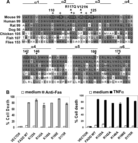
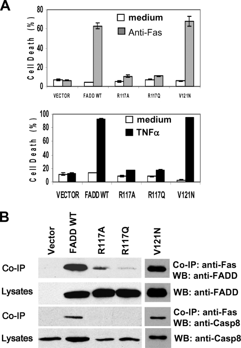
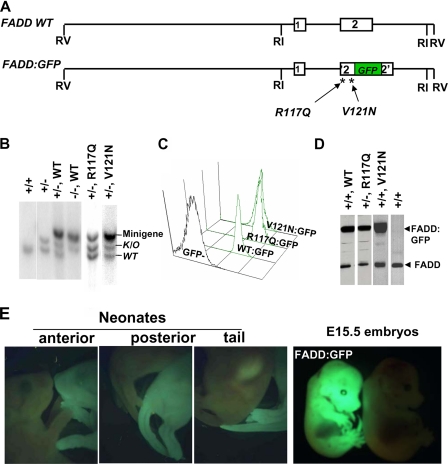
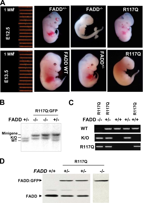
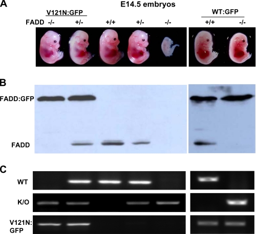
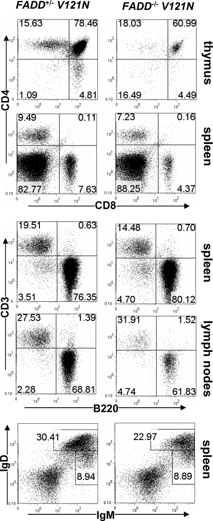
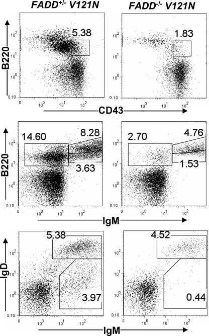
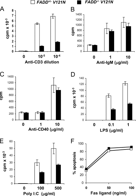
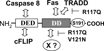
The Fas-associated death domain-containing protein (FADD) is an adaptor for relaying apoptotic signals initiated by death receptors such as Fas. Whereas a lack of death receptors has no effect on mouse development, FADD deficiency results in early embryonic lethality, indicating that FADD has additional functions independent of death receptors. We have previously shown that conditional deletion of FADD not only impairs apoptosis but also leads to defective lymphocyte proliferation. The non-apoptotic signaling mediated by FADD remains poorly understood. Earlier studies have suggested that FADD carboxyl terminal serine phosphorylation likely plays a role in FADD-mediated proliferation signaling in T cells. The FADD death domain is presumably only required for apoptotic signaling, as it interacts with death receptors which are dispensable during embryonic development and lymphocyte proliferation. To test this hypothesis, we have performed mutational analyses of the FADD death domain and identified a mutant, R117Q, which lacks binding to Fas and, thus, is incapable of apoptotic signaling in cell lines. Unexpectedly, this death domain point mutation disrupted mouse embryonic development as shown by in vivo functional reconstitution analyses. Interestingly, a second FADD death domain mutant, V121N, retained normal Fas binding and apoptotic signaling ability but also failed to support mouse development. Furthermore, lymphocyte proliferation responses were impaired by V121N. This reverse genetic study has revealed a previously unappreciated role of the FADD death domain, which likely functions as a molecular switch regulating two distinct signals leading to apoptosis and cell proliferation and is critical for embryogenesis, lymphocyte development, and proliferation.


Figures

FIGURE 1.
FIGURE 1.
Further mutational analyses of the DD of FADD. A, cross-species sequence alignment of the DD of FADD. Conserved resides are shaded. The residues that were mutagenized in previous (+) (21), and current studies (*) are indicated. Six α-helices (α1–α6) of the DD are defined according to previous studies (49, 50). The R117Q and V121N mutations indicated by arrowheads were tested in mice in the current study. B, the DD mutations K125A, R142A, K146A, R166E, and D175R were generated by site-directed mutagenesis, and the resulting full-length mouse FADD mutants were stably expressed in FADD/ MEFs that lack the endogenous FADD. FADD/ MEFs stably transfected with the retroviral vector, murine stem cell virus-CMV-IRES-GFP, or with the viral construct containing WT FADD were used as controls. These reconstituted MEFs were treated with anti-Fas antibodies (left) or TNFα (right) to induce apoptosis. Cell death was determined by propidium iodide staining and flow cytometric analysis at 16 h after stimulation. Untreated cells (medium, unfilled bars) were used as controls.
FIGURE 2.
FIGURE 2.
Mutations targeting Arg-117 in the DD disrupted the apoptosis function of FADD. A, Arg-117 was replaced with glutamine, which has a side chain structure similar to that of arginine. The resulting R117Q FADD mutant was stably expressed in FADD/ MEFs, and apoptosis was induced by anti-Fas antibodies (top) or TNFα (bottom). Similar to MEFs reconstituted with the previously described R117A mutant (21), MEFs expressing the R117Q mutant are highly resistant to apoptosis induced by Fas or TNFα. The Faslpr-like mutant of FADD, V121N, was used as a control, which is functional in apoptosis signaling. B, to detect signal-specific protein interactions, reconstituted MEFs were stimulated with anti-Fas antibodies, and Fas was immunoprecipitated (IP). After SDS/PAGE and blotting, the presence or absence of WT, R117A, R117Q, and V121N mutant FADD proteins as well as Caspase (Casp)8 in the coimmunoprecipitation complex was detected by Western blotting (WB) with anti-FADD or anti-caspase 8 antibodies.
FIGURE 3.
FIGURE 3.
The physiological impact of FADD DD mutations was determined by functional reconstitution of FADD knock-out (FADD/) mice. A, diagrams of the wild type FADD (FADD WT) and FADD:GFP fusion minigenes containing exon 1 and 2. The R117Q and V121N mutations were introduced into FADD:GFP, and the resulting mutant minigenes were injected to B6 embryos to generated transgenic mice. B, genotyping by Southern blotting detected the presence of the endogenous (WT; 4.7 kb) and knock-out (K/O; 5.1 kb) FADD alleles as well as WT and mutant FADD fusion minigenes (5.4 kb). C, WT and mutant FADD:GFP fusion proteins expressed from minigenes were detected by flow cytometry as a single GFP-positive peak in a cytometric histogram. D, Western blotting was performed to detect the WT and mutant FADD proteins (26 kDa) and those fused to GFP (56 kDa). E, ubiquitous expression of the WT and mutant FADD:GFP in whole E15.5 embryos and neonates was visualized by fluorescent stereomicroscopy.
FIGURE 4.
FIGURE 4.
The R117Q mutation resulted in defective embryonic development. A, FADD+/ mice were crossed with FADD+/ R117Q:GFP mice. Pregnancy was timed, and embryos of various developmental stages were isolated for stereomicroscopic analysis. Genotypes of embryos were determined by Southern blotting (B) and PCR (C), proteins were extracted from whole embryos, and wild type and mutant FADD:GFP proteins expression in embryos were confirmed by Western blotting (D). K/O, knock out.
FIGURE 5.
FIGURE 5.
The V121N mutation led to defective embryonic development. A, FADD+/ mice were crossed with FADD+/ V121N:GFP mice. Pregnancy was timed, and embryos of various developmental stages were isolated for stereomicroscopic analysis. The FADD protein expression was determined by Western blotting (B), and embryos were genotyped by PCR (C). K/O, knock out.
FIGURE 6.
FIGURE 6.
The effect of the V121N mutation on peripheral lymphocyte production. Fetal liver cells isolated from FADD+/ V121N control (left) and FADD/ V121N mutant (right) embryos were adoptively transferred to NOG hosts. Twelve weeks post-transfer, thymic, splenic, and lymph nodes T and B cells were analyzed by staining for CD3, CD4, CD8, B220, IgM, and IgD. Numbers indicate the percentages of each population.
FIGURE 7.
FIGURE 7.
Defects in bone marrow B linage cells due to the V121N mutation. Similar to Fig. 6, bone marrow cells were isolated from NOG hosts transferred with FADD+/ V121N control and FADD/ V121N mutant fetal liver cells. After staining for CD43, B220, IgM, and IgD, bone marrow cells were analyzed by flow cytometry.
FIGURE 8.
FIGURE 8.
The effect of the FADD DD mutation, V121N, on lymphocyte proliferation and apoptosis. Peripheral T and B cells were isolated from NOG hosts transferred with FADD+/ V121N control and FADD/ V121N mutant fetal liver cells and treated with the stimulants indicated. At 40 h after stimulation, cells were pulsed with [3H]thymidine (A–E). Proliferation was indicated by the amount of incorporated thymidine. At 16 h after stimulation with FasL, apoptosis was determined by propidium staining and flow cytometry (F).
FIGURE 9.
FIGURE 9.
A model for the functional domains of FADD. The DED at the NH2 terminus binds to the DED of caspase 8 and cFLIP. The DD of FADD interacts with the DD of Fas and TRADD, which is blocked by the R117Q mutation. The sequence in the proximity of Arg-117 and V121N may represent an interface contacting a novel protein (X?), which is critical for mouse development. The phosphorylated Ser-191 is indicated.

Similar articles

Cited by

  • FADD Phosphorylation Modulates Blood Glucose Levels by Decreasing the Expression of Insulin-Degrading Enzyme.
    Lin Y, Liu J, Chen J, Yao C, Yang Y, Wang J, Zhuang H, Hua ZC. Lin Y, et al. Mol Cells. 2020 Apr 30;43(4):373-383. doi: 10.14348/molcells.2020.2198. Mol Cells. 2020. PMID: 32191993 Free PMC article.
  • The role of death effector domain-containing proteins in acute oxidative cell injury in hepatocytes.
    Schattenberg JM, Wörns MA, Zimmermann T, He YW, Galle PR, Schuchmann M. Schattenberg JM, et al. Free Radic Biol Med. 2012 May 1;52(9):1911-7. doi: 10.1016/j.freeradbiomed.2012.02.049. Epub 2012 Mar 8. Free Radic Biol Med. 2012. PMID: 22406316 Free PMC article.
  • Developmental checkpoints guarded by regulated necrosis.
    Dillon CP, Tummers B, Baran K, Green DR. Dillon CP, et al. Cell Mol Life Sci. 2016 Jun;73(11-12):2125-36. doi: 10.1007/s00018-016-2188-z. Epub 2016 Apr 7. Cell Mol Life Sci. 2016. PMID: 27056574 Free PMC article. Review.
  • Increased Expression of Phosphorylated FADD in Anaplastic Large Cell and Other T-Cell Lymphomas.
    Patel S, Murphy D, Haralambieva E, Abdulla ZA, Wong KK, Chen H, Gould E, Roncador G, Hatton C, Anderson AP, Banham AH, Pulford K. Patel S, et al. Biomark Insights. 2014 Sep 3;9:77-84. doi: 10.4137/BMI.S16553. eCollection 2014. Biomark Insights. 2014. PMID: 25232277 Free PMC article.
  • Apoptotic cell death in disease-Current understanding of the NCCD 2023.
    Vitale I, Pietrocola F, Guilbaud E, Aaronson SA, Abrams JM, Adam D, Agostini M, Agostinis P, Alnemri ES, Altucci L, Amelio I, Andrews DW, Aqeilan RI, Arama E, Baehrecke EH, Balachandran S, Bano D, Barlev NA, Bartek J, Bazan NG, Becker C, Bernassola F, Bertrand MJM, Bianchi ME, Blagosklonny MV, Blander JM, Blandino G, Blomgren K, Borner C, Bortner CD, Bove P, Boya P, Brenner C, Broz P, Brunner T, Damgaard RB, Calin GA, Campanella M, Candi E, Carbone M, Carmona-Gutierrez D, Cecconi F, Chan FK, Chen GQ, Chen Q, Chen YH, Cheng EH, Chipuk JE, Cidlowski JA, Ciechanover A, Ciliberto G, Conrad M, Cubillos-Ruiz JR, Czabotar PE, D'Angiolella V, Daugaard M, Dawson TM, Dawson VL, De Maria R, De Strooper B, Debatin KM, Deberardinis RJ, Degterev A, Del Sal G, Deshmukh M, Di Virgilio F, Diederich M, Dixon SJ, Dynlacht BD, El-Deiry WS, Elrod JW, Engeland K, Fimia GM, Galassi C, Ganini C, Garcia-Saez AJ, Garg AD, Garrido C, Gavathiotis E, Gerlic M, Ghosh S, Green DR, Greene LA, Gronemeyer H, Häcker G, Hajnóczky G, Hardwick JM, Haupt Y, He S, Heery DM, Hengartner MO, Hetz C, Hildeman DA, Ichijo H, Inoue S, Jäättelä M, Janic A, Joseph B, Jost PJ, Kanneganti TD, Karin M, Kashkar H, Kaufmann T, Kelly … See abstract for full author list ➔ Vitale I, et al. Cell Death Differ. 2023 May;30(5):1097-1154. doi: 10.1038/s41418-023-01153-w. Epub 2023 Apr 26. Cell Death Differ. 2023. PMID: 37100955 Free PMC article. Review.

References

    1. Tartaglia, L. A., Ayres, T. M., Wong, G. H. W., and Goeddel, D. V. (1993) Cell 74 845–853 - PubMed
    1. Itoh, N., and Nagata, S. (1993) J. Biol. Chem. 268 10932–10937 - PubMed
    1. Nagata, S., and Golstein, P. (1995) Science 267 1449–1455 - PubMed
    1. Fisher, G. H., Rosenberg, F. J., Straus, S. E., Dale, J. K., Middleton, L. A., Lin, A. Y., Strober, W., Lenardo, M. J., and Puck, J. M. (1995) Cell 81 935–946 - PubMed
    1. Hill, L. L., Ouhtit, A., Loughlin, S. M., Kripke, M. L., Ananthaswamy, H. N., and Owen-Schaub, L. B. (1999) Science 285 898–900 - PubMed

Publication types

Substances