. 2009 Apr 3;284(14):9382-93.
doi: 10.1074/jbc.M805694200. Epub 2009 Feb 9.

Regulation of nuclear import and export of negative cofactor 2

Affiliations

Regulation of nuclear import and export of negative cofactor 2

Joerg Kahle et al. J Biol Chem. .

Abstract

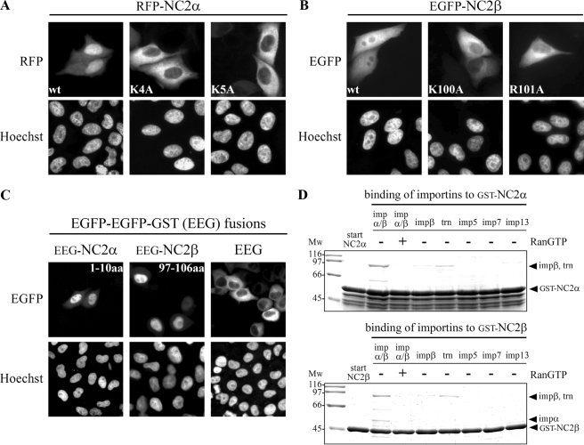
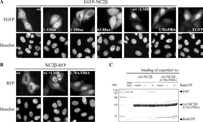
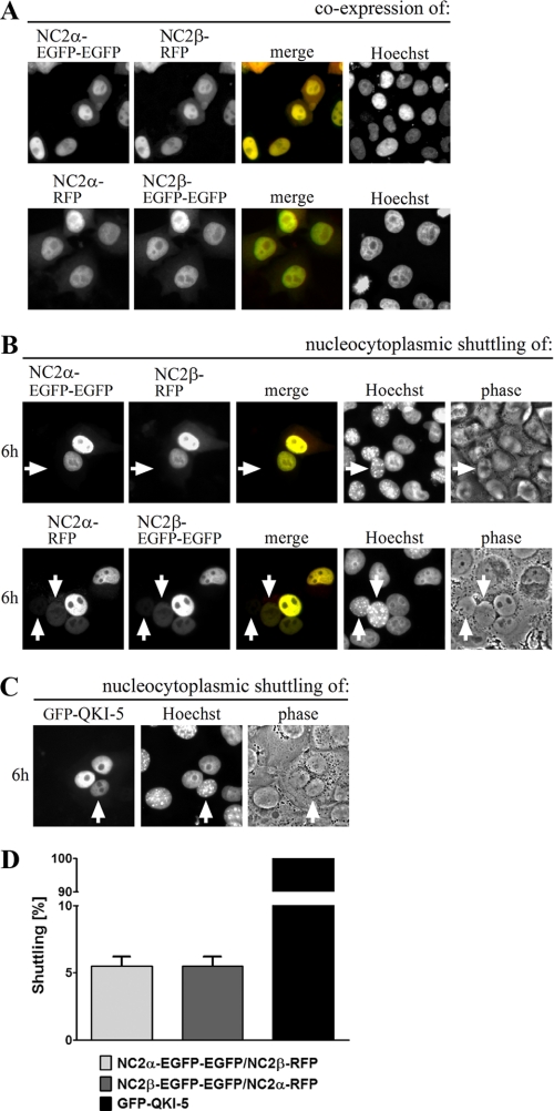
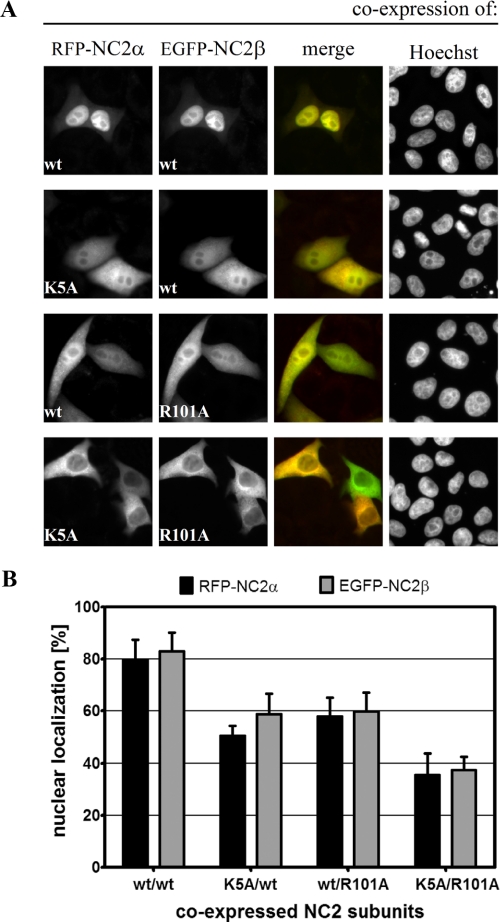
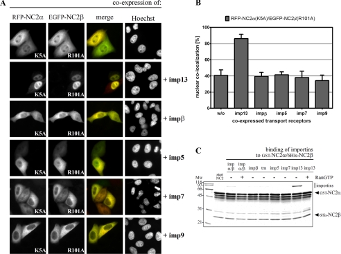
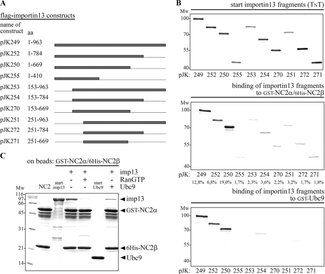
The negative cofactor 2 (NC2) is a protein complex composed of two subunits, NC2alpha and NC2beta, and plays a key role in transcription regulation. Here we investigate whether each subunit contains a nuclear localization signal (NLS) that permits individual crossing of the nuclear membrane or whether nuclear import of NC2alpha and NC2beta depends on heterodimerization. Our results from in vitro binding studies and transfection experiments in cultured cells show that each subunit contains a classical NLS (cNLS) that is recognized by the importin alpha/beta heterodimer. Regardless of the individual cNLSs the two NC2 subunits are translocated as a preassembled complex as co-transfection experiments with wild-type and cNLS-deficient NC2 subunits demonstrate. Ran-dependent binding of the nuclear export receptor Crm1/exportin 1 confirmed the presence of a leucine-rich nuclear export signal (NES) in NC2beta. In contrast, NC2alpha does not exhibit a NES. Our results from interspecies heterokaryon assays suggest that heterodimerization with NC2alpha masks the NES in NC2beta, which prevents nuclear export of the NC2 complex. A mutation in either one of the two cNLSs decreases the extent of importin alpha/beta-mediated nuclear import of the NC2 complex. In addition, the NC2 complex can enter the nucleus via a second pathway, facilitated by importin 13. Because importin 13 binds exclusively to the NC2 complex but not to the individual subunits this alternative import pathway depends on sequence elements distributed among the two subunits.


Figures

FIGURE 1.
FIGURE 1.
Both NC2 subunits exhibit a monopartite cNLS. HeLa P4 cells were transiently transfected with plasmid DNA encoding wild-type (wt) and mutated RFP-NC2α (A), EGFP-NC2β (B), and EGFP-EGFP-GST (EEG), amino acids 1–10 of NC2α or amino acids 97–106 of NC2β fused to EEG (C). The subcellular distribution was examined 24 h after transfection by direct fluorescence. The DNA was counterstained with Hoechst. A, the nuclear accumulation of wild-type RFP-NC2α was blocked when either lysine residue 4 (K4A) or lysine residue 5 (K5A) of NC2α was mutated. B, the homogeneous distribution of wild-type EGFP-NC2β was strongly affected by alanine substitution of either lysine residue 100 (K100A) or arginine residue 101 (R101A) of NC2β. C, the dominant cytoplasmic localization of EEG changed upon fusion to amino acids 1–10 of NC2α (EEG-NC2α-(1–10)) or amino acids 97–106 of NC2β (EEG-NC2β-(97–106)), the localization now becoming nuclear. D, both NC2 subunits bind to importin α/β and the binding is abolished in the presence of RanGTP. Immobilized GST-NC2α and GST-NC2β were incubated with bacterial lysates containing the indicated import receptors. Bound fractions were analyzed by SDS-PAGE and Coomassie stained. Mw, molecular weight; imp, importin; aa, amino acids; trn, transportin 1.
FIGURE 2.
FIGURE 2.
NC2β exposes a leptomycin B-sensitive NES. HeLa P4 cells were transiently transfected with plasmid DNA coding for wild-type (wt), truncated or mutated NC2β, N-terminal fused to EGFP (A) or C-terminal fused to RFP (B). The subcellular distribution of the gene products was examined 24 h post-transfection by direct fluorescence. The DNA was counterstained with Hoechst. A, wild-type EGFP-NC2β and the first 110 amino acids of NC2β fused to EGFP (EGFP-NC2β-(1–110)) show a homogeneous subcellular distribution that becomes nuclear after addition of 10 ng/ml LMB. Additional deletion of amino acids 101–110 (EGFP-NC2β-(1–100)) results in an exclusively cytoplasmic localization of NC2β. Amino acids 63–88 of NC2β fused to EGFP (EGFP-NC2β-(63–88)) are more cytoplasmically localized than EGFP alone. The homogeneous subcellular distribution of wild-type EGFP-NC2β was reduced when the leucine (L) and phenylalanine (F) residues at positions 78 and 80 were mutated (EGFP-NC2β-(L78A/F80A)). B, wild-type NC2β-RFP shows a dominant cytoplasmic localization that becomes nuclear after addition of LMB. Substitution of the two hydrophobic amino acids at positions 78 and 80 (NC2β-RFP-(L78A/F80A) prevented nuclear export of NC2β-RFP leading to a largely nuclear localization. C, NC2β is recognized by exportin 1 in a RanGTP-dependent fashion. GST-NC2β and GST-NC2β-(L78A/F80A) were incubated with exportin 1 (exp1) in the absence or presence of RanGTP (for details see “Experimental Procedures”). For a negative control exportin 1 was omitted (mock). After binding, glutathione-Sepharose bound fractions were analyzed by SDS-PAGE and Coomassie stained. Input of exportin 1 corresponds to 10% of the protein that was used. The hydrophobic amino acids leucine (Leu78) and phenylalanine (Phe80) are necessary for the binding of exportin 1 to NC2β. Mw, molecular weight; aa, amino acids.
FIGURE 3.
FIGURE 3.
One-way transport of the NC2 complex. HeLa P4 cells were transiently co-transfected with plasmid DNA encoding wild-type NC2α and NC2β fused to EGFP-EGFP or RFP (A and B). The shuttling protein QUAKING-5 fused to GFP (GFP-QKI-5) was overexpressed in HeLa P4 cells (C). A, the subcellular distribution of the gene products was examined 24 h post-transfection by direct fluorescence. Co-expression of NC2α and NC2β leads to a nuclear co-localization shown in yellow (merge). The DNA was counterstained with Hoechst. B, 30 h post-transfection, untransfected mouse NIH-3T3 cells were co-seeded onto the HeLa P4 cells, co-cultured for 18 h, and fused with Sigma HybriMax®. The subcellular distribution of the fusion proteins was examined after an additional 6-h incubation in the presence of cycloheximide. To distinguish between HeLa P4 and NIH-3T3 cell nuclei the heterokaryotic cells were counterstained with Hoechst. Co-localized NC2α and NC2β (shown in yellow, merge) could hardly be detected in the nucleus of mouse NIH-3T3 cells (white arrows). Thus, the NES in NC2β does not allow nucleo-cytoplasmic shuttling of the NC2 complex. C, GFP-QKI-5 accumulates in the nucleus of non-transfected mouse NIH-3T3 cells (white arrow) demonstrating that QKI-5 shuttles between the nucleus and cytoplasm. D, quantification of two independent experiments. 100 heterokaryotic cells were analyzed per condition. Only co-transfected HeLa P4 cells were included in the analysis of the NC2 complex. Bars indicate the mean ± S.D.
FIGURE 4.
FIGURE 4.
Nuclear accumulation of the NC2 complex via importin α/β requires the cNLSs of both subunits. A, HeLa P4 cells were transiently co-transfected with plasmid DNA encoding wild-type (wt) and mutated RFP-NC2α and EGFP-NC2β, respectively. The subcellular distribution of the gene products was examined 24 h post-transfection by direct fluorescence. The DNA was counterstained with Hoechst. Co-expression of wild-type RFP-NC2α and EGFP-NC2β results in a nuclear co-localization shown in yellow (merge; top panel). Mutation of either the cNLS of NC2α (K5A) or the cNLS of NC2β (R101A) reduced the nuclear accumulation leading to a homogeneous localization of both subunits (middle panels). Nuclear import of the RFP-NC2α/EGFP-NC2β complex was strongly blocked when the cNLSs of both subunits were mutated (bottom panel). B, for quantitative analysis, the mean red (RFP) and green (EGFP) fluorescence value in the nucleus and cytoplasm of 25 cells that co-expressed RFP-NC2α and EGFP-NC2β was measured using the ImageJ Software (NIH). After subtraction of the background value the percentage of nuclear localization of the different NC2 complexes was calculated. Bars indicate the mean ± S.D.
FIGURE 5.
FIGURE 5.
Importin 13 also mediates nuclear transport of the NC2 complex. A, HeLa P4 cells were transiently co-transfected with plasmid DNA encoding mutated RFP-NC2α (K5A), EGFP-NC2β (R101A), and FLAG-tagged import receptors. The subcellular distribution of the gene products was examined 24 h post-transfection by direct fluorescence of the RFP and EGFP fusion proteins. The overlap between the green EGFP fusion protein and the red RFP fusion protein is shown in yellow (merge). The DNA was counterstained with Hoechst. RFP-NC2α-(K5A) and EGFP-NC2β-(R101A) both predominantly remained in the cytoplasm of co-transfected cells. Although co-expression of importin 13 led to a strong nuclear accumulation of the NC2 complex, co-expressed importin β, importin 5, importin 7, and importin 9 did not affect the subcellular distribution of the NC2 complex. B, quantification of nuclear import of the cNLS-deficient NC2 complex. The mean fluorescence value of co-localized RFP-NC2α-(K5A) and EGFP-NC2β-(R101A) was measured in 15 cells using ImageJ software (NIH). The percentage of nuclear localization in the presence of co-expressed import receptors was calculated. Bars indicate the mean ± S.D. C, immobilized GST-NC2α/His6-NC2β complex was incubated with bacterial lysates containing the indicated import receptors. Bound fractions were analyzed by SDS-PAGE and Coomassie stained. The NC2 complex binds strongly to importin 13 and less pronounced to the importin α/β heterodimer. Both cargo-receptor interactions are reduced in the presence of RanGTP, which was used to simulate nuclear conditions. Mw, molecular weight; imp, importin; trn, transportin 1.
FIGURE 6.
FIGURE 6.
The C terminus of importin 13 is dispensable for recognition of the NC2 complex. A, the names of the importin 13 expression constructs used and the amino acids contained in the constructs are listed. Lines indicate the deleted regions and gray bars represent the different importin 13 fragments. B, the minimal importin 13 fragment that binds to the NC2 complex and UBC9 consists of amino acids 1–669. The two NC2 subunits, GST-NC2α and His6-NC2β, as well as GST-UBC9 were (co-)expressed in E. coli and used as bait after immobilization on glutathione-Sepharose. The immobilized GST-NC2α/His6-NC2β complex and GST-UBC9 were incubated with in vitro transcribed and translated 35S-labeled importin 13 fragments; all from the TnT coupled reticulocyte lysate. Starting material (20% of the importin 13 fragments that were used) and bound fractions were analyzed by SDS-PAGE followed by phosphorimaging (Amersham Biosciences). Among the importin 13 fragments, wild-type importin 13, and amino acids 1–669 of importin 13 showed the highest binding competence for the NC2 complex (see relative binding in percent below the gels, quantified with the program ImageQuant 5.2). A similar binding pattern was observed for UBC9; also here importin 13 fragments lacking the N terminus did not significantly bind to the immobilized substrate. C, the binding sites in importin 13 for the NC2 complex and UBC9 overlap. Importin 13 interacts with the NC2 complex in an UBC9 sensitive manner. Importin 13 was bound specifically to the immobilized GST-NC2α/His6-NC2β complex and this binding was reduced in the presence of equal amounts of UBC9. MW, molecular mass in kilodalton; imp, importin; aa, amino acids.

Similar articles

Cited by

References

    1. Kim, S., Na, J. G., Hampsey, M., and Reinberg, D. (1997) Proc. Natl. Acad. Sci. U. S. A. 94 820-825 - PMC - PubMed
    1. Gadbois, E. L., Chao, D. M., Reese, J. C., Green, M. R., and Young, R. A. (1997) Proc. Natl. Acad. Sci. U. S. A. 94 3145-3150 - PMC - PubMed
    1. Goppelt, A., Stelzer, G., Lottspeich, F., and Meisterernst, M. (1996) EMBO J. 15 3105-3116 - PMC - PubMed
    1. Mermelstein, F., Yeung, K., Cao, J., Inostroza, J. A., Erdjument-Bromage, H., Eagelson, K., Landsman, D., Levitt, P., Tempst, P., and Reinberg, D. (1996) Genes Dev. 10 1033-1048 - PubMed
    1. Kamada, K., Shu, F., Chen, H., Malik, S., Stelzer, G., Roeder, R. G., Meisterernst, M., and Burley, S. K. (2001) Cell 106 71-81 - PubMed

Publication types

MeSH terms

LinkOut - more resources