. 2009 Apr 24;284(17):11405-15.
doi: 10.1074/jbc.M805408200. Epub 2009 Feb 9.

Functional characterization of two secreted SEL1L isoforms capable of exporting unassembled substrate

Affiliations

Functional characterization of two secreted SEL1L isoforms capable of exporting unassembled substrate

Monica Cattaneo et al. J Biol Chem. .

Abstract

SEL1L-A, a transmembrane glycoprotein residing in the endoplasmic reticulum (ER), is a component of the ER-associated degradation (ERAD) pathway. Alternative splicing generates two smaller SEL1L isoforms, -B and -C, that lack the SEL1L-A membrane-spanning region but retain some sel-1-like repeats, known to be involved in multi-protein interactions and signal transduction. In this study the functional characteristics of SEL1L-B and -C were investigated in human cell models. We show that these two isoforms are induced upon ER stress and activation of the unfolded protein response, together with SEL1L-A. Using transient transfection experiments (based on wild-type and mutant SEL1L constructs) combined with several biochemical tests we show that SEL1L-B and, more prominently, SEL1L-C are secreted glycoproteins. Although SEL1L-C is in monomeric form, SEL1L-B is engaged in intramolecular/intermolecular disulfide bonds. Both isoforms localize in secretory and degradative cellular compartments and in areas of cell-cell contact. However, whereas SEL1L-B is mainly associated with membranes, SEL1L-C shows the typical intralumenal localization of soluble proteins and is present in intercellular spaces. Furthermore, because of its peroxisomal domain, SEL1L-C localizes to peroxisomes. Both SEL1L-B and -C are involved in sorting and exporting unassembled Ig-mu(s) but do not affect two other ERAD substrates, the null Hong Kong variant of alpha(1)-antitrypsin, and mutant alpha(1)-AT Z. Overall these findings suggest that SEL1L-B and -C participate to novel molecular pathways that, in parallel with ERAD, contribute to the disposure of misfolded/unfolded or orphan proteins through degradation or secretion.


Figures

FIGURE 1.
FIGURE 1.
A, identification of SEL1L-B and -C transcripts and induction by ER stress. RT-PCR was performed on RNA extracted from 293 FT (panel A.1), KMS11 (panel A.2), and SKBr3 (panel A.3) cells, untreated and treated with DTT (2 mm) for 150 min, or with tunicamycin (T) (2 μg/μl) for 24 h. PCR with isoform-specific primers was as described under “Experimental Procedures,” signals shown here were obtained with 23 cycles for SEL1L-A and 32 cycles for SEL1L-B and -C. HPRT was used as a loading control, CHOP expression and spliced XBP-1 as indicators of ER stress. Panel A.4, down-modulation of SEL1L-B and -C transcripts by SEL1L siRNA. SKBr3 cells were treated with scrambled siRNA (lane 1) or siRNA specific to SEL1L (lane 2) for 72 h. Silencing efficiency was verified by RT-PCR. All of the SEL1L isoforms were down-modulated after SEL1L RNA interference. Densitometric quantifications were normalized relative to the housekeeping HPRT signals, as determined through the Scion imaging program. The data, expressed as fold-decrease relative to untreated, are the averages of two independent experiments, as in the top panel, + S.E. *, p > 0.05 t test. B, identification of SEL1L-B and -C transcriptional start sites. The sequence of the proximal 5′-flanking region of SEL1L-A (-300 to -1) is shown. The numbers indicate nucleotide positions from the translation initiation site (ATG, bold and double-underlined). GC motifs are in gray boxes, and the CAAT box is in open box. B* and C* indicate the transcription start sites predicted by Aceview, and B and C are the transcription start sites identified by 5′-RACE PCR and sequencing.
FIGURE 2.
FIGURE 2.
A, schematic representation of the Myc-tagged constructs used for transfection. SEL1L-B and -C are truncated forms of SEL1L-A. Splicing events at the 3′-terminal ends generate a peroxisomal domain (SRL, circle) in SEL1L-C and a tail of 9 amino acid residues (GIYVSPFTF, hexagon) in SEL1L-B. Four and three sel-1-like repeats, respectively (SEL1), are present in SEL1L-B and -C. A fibronectin domain is present in all of the isoforms. B, SEL1L-B and -C isoforms are secreted glycoproteins. 293 FT cells (4 × 10⁶) transiently transfected with Myc-tagged SEL1L-B and -C constructs were treated with tunicamycin (T), 2 μg/μl, for 24 h; aliquots (50 μg) of untreated lysates were incubated with endoglycosidase H (H) and PNGase F (F). The proteins were fractionated on SDS-PAGE (10%) and blotted with anti-Myc. Note mobility shifts of treated exogenous SEL1L isoforms. Glycosylated and deglycosylated SEL1L-Bmyc and -Cmyc migrate at about 51–47 and 46–41 kDa, respectively (panels B.1 and B.2). Similar results were obtained with anti-SEL1L antibody. For the analysis of exogenous isoforms in the culturing medium, the cells were washed 48 h after transfection and incubated for 24 h in Opti-MEM, and the culturing medium was precipitated with trichloroacetic acid and subjected to endoglycosidase H (H) or PNGase F (F) treatments. Western blot analysis with anti-Myc revealed the presence of PNGase F-sensitive and endoglycosidase H-resistant SEL1L-Bmyc and -Cmyc (panel B.3).
FIGURE 3.
FIGURE 3.
A, stability of exogenous isoforms. 293 FT (4 × 10⁶) transfectants expressing Myc-tagged SEL1L-B or -C were treated for 15 h with cycloheximide (CHX) and incubated for 24 h with Opti-MEM. Aliquots (30 μg) of lysates (left) and of secreted proteins (right) were resolved by 10% SDS-PAGE and probed with anti-SEL1L and anti-actin. The levels of exogenous isoforms in lysates decreased after cycloheximide treatment, concomitantly with increases in the culturing medium. Densitometric quantifications were normalized relative to housekeeping signals by the Scion imaging program. The data are the averages of two independent experiments, as in the top panel, expressed as fold modulation relative to untreated, + S.E. *, p > 0.05 t test. B, SEL1L-B forms dimers. Lysates from 293 FT (4 × 10⁶) transfectants expressing Myc-tagged SEL1L-B or -C were immunoprecipitated with monoclonal anti-Myc and probed with anti-Myc. Under nonredox condition SEL1L-Bmyc immunoprecipitated both as dimer and monomeric forms, whereas SEL1L-Cmyc immunoprecipitated only as monomeric forms. SEL1L-Bmyc dimers were reduced in the redox running condition. Aliquots of lysates (40 μg) were loaded to verify protein expression levels and immunoprecipitation efficiency. C, intra/intermolecular disulfide bonds in SEL1L-B. 293 FT cells (4 × 10⁶) transiently transfected with Myc-tagged SEL1L-B and -C were maintained for 24 h in Opti-MEM. Secreted protein and aliquots of cell lysates (25 μg) were resolved by SDS-PAGE (7%) under reducing (redox) and nonreducing (non redox) conditions and blotted with monoclonal anti-SEL1L (panels C.1, C.2, and C.4) or anti-Myc (panel C.3). The lower bands correspond to the exogenous SEL1L-Bmyc and -Cmyc isoforms. The upper multiple bands revealed by anti-SEL1L in lysates (panel C.2) and culturing medium (panel C.4) and by anti-Myc in lysates (panel C.3) of SEL1L-Bmyc transfectants under nonreducing conditions may correspond to intra/intermolecularly disulfide-bound SEL1L-B. D, overexpression of SEL1L-B and -C isoforms does not affect the UPR response. RNA was extracted from 293 FT (4 × 10⁶) cells transfected with empty vector (PCDN3.1 myc), SEL1L-Bmyc, and SEL1l-Cmyc, respectively, and analyzed by RT-PCR for the UPR response. The exogenous SEL1L isoforms did not modulate SEL1L-A and CHOP expression or XBP-1 splicing. HPRT serves as internal control. On top, aliquots of lysates (30 μg) were loaded to verify transfection efficiencies.
FIGURE 4.
FIGURE 4.
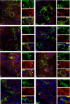
Subcellular localizations of SEL1L-Bmyc and -Cmyc by optical section fluorescence microscopy. The subcellular localizations of SEL1L-Bmyc and SEL1L-Cmyc isoforms in transiently transfected 293 FT kidney cells was analyzed by immunofluorescence double staining using anti-Myc monoclonal antibody (green) and polyclonal antibodies (red) against markers of cellular compartments, including calreticulin (ER), giantin (Golgi), LAMP3 (CD63, late endosomes), and catalase (peroxisomes). Nuclei (blue) are stained with 4′,6-diamido-2-phenylindole. Transfected cells, identified by anti-Myc immunolabeling, were examined with a Zeiss Axiovert 200M microscope fitted with an ApoTome Imaging system. The images shown are from a single representative axial plane. The left column images (A–H) show merged channels obtained by multiplying the red and green channels of a single Apotome optical section. Green, red, and merged details are shown to the right of each image. Indirect immunofluorescence analysis in A and B show that the two SEL1L isoforms are not only present in the ER, identified by calreticulin (arrowheads point to punctate areas of co-localization between SEL1L-Bmyc or -Cmyc and calreticulin), but also in other locations, including the cell periphery (arrows point to green SEL1L-Bmyc signal in an area negative for calreticulin). Both SEL1L-Bmyc and SEL1L-Cmyc co-localize with the Golgi membrane protein giantin and with LAMP3, a marker for multivesicular bodies and late endosomes (C–F, arrowheads). In contrast only SEL1L-Cmyc appears to co-localize with the peroxisomal marker catalase in small punctate structures (G and H, arrowheads). Scale bars, 10 μm.
FIGURE 5.
FIGURE 5.
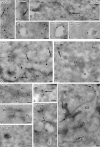
Subcellular localizations of SEL1L-Bmyc and -Cmyc by cryoimmunoelectron microscopy. Cryoimmunoelectron microscopy allowed to define with fine detail the subcellular localizations of SEL1L-Bmyc and -Cmyc in transfected 293 FT cells. SEL1L-Bmyc localizes on the membrane profiles of the ER (A, arrow), identified by ER-resident intraluminal calreticulin (A, arrowhead), on peripheral vesicles near the plasma membrane (B) and at the boundaries of adjacent cells (C, arrow). Perimeter membranes of electronlucent structures, morphologically identifiable as early endosomes (D and E), and LAMP1-positive endosomal compartments (F) are also SEL1L-Bmyc-labeled. SEL1L-Cmyc (G, arrows) co-localizes densely with calreticulin (arrowheads) in ER luminal cisternae and within the nuclear envelope. Gold particles are also localized in Golgi cisternae (H), in peripheral vesicles (H–L), and on both tight (I) and adherent (M) intercellular junctions. Moreover SEL1L-Cmyc decorates vesicles trapped in the lumen of endosomes (O) and small peroxisomes, identified by catalase labeling (N). SEL1L-Cmyc labeling is also present in the intercellular spaces (P), mostly along plasma membranes and in specialized plasma membrane invaginations (P, arrows), often connected with peripheral small endocytic vacuoles (P, asterisk). Bars, 0.1 μm. E, endosomes; er, endoplasmic reticulum; G, Golgi complex; LE, late endosomes; M, mitochondria; Nu, nucleus; Ne, nuclear envelope; P, peroxisomes; PM, plasma membrane; P1-P2, adjacent plasma membranes.
FIGURE 6.
FIGURE 6.
A, exogenous SEL1L isoforms interact with μs. Lysates from 293FT (4 × 10⁶) transfectants expressing μs and combinations of wild-type or mutant Myc-tagged SEL1L isoforms, as indicated, were immunoprecipitated with anti-Myc (IP myc), resolved by SDS-PAGE (10%), and probed with anti-μ. Lysate aliquots (30 μg) were loaded to verify transfection and immunoprecipitation efficiencies. Abundant μs co-immunoprecipitated with each of the tested exogenous SEL1L isoforms. B, analysis of μs stability in presence of SEL1L-Bmyc and SEL1L-20–372KDEL. 293 FT (4 × 10⁶) transfectants expressing Myc-tagged SEL1L-B and SEL1L-20–372KDEL were treated for 4 and 7 h with cycloheximide (CHX, 200 μg/μl). Aliquots of lysates (30 μg) were resolved by SDS-PAGE (10%) and probed with anti-μ, anti-SEL1L, and anti-vinculin, as a control for loading. Although SEL1L-B had no effect on μs stability, SEL1L-20–372KDEL decelerated degradation. WB, Western blot.
FIGURE 7.
FIGURE 7.
A, exogenous SEL1L isoforms promote secretion of unassembled μs. 293 FT (4 × 10⁶) cells were transiently co-transfected for 48 h with μs and various wild-type or mutant SEL1L isoforms including SEL1L-Bmyc, SEL1L-Cmyc, SEL1L-Bmut, SEL1LΔCmyc, and SEL1L-20–372 with or without KDEL, as indicated, and then incubated for 24 h in Opti-MEM. Secreted proteins were precipitated with trichloroacetic acid, resolved by SDS-PAGE, and probed with anti-μs, anti-SEL1L and anti-vinculin as a loading control. Aliquots of lysates (30 μg) were analyzed by Western blotting to monitor the expression levels of the different transgenes. Orphan substrate was secreted only by cells overexpressing SEL1L isoforms, with highest levels in SEL1L-B and SEL1L-20–372 transfectants, and lowest levels in SEL1L-ΔCmyc transfectants. Percentage of secreted proteins over total was normalized and densitometrically quantified by the Scion imaging program. The data are averages of two independent experiments. B, glycosylation analysis of unassembled secreted μs. Secreted μs were subjected to endoglycosidase H (H) and PNGase F (F) treatments, resolved by SDS-PAGE, and probed with anti-μs. Note mobility shift in the presence of PNGase F and endoglycosidase H resistance. C, secretion of unassembled substrate does not affect UPR. RNA was extracted from the same samples described for A and analyzed by RT-PCR for the UPR. Secretion of substrate did not modulate CHOP expression and XBP-1 splicing. Lane 1, un-transfected cells without DTT; lane 2, mock-transfected cells; lane 3, μs transfectants; lane 4, SEL1L-Cmyc and μs co-transfectants; lane 5, SEL1L-Bmyc and μs co-transfectants; lane 6, SEL1L-20–372 and μs co-transfectants; lane 7, SEL1L-20–372 harboring KDEL and μs co-transfectants; lane 8, untransfected DTT-treated cells. D, SEL1L isoforms are unable to promote secretion of α1-antitrypsin variants. 293 FT (4 × 10⁶) cells were transiently co-transfected with the null Hong Kong variant of α1-antitrypsin variant (NHK) or with mutant α1-antitrypsin (α1-AT Z) and with each of various wild-type or mutant SEL1L forms, as indicated, for 48 h and successively incubated for 24 h with Opti-MEM. Secreted proteins were precipitated with trichloroacetic acid, resolved by SDS-PAGE, and probed with anti-α1-antitrypsin (ATT), anti-SEL1L, and anti-actin. Aliquots of lysates (30 μg) were analyzed by Western blotting to monitor expression of the transgenes. The presence of SEL1L isoforms did not enhance secretion of either of the two substrates. The asterisks in the middle left panel indicate the residual signal of the PIZ substrate. WB, Western blot.

Similar articles

Cited by

References

    1. Mueller, B., Lilley, B. N., and Ploegh, H. L. (2006) J. Cell Biol. 175 261-270 - PMC - PubMed
    1. Cattaneo, M., Otsu, M., Fagioli, C., Martino, S., Lotti, L. V., Sitia, R., and Biunno, I. (2008) J. Cell. Physiol. 215 794-802 - PubMed
    1. Christianson, J. C., Shaler, T. A., Tyler, R. E., and Kopito, R. (2008) Nat. Cell Biol. 10 272-282 - PMC - PubMed
    1. Kostova, Z., and Wolf, D. H. (2003) EMBO J. 22 2309-2317 - PMC - PubMed
    1. Modrek, B., and Lee, C. (2002) Nat. Genet. 30 13-19 - PubMed

Publication types