. 2009 Apr 17;284(16):10855-67.
doi: 10.1074/jbc.M804813200. Epub 2009 Feb 9.

Mutant huntingtin N-terminal fragments of specific size mediate aggregation and toxicity in neuronal cells

Affiliations

Mutant huntingtin N-terminal fragments of specific size mediate aggregation and toxicity in neuronal cells

Tamara Ratovitski et al. J Biol Chem. .

Abstract

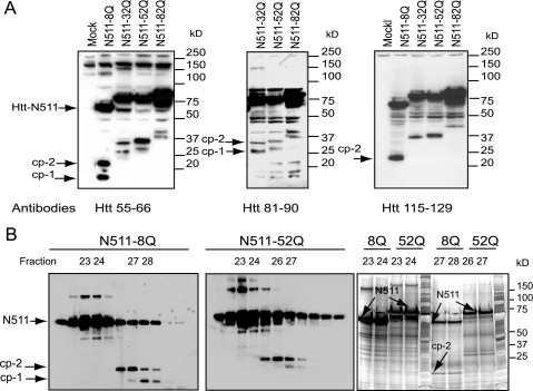
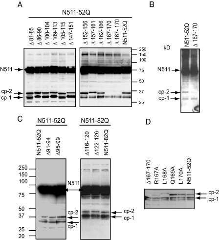
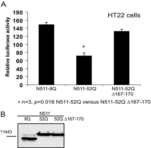
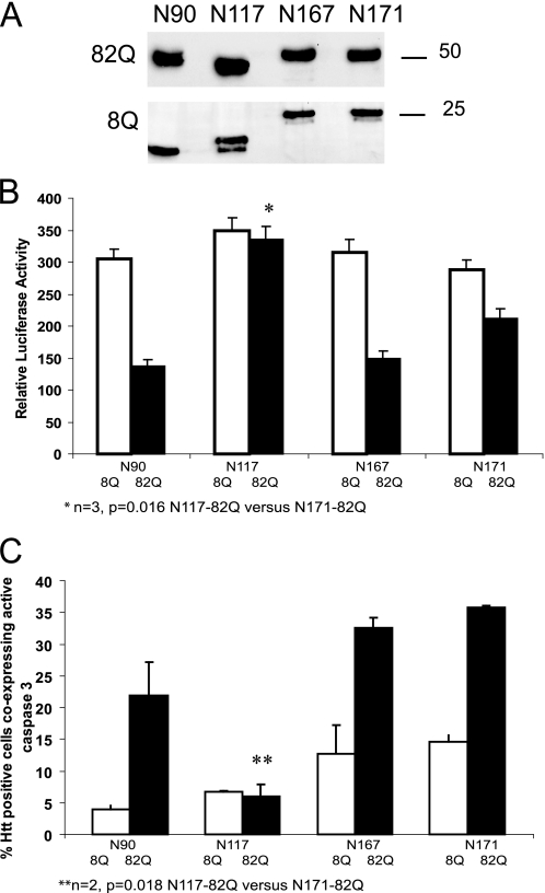
Huntingtin proteolysis is implicated in Huntington disease pathogenesis, yet, the nature of huntingtin toxic fragments remains unclear. Huntingtin undergoes proteolysis by calpains and caspases within an N-terminal region between amino acids 460 and 600. We have focused on proteolytic steps producing shorter N-terminal fragments, which we term cp-1 and cp-2 (distinct from previously described cp-A/cp-B). We used HEK293 cells to express the first 511 residues of huntingtin and further define the cp-1 and cp-2 cleavage sites. Based on epitope mapping with huntingtin-specific antibodies, we found that cp-1 cleavage occurs between residues 81 and 129 of huntingtin. Affinity and size exclusion chromatography were used to further purify huntingtin cleavage products and enrich for the cp-1/cp-2 fragments. Using mass spectrometry, we found that the cp-2 fragment is generated by cleavage of huntingtin at position Arg(167). This site was confirmed by deletion analysis and specific detection with a custom-generated cp-2 site neo-epitope antibody. Furthermore, alterations of this cleavage site resulted in a decrease in toxicity and an increase in aggregation of huntingtin in neuronal cells. These data suggest that cleavage of huntingtin at residue Arg(167) may mediate mutant huntingtin toxicity in Huntington disease.


Figures

FIGURE 1.
FIGURE 1.
The schematics of Htt proteolysis. HEAT repeats (blue boxes) are indicated within the Htt sequence, N-terminal and caspase/calpain putative proteolytic domains are shown (yellow boxes). Putative cp-1 and confirmed cp-2 sites are indicated by arrows. Epitopes recognized by Htt antibodies are marked with red lines.
FIGURE 2.
FIGURE 2.
Expression and purification of Htt fragments in HEK293 cells. A, epitope mapping of the cp-1/cp-2 fragments. Western blot of total cell extracts (30 μg/lane) from HEK293 cells transfected with either N511-Htt constructs with different poly(Q) lengths, or cells transfected with empty vector (Mock). Htt-N511 and cp-1/cp-2 fragments (arrows) were detected with an antibody to residues 55-66 of Htt (left panel), with antibody to residues 81-90 of Htt (middle panel), or with antibody to residues 115-129 of Htt (right panel). HEK293 cells transfected with expanded Htt constructs produce fragments similar to the cp-1/cp-2 fragments observed previously in PC12 cells (30). B, purification of the cp-1/cp-2 fragments for mass spectrometry. Western blotting analysis of 1/10 aliquots of fractions from size chromatography of FLAG immunoprecipitates of either Htt-N511-8Q (left panel), or Htt-N511-52Q (middle panel) are expressed in HEK293 cells. Htt proteins, marked by arrows, were detected with antibody to FLAG. Indicated fractions containing either the cp-1/cp-2 fragments or in Htt-N511, were separated on SDS-PAGE, and stained with silver protein stain (right panel). Htt bands (indicated with arrows) corresponding to cp-1/cp-2 fragments, and bands containing only Htt-N511 were excised from the gel for mass spectrometry.
FIGURE 3.
FIGURE 3.
Identification of the cp-2 cleavage site by mass spectrometry. A, peptide coverage obtained from chymotrypsin digest of huntingtin bands (N511 and cp-2). B, extracted ion chromatogram of MDSNPLR ion (m/z 416.7) and full spectrum at 17.7 min for fragment cp-2 (showing the presence of the peptide). C, confirmation of sequence identification of peptide MDSNLPR (comparison of fragmentation spectra obtained from the cp-2 fragment, produced from Htt-N511-8Q (top), and from synthesized peptide MDSNLPR (bottom)). The tables below show the matched fragments in bold. The two fragmentation spectra are very similar, which confirms that the peptide detected in the cp-2 fragment digest is indeed MDSNLPR.
FIGURE 4.
FIGURE 4.
Mapping of the cp-2 cleavage site by deletion analysis. A, C, D, Htt constructs with indicated deletions (A and C) or point mutations (D), or unaltered Htt with expanded poly(Q) (N511-52Q or N511-82Q) were expressed in HEK293 cells, and Htt-FLAG fusion proteins (indicated by arrows) were detected by Western blotting with an antibody to FLAG. B, Htt constructs with either Δ167-170 deletion or unaltered Htt with expanded poly(Q) (N511-52Q) were expressed in neuronal HT22 cells, FLAG-Htt fusion proteins (indicated by arrows) were immunoprecipitated with FLAG-agarose (∼2 mg of protein per immunoprecipitation), and detected with an antibody to exon 1 of Htt. Deletion of residues 167-170 greatly reduces generation of the cp-2 fragment from Htt-N511-52Q construct expressed in HEK293 or HT22 cells.
FIGURE 5.
FIGURE 5.
Detection of the cp-2 fragment by a specific antibody to the neo-epitope Arg167 of Htt in HEK293 cells. A, Western blot of total cell extracts (30 μg/lane) from HEK293 cells transfected with the indicated Htt constructs, or an empty vector (Mock). Truncated FLAG-Htt fusion proteins and cp-1/cp-2 fragments (marked by arrows) were detected with antibodies to FLAG (left panel). The Htt-N167-82Q fragment migrates on a gel more slowly than the cp-2 fragment derived from Htt-N511-52Q, consistent with the length of poly(Q). Antibody to Arg167 neo-epitope detects the Htt-N167 fragment and the cp-2 fragment, has low cross-reactivity with the Htt-N171 fragment, and fails to detect Htt-N511 fragment or any fragments of Htt-Δ167-170 (right panel, data shown for NE167 antibody). B, Western blot of total cell extracts from HEK293 cells transfected with the indicated Htt constructs or an empty vector (Mock). Htt proteins (indicated by arrows) were detected with an antibody to exon 1 of Htt (left panel, 20 μg per lane). The cp-2 fragments produced from Htt-N511 or Htt-N586 were specifically detected by an antibody to the Arg167 neo-epitope (right panel, 40 μg/lane). Blots, shown in A and B, were re-probed with an antibody to actin for loading control. C, Western blot of formic acid-soluble aggregate fractions from HEK293 cells transfected with indicated constructs or an empty vector (Mock). Htt fragments were detected with antibodies to exon 1 (left panel) or to the Arg167 neo-epitope (right panel) of Htt.
FIGURE 6.
FIGURE 6.
Immunofluorescent detection of the cp-2 fragment by a specific antibody to the neo-epitope Arg167 of Htt. HEK293 (A) or HT22 (B) cells were transfected with indicated Htt constructs. Epitope between residues 115-129 of Htt is shown in green (FITC); Arg167 neo-epitope is shown in red (Cy 3); the nuclear staining (4′,6-diamidino-2-phenylindole) is shown in blue. Yellow staining in merged images (marked by arrows) demonstrates the presence of cp-2.
FIGURE 7.
FIGURE 7.
Alteration of cp-2 cleavage site results in an increase in aggregation of Htt in neuronal cells. A, aggregate formation in HT22 cells expressing either Htt-N511-52Q or Htt-N511-52Q-Δ167-170. Cells were labeled with an antibody to FLAG, followed by anti-mouse FITC-conjugated antibody. B, aggregate formation in N2a and HT22 cells expressing Htt-N511-8Q, Htt-N511-52Q, or Htt-N511-52Q-Δ167-170. Cells were labeled with Htt-(1-82) antibody, followed by anti-mouse FITC-conjugated antibody and the aggregates were counted in 24-well plates in ∼200-400 cells for each construct (n = 2, *, p = 0.02 N511-52Q versus N511-52Q-Δ167-170 for N2a cells; **, n = 3, p = 0.07 N511-52Q versus N511-52Q-Δ167-170 for HT22 cells). One representative experiment of two is shown.
FIGURE 8.
FIGURE 8.
Alteration of cp-2 cleavage site changes biochemical properties of Htt protein. A, the procedure used to fractionate and dissociate Htt proteins (see “Experimental Procedures”). B, Western blots of subcellular fractions from HEK293 cells transfected with the indicated constructs: 1, native PAGE of soluble cytoplasmic fractions; 2, SDS-PAGE of soluble cytoplasmic fractions; 3, SDS-PAGE of the pellet, with SDS-insoluble material detected on top of the gel; 4, SDS-PAGE of formic acid-soluble aggregate fractions. Htt fragments were detected with antibodies to exon 1. *, minimal immunoreactivity is observed for Htt-N511-52Q-Δ167-170 following formic acid treatment. **, less SDS-insoluble material is detected for the Δ167-170 mutant than for unaltered N511-52Q. ***, new high molecular weight soluble complexes are detected for Htt-N511-52Q-Δ167-170 in native conditions.
FIGURE 9.
FIGURE 9.
Alteration of cp-2 cleavage site results in a decrease in toxicity of Htt protein in neuronal cells. A, cell viability (measured by luciferase assay, see “Experimental Procedures”) of HT22 cells, expressing the indicated Htt constructs co-transfected with the luciferase construct (n = 3, **, p = 0.018 N511-52Q versus N511-52Q-Δ167-170). One representative experiment of three for each set of constructs is shown. B, the expression levels of the indicated constructs were verified by Western blotting with an antibody to FLAG.
FIGURE 10.
FIGURE 10.
Htt N-terminal fragments of specific size have different toxic properties. A, Western blot of total cell extracts from HT22 cells transfected with the indicated Htt constructs. Htt proteins were detected with an antibody to exon 1 of Htt. B, cell viability, measured by luciferase assay (see “Experimental Procedures”) of HT22 cells, expressing the indicated Htt construct co-transfected with the luciferase construct (n = 3, *, p = 0.016 N117-82Q versus N171-82Q, p < 0.05 for all normal repeat versus expanded repeat constructs). One representative experiment of three for each set of constructs is shown. C, cytotoxicity, measured by caspase 3 activation (see “Experimental Procedures”) of HT22 cells, expressing the indicated Htt construct. Results are presented as a percentage of Htt positive cells co-expressing active caspase 3 (n = 2, over 100 cells were counted for each condition, **, p < 0.05 N117-82Q versus N171-82Q). One representative experiment of two is shown.

References

    1. The Huntington's Disease Collaborative Research Group (1993) Cell 72 971-983 - PubMed
    1. Ross, C. A., Margolis, R. L., Rosenblatt, A., Ranen, N. G., Becher, M. W., and Aylward, E. (1997) Medicine (Baltimore) 76 305-338 - PubMed
    1. Becher, M. W., Kotzuk, J. A., Sharp, A. H., Davies, S. W., Bates, G. P., Price, D. L., and Ross, C. A. (1998) Neurobiol. Dis. 4 387-397 - PubMed
    1. DiFiglia, M., Sapp, E., Chase, K. O., Davies, S. W., Bates, G. P., Vonsattel, J. P., and Aronin, N. (1997) Science 277 1990-1993 - PubMed
    1. Lunkes, A., Lindenberg, K. S., Ben-Haiem, L., Weber, C., Devys, D., Land-wehrmeyer, G. B., Mandel, J. L., and Trottier, Y. (2002) Mol. Cell 10 259-269 - PubMed

Publication types

MeSH terms

LinkOut - more resources