. 2009 Jun;8(6):1192-205.
doi: 10.1074/mcp.M800443-MCP200. Epub 2009 Feb 9.

Exosomal secretion of cytoplasmic prostate cancer xenograft-derived proteins

Affiliations

Exosomal secretion of cytoplasmic prostate cancer xenograft-derived proteins

Flip H Jansen et al. Mol Cell Proteomics. 2009 Jun.

Abstract

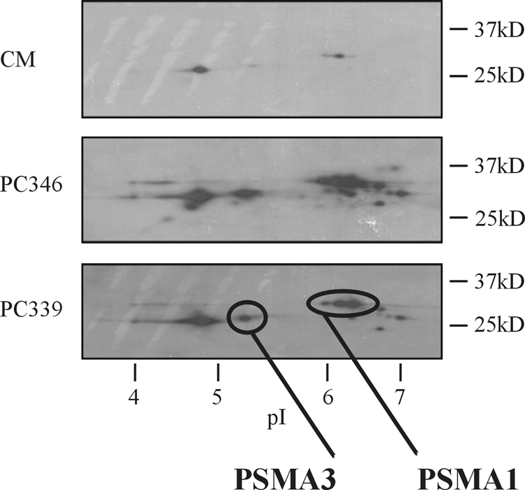
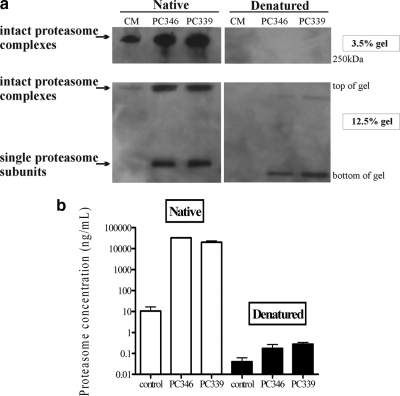
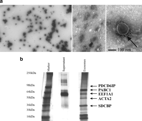
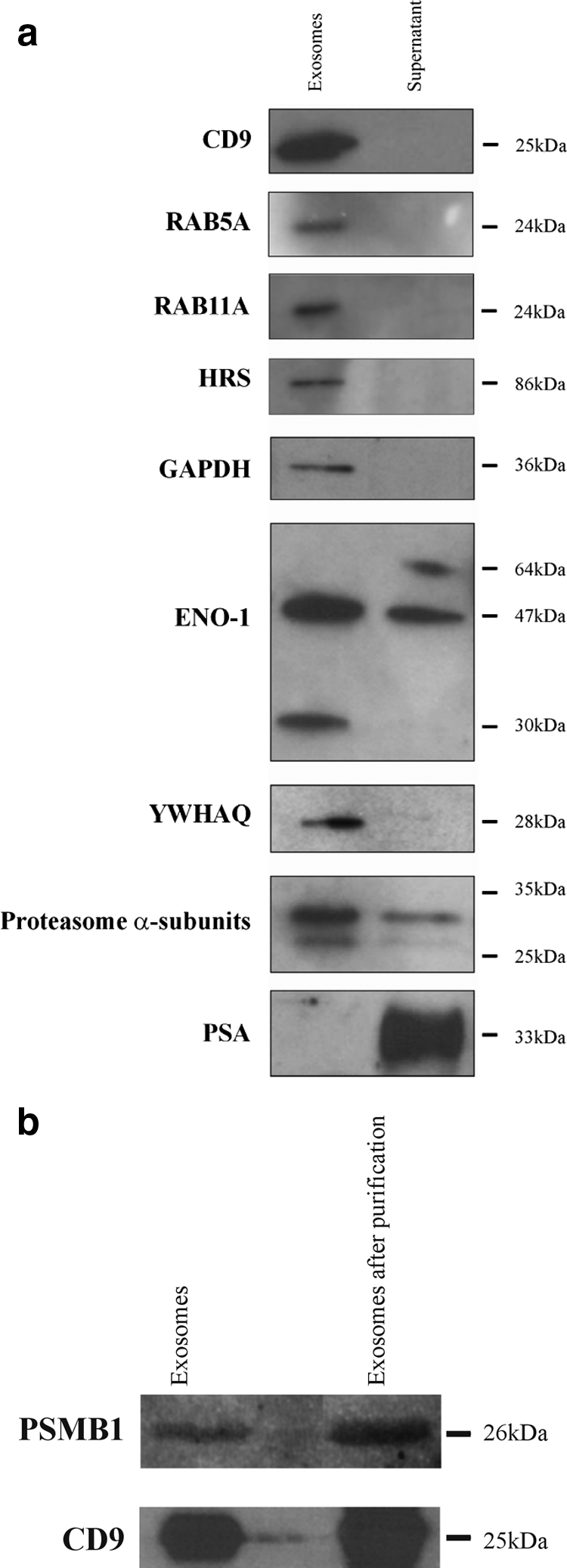
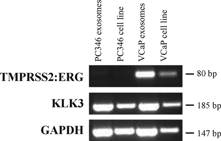
Novel markers for prostate cancer (PCa) are needed because current established markers such as prostate-specific antigen lack diagnostic specificity and prognostic value. Proteomics analysis of serum from mice grafted with human PCa xenografts resulted in the identification of 44 tumor-derived proteins. Besides secreted proteins we identified several cytoplasmic proteins, among which were most subunits of the proteasome. Native gel electrophoresis and sandwich ELISA showed that these subunits are present as proteasome complexes in the serum from xenograft-bearing mice. We hypothesized that the presence of proteasome subunits and other cytoplasmic proteins in serum of xenografted mice could be explained by the secretion of small vesicles by cancer cells, so-called exosomes. Therefore, mass spectrometry and Western blotting analyses of the protein content of exosomes isolated from PCa cell lines was performed. This resulted in the identification of mainly cytoplasmic proteins of which several had previously been identified in the serum of xenografted mice, including proteasome subunits. The isolated exosomes also contained RNA, including the gene fusion TMPRSS2-ERG product. These observations suggest that although their function is not clearly defined cancer-derived exosomes offer possibilities for the identification of novel biomarkers for PCa.


Figures

F<sc>ig</sc>. 1.
Fig. 1.
The selection procedure followed to annotate identified proteins as tumor-derived proteins.
F<sc>ig</sc>. 2.
Fig. 2.
2D PAGE Western blotting analysis of depleted serum from control mice (CM), PC346 xenograft-bearing mice (PC346), and PC339 xenograft-bearing mice (PC339). Spots were detected by using a monoclonal antibody to proteasome α subunits 6, 2, 4, 5, 1, and 3. The presence of PSMA1 and PSMA3 proteasome subunits was confirmed with specific monoclonal antibodies. Strong signals are visible in the serum from both the PC346 and PC339 mice. Also faint signals were detected in the control serum because of cross-reactivity with mouse proteasome subunits, which are present under normal conditions in the mouse serum.
F<sc>ig</sc>. 3.
Fig. 3.
a, native 1D gel electrophoresis of serum from control mice (CM) and PC346 or PC339 xenograft-bearing mice showing the presence of intact proteasome complexes. High molecular weight complexes are visible under native conditions in both 12.5 and 3.5% gels. After denaturation, mostly single subunits are visible in the 12.5% gel, and the high molecular weight complexes in the 3.5% gel have disappeared. Bands were detected by a monoclonal antibody directed against the PSMA1 subunit of the proteasome. b, proteasome concentrations in xenograft-bearing mice and control mice serum samples under native (left) and denatured (right) conditions as measured by sandwich ELISA. After serum denaturation proteasome levels are strongly diminished, indicating the existence of proteasome complexes in xenograft and control serum samples. Error bars represent standard deviations.
F<sc>ig</sc>. 4.
Fig. 4.
a, electron microscopy of exosomes isolated from the PC346C cell line. Left, electron micrograph of negatively stained exosomes showing a homogenous mixture of isolated vesicles; middle, ImmunoGold labeling of exosomes with the exosomal marker CD9 (Tetraspanin 29); right, increased magnification of the middle image shows positive CD9 membrane staining of exosomes (see arrow). b, separation of PC346C cell-derived exosomal proteins by 1D SDS-PAGE followed by silver staining. The indicated protein bands were excised and identified by LTQ-Orbitrap. SDCBP, syndecan-binding protein.
F<sc>ig</sc>. 5.
Fig. 5.
a, 1D PAGE and Western blotting analysis comparing the exosome and supernatant fractions of the PC346C cell line for CD9, the members of the RAS oncogene family RAB5A and RAB11A, HRS, GAPDH, ENO1, YWHAQ (14-3-3 protein θ), proteasome α subunits, and PSA. CD9, RAB5A, RAB11A, HRS, GAPDH, and YWHAQ were uniquely identified in the isolated exosomes, whereas PSA could only be detected in the supernatant of the PC346C cell line. b, 1D PAGE and Western blotting analysis of the exosome fraction after purification with magnetic beads. This figure shows that the CD9 and proteasome β1 (PSMB1) signals are visible in the exosome fraction both before and after purification with magnetic beads, indicating that proteasome subunits are present inside exosomes or exosomal membranes.
F<sc>ig</sc>. 6.
Fig. 6.
RT-PCR analysis of VCaP and PC346C cell lines and exosomes. The TMPRSS2-ERG fusion gene is exclusively expressed in both the VCaP cell line and exosomes, whereas both cell lines and exosomes express KLK3 (PSA) and GAPDH. H2O and −RT controls were negative (data not shown).

Similar articles

Cited by

References

    1. Stamey, T. A., Yang, N., Hay, A. R., McNeal, J. E., Freiha, F. S., and Redwine, E. ( 1987) Prostate-specific antigen as a serum marker for adenocarcinoma of the prostate. N. Engl. J. Med. 317, 909–916 - PubMed
    1. McDavid, K., Lee, J., Fulton, J. P., Tonita, J., and Thompson, T. D. ( 2004) Prostate cancer incidence and mortality rates and trends in the United States and Canada. Public Health Rep. 119, 174–186 - PMC - PubMed
    1. Thompson, I. M., Pauler, D. K., Goodman, P. J., Tangen, C. M., Lucia, M. S., Parnes, H. L., Minasian, L. M., Ford, L. G., Lippman, S. M., Crawford, E. D., Crowley, J. J., and Coltman, C. A., Jr. ( 2004) Prevalence of prostate cancer among men with a prostate-specific antigen level < or =4.0 ng per milliliter. N. Engl. J. Med. 350, 2239–2246 - PubMed
    1. Mikolajczyk, S. D., and Rittenhouse, H. G. ( 2004) Tumor-associated forms of prostate specific antigen improve the discrimination of prostate cancer from benign disease. Rinsho Byori 52, 223–230 - PubMed
    1. Paul, B., Dhir, R., Landsittel, D., Hitchens, M. R., and Getzenberg, R. H. ( 2005) Detection of prostate cancer with a blood-based assay for early prostate cancer antigen. Cancer Res. 65, 4097–4100 - PubMed

Publication types

Substances

LinkOut - more resources