. 2009 Apr 1;587(Pt 7):1481-98.
doi: 10.1113/jphysiol.2009.169243. Epub 2009 Feb 9.

{beta} subunit-specific modulations of BK channel function by a mutation associated with epilepsy and dyskinesia

Affiliations

{beta} subunit-specific modulations of BK channel function by a mutation associated with epilepsy and dyskinesia

Urvi S Lee et al. J Physiol. .

Abstract

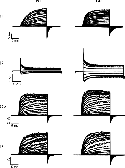
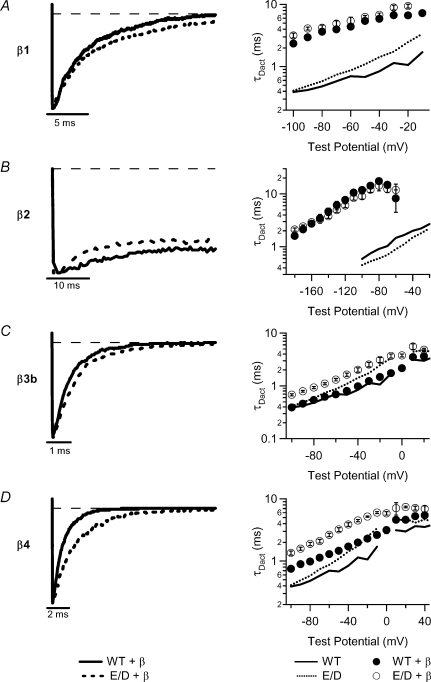
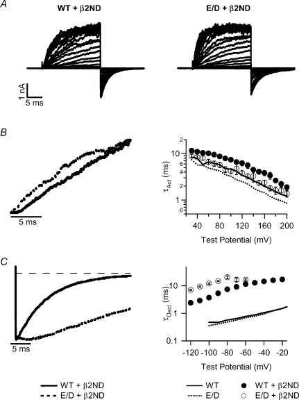
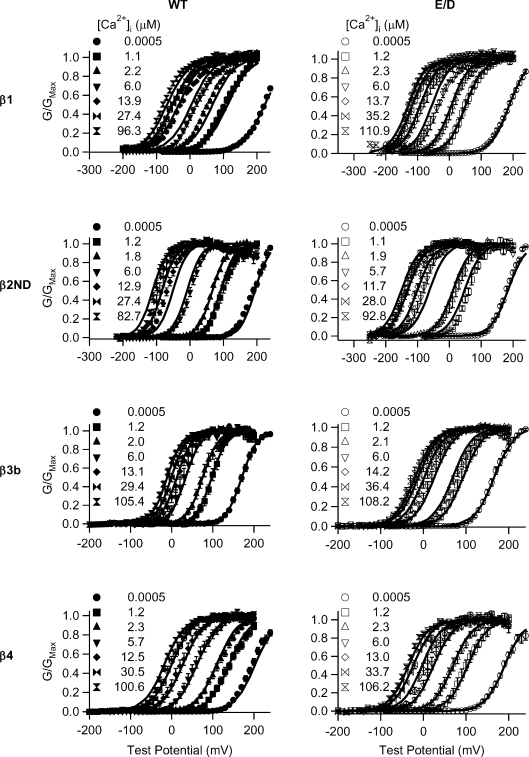
Large conductance Ca(2+)-activated K(+) (BK) channels modulate many physiological processes including neuronal excitability, synaptic transmission and regulation of myogenic tone. A gain-of-function (E/D) mutation in the pore-forming alpha subunit (Slo1) of the BK channel was recently identified and is linked to human neurological diseases of coexistent generalized epilepsy and paroxysmal dyskinesia. Here we performed macroscopic current recordings to examine the effects of the E/D mutation on the gating kinetics, and voltage and Ca(2+) dependence of the BK channel activation in the presence of four different beta subunits (beta1-4). These beta subunits are expressed in a tissue-specific pattern and modulate BK channel function differently, providing diversity and specificity for BK channels in various physiological processes. Our results show that in human (h) Slo1-only channels, the E/D mutation increased the rate of opening and decreased the rate of closing, allowing a greater number of channels to open at more negative potentials both in the presence and absence of Ca(2+) due to increased Ca(2+) affinity and enhanced activation compared with the wild-type channels. Even in the presence of beta subunits, the E/D mutation exhibited these changes with the exception of beta3b, where Ca(2+) sensitivity changed little. However, quantitative examination of these changes shows the diversity of each beta subunit and the differential modulation of these subunits by the E/D mutation. For example, in the presence of the beta1 subunit the E/D mutation increased Ca(2+) sensitivity less but enhanced channel activation in the absence of Ca(2+) more than in hSlo1-only channels, while in the presence of the beta2 subunit the E/D mutation also altered inactivation properties. These findings suggest that depending on the distribution of the various beta subunits in the brain, the E/D mutation can modulate BK channels differently to contribute to the pathophysiology of epilepsy and dyskinesia. Additionally, these results also have implications on physiological processes in tissues other than the brain where BK channels play an important role.


Figures

Figure 1
Figure 1. Functional differences between the WT and E/D mutant hSlo1 channels
A, macroscopic current traces of WT and E/D mutation in ∼2 μm[Ca2+]i. Voltage ranges from −80 to +200 mV with 10 mV increments and the repolarizing potential at −50 mV. B, activation kinetics of WT and E/D mutation in ∼2 μm[Ca2+]i. τAct in this and other figures was obtained by fitting current traces with a single exponential function. C, deactivation kinetics of WT and E/D mutation in ∼2 μm[Ca2+]i. In this and other figures, tail currents at various voltages after 20 ms prepulse were fitted with a single exponential function to obtain τDact. Prepulse potential =+200 mV. D, mean G–V relationship of WT and E/D mutant in ∼0, 2 and 100 μm[Ca2+]i fitted with Boltzmann equation. E, V1/2vs.[Ca2+]i plot of WT and E/D mutant hSlo1. F, mean G–V relationship of WT and E/D mutant in different [Ca2+]i are fitted using the MWC model. The specific [Ca2+]i for each symbol is shown. In this and other figures, error bars represent s.e.m.
Figure 2
Figure 2. Macroscopic currents of WT and E/D mutant hSlo1 with β subunits
All measurements were made in ∼2 μm[Ca2+]i except for those with the β2 subunit, which were in ∼10 μm[Ca2+]i. The voltage protocols were: for hSlo1 +β1: −80 to +200 mV with 10 mV increments and the repolarization potential at −120 mV; for hSlo1 +β2: −100 to +140 mV with 20 mV increments and the repolarization potential at −80 mV; for hSlo1 +β3b: −80 to +200 mV with 10 mV increments and the repolarization potential at −50 mV; and for hSlo1 +β4: −80 to +200 mV with 10 mV increments and the repolarization potential at −80 mV. In this and other figures, hSlo1 splice variant U11058 was coexpressed with the β2/β2ND subunits.
Figure 3
Figure 3. Activation kinetics of WT and E/D mutant hSlo1 with β subunits
All measurements were made in ∼2 μm[Ca2+]i except for those with the β2 subunit, which were in ∼10 μm[Ca2+]i. Left column: overlay of normalized current traces for hSlo1 +β1 at +40 mV (A), for hSlo1 +β2 at +40 mV (B), for hSlo1 +β3b at +80 mV (C), and for hSlo1 +β4 at +100 mV (D). Right column: τActV relations. The descriptions of the symbols are shown at the bottom.
Figure 4
Figure 4. Deactivation kinetics of WT and E/D mutant hSlo1 with β subunits in ∼2 μm[Ca2+]i
Left column: overlay of normalized current traces at −70 mV. Prepulse potential =+200 mV for hSlo1 +β1 (A), β3b (C), and β4 (D). Prepulse potential =+140 mV for hSlo1 +β2 (B). Right column: τDactV relations. The descriptions of the symbols are shown at the bottom.
Figure 5
Figure 5. Activation gating of WT and E/D mutant hSlo1 with the β2ND subunit in ∼2 μm[Ca2+]i
A, macroscopic currents: voltage ranges from −150 to +200 mV for WT +β2ND and −200 to +200 mV for E/D +β2ND with the repolarization potential at −120 mV. B, activation and C, deactivation kinetics: left panels, overlay of normalized current traces at +60 mV (B) and −70 mV (C); right panels, τActV (B) and τDactV (C) relations. The descriptions of the symbols are shown at the bottom.
Figure 6
Figure 6. V1/2/Z vs.[Ca2+]i of WT and E/D mutant hSlo1 with and without β subunits
Left column shows V1/2vs.[Ca2+]i plots and right column shows Z vs.[Ca2+]i plots for β1, β2ND, β3b and β4 subunits. The descriptions of symbols are shown at the bottom.
Figure 7
Figure 7. MWC model fits of WT and E/D mutant hSlo1 with β subunits in different [Ca2+]i values
Mean G–V relationships for WT and E/D mutant are fitted using the MWC model. The specific [Ca2+]i for each symbol is shown.
Figure 8
Figure 8. Inactivation properties of WT and E/D mutant hSlo1 with β2 in ∼10 μm[Ca2+]i
A, inactivation kinetics. Left: voltage protocol (upper) and macroscopic currents (lower two). Right: voltage dependence of the inactivation time. τInact was measured by fitting the inactivating current traces with a single exponential function from the peak amplitude to steady-state value. B, inactivation is not complete. Left: normalized current traces at +40 mV. Right: the fraction of non-inactivating current as the ratio of steady-state current (ISS) to peak current (IP) at different voltages. C, steady-state inactivation. Left: voltage protocol (upper) and macroscopic currents (lower two). Right: inactivation was measured as the ratio of the peak current at the +140 mV test pulse with different prepulses (I) versus the peak current at the +140 mV test pulse with the prepulse of −210 mV (I0). The plot was fitted using a sigmoidal function: formula image where base is the minimum value of I/I0, a is the I/I0 (max) −I/I0 (min), V1/2 is the half I/I0 voltage and rate is slope of the curve.
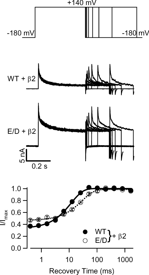
Figure 9
Figure 9. Recovery from inactivation in WT and E/D mutant hSlo1 with the β2 subunit at ∼10 μm[Ca2+]i
Top three panels show the paired-pulse recovery protocol (upper) and current traces (middle two). Channels inactivated during the +140 mV prepulse (400 ms), were allowed to recover from inactivation during various intervals (0.4, 0.8, 1.6, 3.2, 6.4, 12.8, 25.6, 51.2, 102.4 and 204.8 ms) at −180 mV, and then were activated again by a test pulse to +140 mV (200 ms). Bottom graph plots the ratio of the peak current at each of the test pulses (I) to the peak current at the test pulse following the longest interval between the prepulse and the test pulse (Imax). The continuous and dashed lines are fits using a single exponential function with the time constants of 11.7 ms for WT +β2 and 28.8 ms for E/D +β2.

Similar articles

Cited by

References

    1. Adelman JP, Shen KZ, Kavanaugh MP, Warren RA, Wu YN, Lagrutta A, Bond CT, North RA. Calcium-activated potassium channels expressed from cloned complementary DNAs. Neuron. 1992;9:209–216. - PubMed
    1. Atkinson NS, Robertson GA, Ganetzky B. A component of calcium-activated potassium channels encoded by the Drosophila slo locus. Science. 1991;253:551–555. - PubMed
    1. Bao L, Cox DH. Gating and ionic currents reveal how the BKCa channel's Ca2+ sensitivity is enhanced by its β1 subunit. J Gen Physiol. 2005;126:393–412. - PMC - PubMed
    1. Behrens R, Nolting A, Reimann F, Schwarz M, Waldschutz R, Pongs O. hKCNMB3 and hKCNMB4, cloning and characterization of two members of the large-conductance calcium-activated potassium channel β subunit family. FEBS Lett. 2000;474:99–106. - PubMed
    1. Benzinger GR, Xia XM, Lingle CJ. Direct observation of a preinactivated, open state in BK channels with β2 subunits. J Gen Physiol. 2006;127:119–131. - PMC - PubMed

Publication types

MeSH terms

Substances