Stimulation of orexin/hypocretin neurones by thyrotropin-releasing hormone
- PMID: 19204048
- PMCID: PMC2674990
- DOI: 10.1113/jphysiol.2008.167940
Stimulation of orexin/hypocretin neurones by thyrotropin-releasing hormone
Abstract
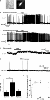
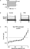
Central orexin/hypocretin neurones are critical for sustaining consciousness: their firing stimulates wakefulness and their destruction causes narcolepsy. We explored whether the activity of orexin cells is modulated by thyrotropin-releasing hormone (TRH), an endogenous stimulant of wakefulness and locomotor activity whose mechanism of action is not fully understood. Living orexin neurones were identified by targeted expression of green fluorescent protein (GFP) in acute brain slices of transgenic mice. Using whole-cell patch-clamp recordings, we found that TRH robustly increased the action potential firing rate of these neurones. TRH-induced excitation persisted under conditions of synaptic isolation, and involved a Na(+)-dependent depolarization and activation of a mixed cation current in the orexin cell membrane. By double-label immunohistochemistry, we found close appositions between TRH-immunoreactive nerve terminals and orexin-A-immunoreactive cell bodies. These results identify a new physiological modulator of orexin cell firing, and suggest that orexin cell excitation may contribute to the arousal-enhancing actions of TRH.
Figures


Similar articles
-
Thyrotropin-releasing hormone increases behavioral arousal through modulation of hypocretin/orexin neurons.J Neurosci. 2009 Mar 25;29(12):3705-14. doi: 10.1523/JNEUROSCI.0431-09.2009. J Neurosci. 2009. PMID: 19321767 Free PMC article.
-
Experience-dependent plasticity in hypocretin/orexin neurones: re-setting arousal threshold.Acta Physiol (Oxf). 2010 Mar;198(3):251-62. doi: 10.1111/j.1748-1716.2009.02047.x. Epub 2009 Sep 24. Acta Physiol (Oxf). 2010. PMID: 19785627 Free PMC article. Review.
-
Direct excitation of hypocretin/orexin cells by extracellular ATP at P2X receptors.J Neurophysiol. 2005 Sep;94(3):2195-206. doi: 10.1152/jn.00035.2005. Epub 2005 Jun 15. J Neurophysiol. 2005. PMID: 15958604
-
Acute optogenetic silencing of orexin/hypocretin neurons induces slow-wave sleep in mice.J Neurosci. 2011 Jul 20;31(29):10529-39. doi: 10.1523/JNEUROSCI.0784-11.2011. J Neurosci. 2011. PMID: 21775598 Free PMC article.
-
Activation of the basal forebrain by the orexin/hypocretin neurones.Acta Physiol (Oxf). 2010 Mar;198(3):223-35. doi: 10.1111/j.1748-1716.2009.02036.x. Epub 2009 Sep 1. Acta Physiol (Oxf). 2010. PMID: 19723027 Free PMC article. Review.
Cited by
-
Hypothyroidism compromises hypothalamic leptin signaling in mice.Mol Endocrinol. 2013 Apr;27(4):586-97. doi: 10.1210/me.2012-1311. Epub 2013 Mar 21. Mol Endocrinol. 2013. PMID: 23518925 Free PMC article.
-
The Thyrotropin-Releasing Hormone-Degrading Ectoenzyme, a Therapeutic Target?Front Pharmacol. 2020 May 8;11:640. doi: 10.3389/fphar.2020.00640. eCollection 2020. Front Pharmacol. 2020. PMID: 32457627 Free PMC article. Review.
-
Thyrotropin-releasing hormone increases behavioral arousal through modulation of hypocretin/orexin neurons.J Neurosci. 2009 Mar 25;29(12):3705-14. doi: 10.1523/JNEUROSCI.0431-09.2009. J Neurosci. 2009. PMID: 19321767 Free PMC article.
-
Regulation of the hypothalamic thyrotropin releasing hormone (TRH) neuron by neuronal and peripheral inputs.Front Neuroendocrinol. 2010 Apr;31(2):134-56. doi: 10.1016/j.yfrne.2010.01.001. Epub 2010 Jan 13. Front Neuroendocrinol. 2010. PMID: 20074584 Free PMC article. Review.
-
Experience-dependent plasticity in hypocretin/orexin neurones: re-setting arousal threshold.Acta Physiol (Oxf). 2010 Mar;198(3):251-62. doi: 10.1111/j.1748-1716.2009.02047.x. Epub 2009 Sep 24. Acta Physiol (Oxf). 2010. PMID: 19785627 Free PMC article. Review.
References
-
- Agnati LF, Bjelke B, Fuxe K. Volume versus wiring transmission in the brain: a new theoretical frame for neuropsychopharmacology. Med Res Rev. 1995;15:33–45. - PubMed
-
- Broberger C, Visser TJ, Kuhar MJ, Hökfelt T. Neuropeptide Y innervation and neuropeptide-Y-Y1-receptor-expressing neurones in the paraventricular hypothalamic nucleus of the mouse. Neuroendocrinology. 1999;70:295–305. - PubMed
Publication types
MeSH terms
Substances
Grants and funding
LinkOut - more resources
Full Text Sources

