. 2009 Apr;77(4):1324-36.
doi: 10.1128/IAI.01318-08. Epub 2009 Feb 9.

Francisella tularensis genes required for inhibition of the neutrophil respiratory burst and intramacrophage growth identified by random transposon mutagenesis of strain LVS

Affiliations

Francisella tularensis genes required for inhibition of the neutrophil respiratory burst and intramacrophage growth identified by random transposon mutagenesis of strain LVS

Grant S Schulert et al. Infect Immun. 2009 Apr.

Abstract

Francisella tularensis is a facultative intracellular pathogen and the causative agent of tularemia. We have shown that F. tularensis subspecies holarctica strain LVS prevents NADPH oxidase assembly and activation in human neutrophils, but how this is achieved is unclear. Herein, we used random transposon mutagenesis to identify LVS genes that affect neutrophil activation. Our initial screen identified carA, carB, and pyrB, which encode the small and large subunits of carbamoylphosphate synthase and aspartate carbamoyl transferase, respectively. These strains are uracil auxotrophs, and their growth was attenuated on cysteine heart agar augmented with sheep blood (CHAB) or in modified Mueller-Hinton broth. Phagocytosis of the uracil auxotrophic mutants triggered a respiratory burst in neutrophils, and ingested bacteria were killed and fragmented in phagosomes that contained superoxide. Conversely, phagocytosis did not trigger a respiratory burst in blood monocytes or monocyte-derived macrophages (MDM), and phagosomes containing wild-type or mutant bacteria lacked NADPH oxidase subunits. Nevertheless, the viability of mutant bacteria declined in MDM, and ultrastructural analysis revealed that phagosome egress was significantly inhibited despite synthesis of the virulence factor IglC. Other aspects of infection, such as interleukin-1beta (IL-1beta) and IL-8 secretion, were unaffected. The cultivation of carA, carB, or pyrB on uracil-supplemented CHAB was sufficient to prevent neutrophil activation and intramacrophage killing and supported escape from MDM phagosomes, but intracellular growth was not restored unless uracil was added to the tissue culture medium. Finally, all mutants tested grew normally in both HepG2 and J774A.1 cells. Collectively, our data demonstrate that uracil auxotrophy has cell type-specific effects on the fate of Francisella bacteria.


Figures

FIG. 1.
FIG. 1.
Identification of LVS mutants defective for inhibition of neutrophil NADPH oxidase activity. (A) ROS production by untreated PMN (UN) or cells infected with formalin-killed (FK) or live LVS (WT) or Tn5 mutant strain 1C9, 3E10, or 5H9 at an MOI of 50:1. Data indicate luminol CL in counts per second (cps) generated over 60 min at 37°C and are the means ± standard errors of the means of the results for triplicate samples from a representative experiment. (B) Southern blot of MfeI-digested genomic DNA probed to detect the kanamycin resistance cassette of the transposon, ahpA3. Lanes indicate molecular size standards (23.1, 9.4, 6.1, 4.3, 2.3, and 2.0 kbp; lane 1), LVS (lane 2), and mutants 3E10 (lane 3), 5H9 (lane 4), and 9H5 (lane 5). (C) Growth curves for LVS (WT), 3E10, and 5H9 in MHB or MHB supplemented with 200 μg/ml uracil (+Ura), 200 μg/ml arginine (+Arg) or both uracil and arginine (+/+). Data indicate the means ± standard errors of the means of the results for triplicate samples from one experiment that is representative of six. Where not visible, error bars are smaller than symbols. WT, wild type.
FIG. 2.
FIG. 2.
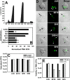
Uracil auxotrophs activate PMN, and intracellular survival is impaired. (A) PMN were left untreated (UN) or infected with wild-type LVS (WT) or mutant 3E10 (carB), 5H9 (carA), or 9H5 (single-iglC mutant). Where indicated, mutants 3E10 and 5H9 were grown on CHAB supplemented with uracil (3U and 5U, respectively) or uracil and arginine (3UR and 5UR, respectively) prior to use. Data indicate luminol CL in counts per second and are the averages ± standard errors of the means (SEM) of the results for triplicate samples from one experiment that is representative of four. (B) Same as described for panel A except that neutrophils were stimulated with 200 nM PMA 10 min after infection with the indicated strain of bacteria. “+U” indicates 3E10 and 5H9 that were grown on uracil-CHAB. Data are normalized to the signal obtained for neutrophils stimulated with 200 nM PMA after pretreatment with buffer alone and are the averages ± SEM from three determinations. *, P < 0.05 versus results for PMA control. (C) Viability of opsonized bacteria prior to infection of neutrophils. Opsonized bacteria were washed, diluted, and then plated on CHAB or CHAB supplemented with uracil (CHAB+U) for enumeration of CFU. Data from a representative experiment are shown. (D) PMN were infected with the indicated strain of bacteria (shown in green), and intraphagosomal superoxide was detected by NBT staining. Arrows and arrowheads indicate NBT-positive and NBT-negative phagosomes, respectively. Note that bacteria in NBT-positive phagosomes appear fragmented. (E) Viability of bacteria inside PMN was quantified 15 min and 2 h after uptake by measurement of CFU. Data are the averages ± SEM of the results for triplicate samples from one experiment that is representative of four.
FIG. 3.
FIG. 3.
Differential growth and survival of uracil auxotrophs in MDM, HepG2, and J774 cells. Bacteria were grown on CHAB (A to C) or CHAB plus uracil (D) prior to infection of host cells. (A) Intracellular growth and survival of unopsonized LVS, 5H9 (carA), and 3E10 (carB) in HepG2 cells over 48 h at 37°C. (B) Similar to experiment described for panel A except that J774 cells were infected with unopsonized LVS, 5H9, 3E10, or 9H5 (single-iglC mutant). (C, D) Extent of opsonized LVS, 5H9, 3E10, and 9H5 growth and survival in MDM over 48 h at 37°C. In panel C, “+UM” indicates the addition of uracil to RPMI at the time of infection with 5H9. In the experiment whose results are shown in panel D, all bacteria were cultivated on uracil-CHAB prior to infection of MDM in normal medium. For panels A to D, data indicate the means ± standard errors of the means of the results for triplicate samples from one experiment that is representative of three. Where not visible, error bars are smaller than symbols.
FIG. 4.
FIG. 4.
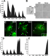
Neither wild-type nor mutant bacteria trigger a respiratory burst in primary human monocytes and MDM. (A) Superoxide production by resting MDM (Un) or cells stimulated with 200 nM PMA, zymosan (Zymo., MOI of 5:1), LVS, the carB mutant, or formalin-killed LVS (FK-LVS) (each at an MOI of 50:1) was quantified at 30-s intervals over 60 min at 37°C as lucigenin CL. Data indicate the means of the results for triplicate samples from one experiment that is representative of four. (B) NBT staining of MDM demonstrates accumulation of superoxide inside zymosan phagosomes (Zym, black arrows) but not compartments containing LVS or the carB mutant (white arrowheads). Data shown are representative of three independent experiments. (C) Confocal sections of MDM stained to show gp91phox/p22phox heterodimers in green and LVS in red. Left panel, distribution of gp91phox/p22phox in uninfected MDM. Arrows indicate the plasma membrane, and the arrowhead indicates the biosynthetic-secretion pathway. Middle panel, MDM infected with zymosan for 15 min. Arrows indicate phagosomes. Right panel, MDM infected with LVS (red) for 15 min. Arrows indicate LVS phagosomes. (D) Superoxide production by resting monocytes (Un) or cells stimulated with zymosan (Zymo.; MOI, 5:1), LVS, the carA or carB mutant, or the single-iglC mutant (each at an MOI of 50:1) was quantified over 60 min at 37°C as lucigenin CL. Data indicate the means of the results for triplicate samples from one experiment that is representative of four. (E) Same as described for panel D except that monocyte ROS were detected by using luminol. Un, untreated.
FIG. 5.
FIG. 5.
Quantitation of phagosome escape and IglC. (A) MDM were infected with LVS, the carA mutant, or the carA mutant that had been grown on uracil-CHAB (carA+U). After 9 h at 37°C, samples were fixed and processed for electron microscopy. Black arrows and white arrows indicate intraphagosomal and cytosolic bacteria, respectively. (B) Pooled data indicate the extent of bacterial escape from phagosomes in MDM. Error bars indicate means ± standard errors of the means. *, P < 0.05 versus results for LVS; **, P < 0.01 versus results for LVS and carA+U. (C) Bacteria were grown on CHAB plates (control) or CHAB supplemented with uracil (+ uracil) at 37°C. Immunoblots show IglC and the GroEL loading control in normalized bacterial lysates. Lanes 1 to 4 show LVS, 3E10 (carB), 5H9 (carA), and 9H5 (single-iglC mutant), respectively. Data shown are representative of two independent determinations.
FIG. 6.
FIG. 6.
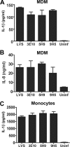
Both wild-type and mutant bacteria trigger secretion of IL-1β and IL-8 from mononuclear phagocytes. MDM (A and B) and monocytes (C) were infected with LVS, the carA mutant (5H9), the carB mutant (3E10), or the single-iglC mutant (9H5) as described in Materials and Methods, and at 24 hpi, the amount of mature IL-1β (A and C) or IL-8 (B) present in the tissue culture medium was quantified by ELISA. In each case, error bars indicate the means ± standard errors of the means of the results for triplicate samples from one experiment that is representative of three. Uninf, uninfected.

Similar articles

Cited by

References

    1. Allen, L.-A. H., and A. Aderem. 1995. Protein kinase C regulates MARCKS cycling between the plasma membrane and lysosomes in fibroblasts. EMBO J. 141109-1121. - PMC - PubMed
    1. Allen, L.-A. H., B. R. Beecher, J. T. Lynch, O. V. Rohner, and L. M. Wittine. 2005. Helicobacter pylori disrupts NADPH oxidase targeting in human neutrophils to induce extracellular superoxide release. J. Immunol. 1743658-3667. - PubMed
    1. Allen, L.-A. H., and R. L. McCaffrey. 2007. To activate or not to activate: distinct strategies used by Helicobacter pylori and Francisella tularensis to modulate the NADPH oxidase and survive in human neutrophils. Immunol. Rev. 219103-117. - PubMed
    1. Allen, L.-A. H., L. S. Schlesinger, and B. Kang. 2000. Virulent strains of Helicobacter pylori demonstrate delayed phagocytosis and stimulate homotypic phagosome fusion in macrophages. J. Exp. Med. 191115-127. - PMC - PubMed
    1. Astarie-Dequeker, C., E. N. N′Diaye, V. Le Cabec, M. G. Rittig, J. Prandi, and I. Maridonneau-Parini. 1999. The mannose receptor mediates uptake of pathogenic and nonpathogenic mycobacteria and bypasses bactericidal responses in human macrophages. Infect. Immun. 67469-477. - PMC - PubMed

Publication types

MeSH terms

LinkOut - more resources