. 2009 Apr;77(4):1689-99.
doi: 10.1128/IAI.01331-08. Epub 2009 Feb 9.

Polymorphisms in erythrocyte binding antigens 140 and 181 affect function and binding but not receptor specificity in Plasmodium falciparum

Affiliations

Polymorphisms in erythrocyte binding antigens 140 and 181 affect function and binding but not receptor specificity in Plasmodium falciparum

Alexander G Maier et al. Infect Immun. 2009 Apr.

Abstract

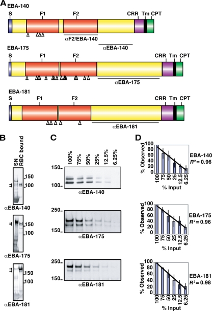
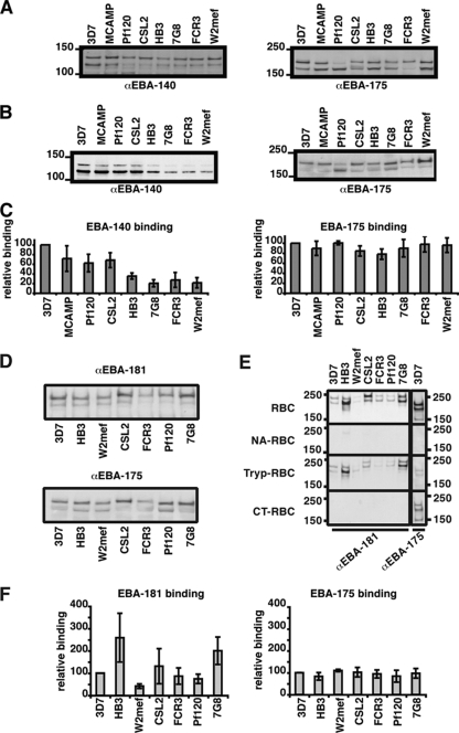
Invasion of human erythrocytes by the malaria parasite Plasmodium falciparum utilizes multiple ligand-receptor interactions involving erythrocyte receptors and parasite erythrocyte binding proteins of the Duffy binding-like family. Erythrocyte binding antigen 175 (EBA-175) binds to glycophorin A, the most abundant protein on the human erythrocyte surface and EBA-140 (also known as BAEBL) binds to glycophorin C, while the receptor for EBA-181 (also known as JESEBL) remains unknown. EBA binding is mediated via region II, a highly structured extracellular domain that shows a degree of sequence variability between different laboratory strains/isolates. Here, we determined the influence of region II polymorphisms on host cell receptor binding and overall function during invasion of EBA-140, EBA-175, and EBA-181. Polymorphisms in the binding domains of EBA-140 and EBA-181 have been suggested previously to alter their respective receptor specificities. In our hands, these polymorphisms affected the levels of EBA-140 and EBA-181 binding to receptors but, critically, not the receptor specificities of these proteins. The degree of EBA-140 binding to glycophorin C correlates with the level of function for this ligand-receptor interaction in merozoite invasion. In contrast, EBA-175, which is highly polymorphic in region II, shows no variability in its ability to bind to its receptor, glycophorin A. Combined, these data highlight the importance of sequence variability in EBAs as driven by immune selection but not by receptor specificity.


Figures

FIG. 1.
FIG. 1.
Schematic representation of the domain structures and quantitative erythrocyte binding assays for EBA-140, EBA-175, and EBA-181. (A) The signal sequence (S), F1 and F2 domains, C-terminal cysteine-rich region (CRR), transmembrane domain (Tm), and cytoplasmic tail (CPT) are shown. The regions against which antibodies were raised are indicated as αF2/EBA-140 (27), αEBA-140 (27), αEBA-175, and αEBA-181. Open triangles indicate the positions of strain-specific mutations within the F1/F2 domain. (B) Supernatant obtained from a culture of ruptured schizonts (SN) was incubated with erythrocytes, unbound supernatant was removed, the cells were washed, and EBAs bound to the erythrocytes were eluted with high-salt medium (RBC bound). Supernatant and eluted EBAs were subjected to Western blotting and probed with antibodies against EBA-140, EBA-175, and EBA-181 to compare binding patterns. Molecular mass markers in kDa are indicated. The arrows for each gel show the two specific bands for each ligand used for quantitation of erythrocyte binding. (C) Titration of different amounts of culture supernatant used in erythrocyte binding assays. Culture supernatant was diluted with culture media before an erythrocyte binding assay was performed on erythrocytes. Obtained eluants were subjected to Western blotting and probed with antibodies against different EBAs. Percentages indicate the amounts of culture supernatant used. The two bands shown in panel C (arrows in panel B) were used throughout this study for quantitation in densitometry, as these were the major bands observed. The ratio of these two bands can vary in different supernatant preparations; however, the sum totals of binding remain the same. This occurs due to different amounts of processing of the larger band that yields the smaller protein band. Molecular mass markers (in kDa) are indicated. α, anti. (D) Densitometric analysis of three titration experiments. Error bars indicate standard deviations. The diagonal lines indicate linear regressions with an R2 coefficient of determination of >0.95.
FIG. 2.
FIG. 2.
EBA-140 and EBA-181 from different strains bind with different affinities to whole erythrocytes. Molecular mass markers (in kDa) are indicated to the left of the gels. (A) For loading controls, supernatants used for the erythrocyte binding assays were probed for the presence of EBA-140 and EBA-175. Differences in EBA-140 contents were adjusted by adding culture medium. The same supernatants were used for EBA-140 and EBA-175 erythrocyte binding assays. α, anti. (B) The amount of EBA-140 present in eluants from erythrocyte binding assays performed with strain-specific culture supernatant was determined via Western blotting, and this was probed with anti-EBA-140 antibodies (left panel). In this panel, the supernatants are ordered by descending ability of EBA-140 of each strain to bind to red blood cells. This order was then used for the panels presented for EBA-181 binding. The same filer was used for the blot shown for anti-EBA-175 (αEBA-175), but the tracks have been reordered digitally so that they can be easily compared with the corresponding blots for αEBA-140 and αEBA-181. For comparison, the same samples were probed for the presence of EBA-175 (right panel). (C) Densitometric analysis of erythrocyte binding assays. The levels of binding of EBA-140 (left) and EBA-175 (right) were quantified by densitometry and expressed as percentages relative to the levels of binding of 3D7 EBA-140 and EBA-175, respectively (which were set to 100%). Error bars indicate standard deviations for three experiments. (D) For loading controls, supernatants used for the erythrocyte binding assays were probed for the presence of EBA-181 and EBA-175. Differences in EBA-181 contents were adjusted by adding culture medium. The same supernatants were used for EBA-181 and EBA-175 erythrocyte binding assays. The apparent differences in mobility of EBA-181 were probably due to different amounts of EBA-181 eluted from the red blood cells in some. These experiments were done at least three times using different supernatant preparations, and the EBA-181 bands have the same molecular mass in independent experiments. (E) Binding of EBA-181 from culture supernatant of different strains to normal red blood cells (RBC) and red blood cells which were treated with neuraminidase (NA-RBC), trypsin (Tryp-RBC), and chymotrypsin (CT-RBC). For a comparison, the binding of 3D7 EBA-175 to these red blood cells is shown. (F) Quantification of EBA-181 and EBA-175 binding from different strains. Western blots detecting the binding of the EBAs were densitometrically analyzed, and binding was expressed as a percentage relative to binding of 3D7 EBA-181 (left) or 3D7 EBA-175 (right). Error bars show standard deviations for three experiments.
FIG. 3.
FIG. 3.
Binding of EBA-140 to enzyme-treated and mutant erythrocyte (erythroc.) membranes. (Left panel) Binding of EBA-140 to intact erythrocytes determined by using erythrocyte binding assays. The erythrocytes were left untreated (nRBC) or treated with neuraminidase (NA-RBC), trypsin (Try-RBC), or chymotrypsin (CT-RBC) and incubated with supernatants from different parasite strains (3D7, MCAMP, Pf120, HB3, and W2mef). Parasite proteins that bound to the intact erythrocytes were eluted off with a salt solution, separated on a SDS-PAGE gel, and analyzed via Western blotting using anti-EBA-140 (αEBA-140) antibodies. The strain-specific variations in the F1 domain of EBA-140 are indicated underneath the strain name (Table 1). (Middle panel) Binding of EBA-140 from these culture supernatants to ghost erythrocyte membranes derived from NA-, Try-, or CT-treated or normal red blood cells by using overlay assays. (Right panel) Binding of EBA-140 from 3D7, MCAMP, Pf120, HB3, and W2mef culture supernatant to ghost erythrocyte membranes from normal, Yus (Ge −2, 3, 4), Gerbich (Ge −2, −3, 4), and Leach (Ge −2, −3, −4) homozygote cells and Yus/Gerbich heterozygote erythrocytes by using overlay assays. Binding of EBA-175 to the same solubilized membranes and detection of GYP A/B with anti-GYP A/B (αGYP A/B) as a positive control are shown at the bottom of the figure.
FIG. 4.
FIG. 4.
Invasion pathways in EBA-deficient cell lines. (A) Binding of EBA-140 to GYP C from erythrocyte ghost membranes (RBC), from purified glycophorins (GYP), or from Gerbich erythrocyte ghost membranes (Ge) by using overlay assays. The erythrocyte membranes or purified glycophorins after SDS-PAGE and transfer to nitrocellulose membrane were incubated with culture supernatant, and bound EBA-140 was detected with anti-EBA-140 (αEBA-140) as indicated. The GYP C was detected in nitrocellulose membranes with anti-GYP C (αGYP C) antibodies as a control. Supernatants were derived from parental or EBA-140- and EBA-175-deficient cell lines from both the 3D7 and W2mef backgrounds. The parasite strains from which the culture supernatant was taken for incubation with the filter are indicated on top of each panel. PBST lanes are negative controls in which PBS-Tween 20 rather than culture supernatant was incubated with the nitrocellulose membrane. EBA-140 does not bind to Ge-negative red blood cells as has been shown previously (28). 3D7Δ140 and W2mefΔ140 lack expression of EBA-140 and therefore show no binding to GYP C. The far-right column corresponds to antibodies against GYP C after incubation with PBS-Tween 20 (PBST) as a control. trunc. C, truncated GYP C in Ge-negative red blood cells. (B) Binding of EBA-140 to GYP A from erythrocyte ghost membranes (RBC) or from purified glycophorins (GYP). The lanes are identical to those in panel A except that binding to GYP A is shown. Binding of EBA-140 to GYP A can be detected only when the supernatant used contained EBA-140, i.e., not with supernatant from 3D7Δ140 or W2mefΔ140. The binding of EBA-140 in W2mef and W2mefΔEBA-175 is barely visible, consistent with its decreased binding affinity. (C) Binding of EBA-175 to GYP A. The lanes are identical to those in panel A except that the nitrocellulose membranes were probed with anti-EBA-175 (αEBA-175) or anti-GYP A/B (αGYP A/B) antibodies and detect binding of EBA-175 to GYP A. A/A, GYP A homodimer; A/B, GYP A/B heterodimer.
FIG. 5.
FIG. 5.
Invasion of P. falciparum into human erythrocytes. Inhibition of merozoite invasion by an anti-EBA-140 F2 antibody (Fig. 1). Levels of invasion inhibition in the presence of nonspecific antibodies (immunoglobulin G [IgG]) from normal rabbit serum (Pre IgG) and in the presence of EBA-140 F2 antibodies were compared. Error bars indicate confidence levels (α = 0.1) of three independent experiments performed in triplicate. The anti-EBA-140 F2 antibodies have been described previously, and they have been proven to specifically inhibit the function of EBA-140 in these experiments (27).
FIG. 6.
FIG. 6.
Comparison of the structures of EBA-175, EBA-140, and EBA-181. The X-ray crystal structure of EBA-175 (yellow) and homology models of EBA-140 (light blue) and EBA-181 (light green) are presented in ribbon form. Arrows point to the beta-hairpin loops on the F1 and F2 domains of EBA-175. The molecular surfaces of polymorphic residues are shown in red, while the molecular surface of the putative glycan contact residues on EBA-175 are shown in orange. Circled on the structures of EBA-140 and EBA-181 are the molecular surfaces (red) resulting from the polymorphisms listed in Table 1.

Similar articles

Cited by

References

    1. Adams, J. H., P. L. Blair, O. Kaneko, and D. S. Peterson. 2001. An expanding ebl family of Plasmodium falciparum. Trends Parasitol. 17297-299. - PubMed
    1. Adams, J. H., B. K. L. Sim, S. A. Dolan, X. Fang, D. C. Kaslow, and L. H. Miller. 1992. A family of erythrocyte binding proteins of malaria parasites. Proc. Natl. Acad. Sci. USA 897085-7089. - PMC - PubMed
    1. Barnwell, J. W., and M. R. Galinski. 1998. Invasion of vertebrate cells: erythrocytes, p. 93-120. In I. W. Sherman (ed.), Malaria: parasite biology, pathogenesis and protection. ASM Press, Washington DC.
    1. Baum, J., A. G. Maier, R. T. Good, K. M. Simpson, and A. F. Cowman. 2005. Invasion by P. falciparum merozoites suggests a hierarchy of molecular interactions. PLoS Pathog. 1e37. - PMC - PubMed
    1. Baum, J., M. Pinder, and D. J. Conway. 2003. Erythrocyte invasion phenotypes of Plasmodium falciparum in The Gambia. Infect. Immun. 711856-1863. - PMC - PubMed

Publication types

MeSH terms

Associated data

LinkOut - more resources